. 2016 Aug 10;8(8):237.
doi: 10.3390/toxins8080237.

Chloroquine Analog Interaction with C2- and Iota-Toxin in Vitro and in Living Cells

Affiliations

Chloroquine Analog Interaction with C2- and Iota-Toxin in Vitro and in Living Cells

Angelika Kronhardt et al. Toxins (Basel). .

Abstract

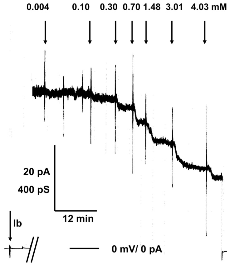
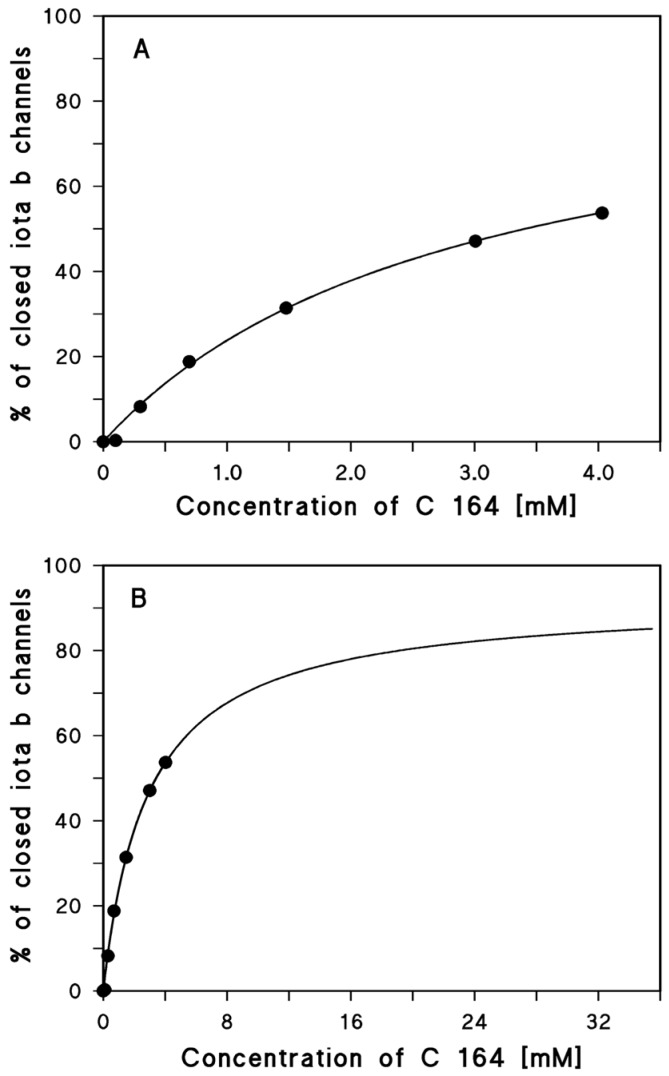
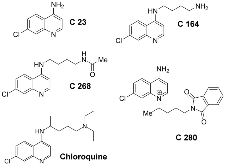
C2-toxin from Clostridium botulinum and Iota-toxin from Clostridium perfringens belong both to the binary A-B-type of toxins consisting of two separately secreted components, an enzymatic subunit A and a binding component B that facilitates the entry of the corresponding enzymatic subunit into the target cells. The enzymatic subunits are in both cases actin ADP-ribosyltransferases that modify R177 of globular actin finally leading to cell death. Following their binding to host cells' receptors and internalization, the two binding components form heptameric channels in endosomal membranes which mediate the translocation of the enzymatic components Iota a and C2I from endosomes into the cytosol of the target cells. The binding components form ion-permeable channels in artificial and biological membranes. Chloroquine and related 4-aminoquinolines were able to block channel formation in vitro and intoxication of living cells. In this study, we extended our previous work to the use of different chloroquine analogs and demonstrate that positively charged aminoquinolinium salts are able to block channels formed in lipid bilayer membranes by the binding components of C2- and Iota-toxin. Similarly, these molecules protect cultured mammalian cells from intoxication with C2- and Iota-toxin. The aminoquinolinium salts did presumably not interfere with actin ADP-ribosylation or receptor binding but blocked the pores formed by C2IIa and Iota b in living cells and in vitro. The blocking efficiency of pores formed by Iota b and C2IIa by the chloroquine analogs showed interesting differences indicating structural variations between the types of protein-conducting nanochannels formed by Iota b and C2IIa.

Keywords: C2-toxin; aminoquinolinium salts; binding components; black lipid bilayer; chloroquine; iota-toxin.


Figures

Figure 1
Figure 1
Titration experiment of Iota b-induced membrane conductance with C 164. The membrane was formed from diphytanoyl phosphatidylcholine/n-decane. The aqueous phase contained 20 ng/mL Iota b (Ib) protein (added to the cis-side of the membrane at the time of the left side arrow), 150 mM KCl, 10 mM MES, pH 6.0. The temperature was constantly 20 °C and the applied voltage was 50 mV. The two bars at the base line indicate the time interval of about 1h and 50 min between the addition of Ib and the start of the titration experiment. The membrane contained about 70 Iota b-channels (single channel conductance G = 15 pS) when C 164 was added at the indicated concentrations to the aqueous phase. The bottom line represents zero level of conductance (at begin of the experiment) or also zero level of current when voltage was switched off (at the end of titrations). Note that the high noise of the current recording during the titration experiment was caused by stirring in the membrane cell to allow rapid equilibration of C 164 in the aqueous phase.
Figure 2
Figure 2
(A) Langmuir isotherm of the inhibition of Iota b-induced membrane conductance (about 70 Iota b-channels) by the aminoquinolinium salt C 164. The fit line corresponds to the data points taken from the titration experiment in Figure 1. The fit of the data was performed using Equation (2). The stability constant, K, for binding of C 164 to the Iota b-channels was (348 ± 48) 1/M (The channel block was at maximum 92% ± 7%; KS = 2.9 mM (r2 = 0.997645)); (B) Because of the low degree of inhibition in C 164 concentration range, we extrapolated its concentration to about 33 mM and used the same fit parameters as in A. The fit curve indicates that high concentration of C 164 almost fully blocked the Iota b channels.
Figure 3
Figure 3
Structures of chloroquine and the chloroquine analogs (aminoquinolinium salts) used in this study). The chloroquine analogs were designated as suggested by Lödige (2013) [46].
Figure 4
Figure 4
Inhibitory effect of C 280 on the pH-dependent trans-membrane transport of the C2 and iota toxins in living cells. (A) Baf A1-treated Vero cells were incubated for 30 min at 4 °C with C2 toxin (100 ng/mL C2I + 200 ng/mL C2IIa) to enable toxin binding. Noteworthy, Baf A1 was present to inhibit in a later step the “normal” transport of the A components of the internalized toxins into the cytosol via acidified endosomes, which is a prerequisite to investigate the toxin transport across the cytoplasmic membrane in this approach. For control (con), cells were incubated without toxin. Subsequently, cells were exposed for 5 min at 37 °C to acidic medium (pH 4.5 or to neutral medium pH 7.5 for control) and subsequently incubated at 37 °C in neutral medium containing Baf A1. During the acidic pulse, the B components insert as pores into the cytoplasmic membrane and the A components translocate through these pores into the cytosol of the cells and induce ADP-ribosylation of actin and cell-rounding. In this approach, the toxin-induced cell-rounding serves as an established specific and sensitive endpoint to monitor the uptake of the A components into the cytosol in the presence and absence of the inhibitor. To test the effect of C 280 on toxin translocation, the indicated concentrations of C 280 were present in the medium during acidic pulse and the subsequent incubation periods. Pictures were taken after 1 and 2 h to document cell rounding, i.e., intoxication of cells (shown in A for 2 h and 1 mM C 280); (B) The percentage of intoxicated cells was determined after 1 h, and values are given as mean ± S.D. (n = 3). Significance was tested between cells, which have been treated with C2 toxin either in the absence or presence of C 280 by using the student’s t-test (*** p < 0.0005, ** p < 0.005); (C) To test the influence of C 280 on membrane translocation of iota toxin, Baf A1-treated Vero cells were exposed for 15 min at ph 4.0 to Iota toxin (100 ng/mL Ia + 200 ng/mL Ib) in the presence or absence of 1 mM C 280. Cells were incubated for further 2 h at 37 °C in neutral medium containing Baf A1 in the presence or absence of C 280. Pictures were taken and the percentage of round cells was determined (D) Values are given as mean ± S.D. (n = 3). Significance was tested between cells, which have been treated with iota toxin either in the absence or presence of C 280 by using the student’s t-test (*** p < 0.0005, ** p < 0.005).

References

    1. Barth H., Aktories K., Popoff M.R., Stiles B.G. Binary bacterial toxins: Biochemistry, biology, and applications of common Clostridium and Bacillus proteins. Microbiol. Mol. Biol. Rev. 2004;68:373–402. doi: 10.1128/MMBR.68.3.373-402.2004. - DOI - PMC - PubMed
    1. Aktories K., Barth H. New insights into the mode of action of the actin ADP-ribosylating virulence factors Salmonella enterica SpvB and Clostridium botulinum C2 toxin. Eur. J. Cell Biol. 2011;90:944–950. - PubMed
    1. Knapp O., Benz R., Popoff M.R. Pore-forming activity of clostridial binary toxins. Biochim. Biophys. Acta. 2016;1858:512–525. doi: 10.1016/j.bbamem.2015.08.006. - DOI - PubMed
    1. Friedlander A.M. Macrophages are sensitive to anthrax lethal toxin through an acid-dependent process. J. Biol. Chem. 1986;261:7123–7126. - PubMed
    1. Mock M., Fouet A. Anthrax. Annu. Rev. Microbiol. 2001;55:647–671. doi: 10.1146/annurev.micro.55.1.647. - DOI - PubMed

MeSH terms

LinkOut - more resources