. 2017 Jan;24(1):26-37.
doi: 10.1038/cdd.2016.78. Epub 2016 Aug 12.

Coordinated ubiquitination and phosphorylation of RIP1 regulates necroptotic cell death

Affiliations

Coordinated ubiquitination and phosphorylation of RIP1 regulates necroptotic cell death

M Cristina de Almagro et al. Cell Death Differ. 2017 Jan.

Abstract

Proper regulation of cell death signaling is crucial for the maintenance of homeostasis and prevention of disease. A caspase-independent regulated form of cell death called necroptosis is rapidly emerging as an important mediator of a number of human pathologies including inflammatory bowel disease and ischemia-reperfusion organ injury. Activation of necroptotic signaling through TNF signaling or organ injury leads to the activation of kinases receptor-interacting protein kinases 1 and 3 (RIP1 and RIP3) and culminates in inflammatory cell death. We found that, in addition to phosphorylation, necroptotic cell death is regulated by ubiquitination of RIP1 in the necrosome. Necroptotic RIP1 ubiquitination requires RIP1 kinase activity, but not necroptotic mediators RIP3 and MLKL (mixed lineage kinase-like). Using immunoaffinity enrichment and mass spectrometry, we profiled numerous ubiquitination events on RIP1 that are triggered during necroptotic signaling. Mutation of a necroptosis-related ubiquitination site on RIP1 reduced necroptotic cell death and RIP1 ubiquitination and phosphorylation, and disrupted the assembly of RIP1 and RIP3 in the necrosome, suggesting that necroptotic RIP1 ubiquitination is important for maintaining RIP1 kinase activity in the necrosome complex. We also observed RIP1 ubiquitination in injured kidneys consistent with a physiological role of RIP1 ubiquitination in ischemia-reperfusion disease. Taken together, these data reveal that coordinated and interdependent RIP1 phosphorylation and ubiquitination within the necroptotic complex regulate necroptotic signaling and cell death.


Conflict of interest statement

MCdA and MK are former and all other authors are current employees of Genentech. The authors declare no conflict of interest.

Figures
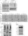
Figure 1
Figure 1
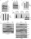
RIP1 is ubiquitinated during necroptotic signaling independently of RIP3 and MLKL. (a) HT29 cells were treated for the indicated periods of time with TNFα 20 ng/ml (T), BV6 2 μM (B) and zVAD 20 μM (Z). Cell lysates were immunoprecipitated with caspase-8 antibody. The pull-downs and lysates were analyzed by western blotting with the indicated antibodies. (b) HT29 cells were treated with TBZ as in (a) for the indicated times. Lysates were fractionated on a Superose 6 10/300 GL column and the resulting fractions as well as total cell lysates were separated by SDS-PAGE and probed with the indicated antibodies. Asterisks denote nonspecific bands. Fraction numbers are indicated below and molecular weight markers on top of western blots. (c) WT, MLKL KO or RIP3 KO MEFs were treated for 2 h with TNFα 100 ng/ml (T), BV6 2 μM (B) and zVAD 20 μM (Z) as indicated. Cells were lysed in 6 M urea buffer, immunoprecipitated using linear and K63 linkage-specific anti-ubiquitin antibodies and analyzed by western blotting with the indicated antibodies. (d) HT29 cells were treated for 4 h with TNFα 10 ng/ml (T), BV6 0.5 μM (B) and zVAD 20 μM (Z) in the absence or presence of GSK843 (10 μM). Cell lysates were analyzed by western blotting with the indicated antibodies. Asterisk denotes lower nonspecific band
Figure 2
Figure 2
RIP1 kinase activity regulates RIP1 ubiquitination. (a) HT29 cells were treated for 3 h with TNFα 10 ng/ml (T), BV6 0.5 μM (B), zVAD 20 μM (Z) and necrostatin compounds 30μM (N) as indicated. Cell lysates were analyzed by western blotting with the indicated antibodies. (b) Primary BMDMs were treated for 2 h with TNFα 100ng/ml (T), BV6 2 μM (B) and zVAD 20 μM (Z) as indicated. Cell lysates were analyzed by western blotting with the indicated antibodies. (c) Primary BMDMs were treated for 24 h with TNFα 100 ng/ml (T), BV6 2 μM (B), zVAD 20 μM (Z) and Nec-1 30 μM (N) as indicated. Cell viability was assessed by CellTiter-Glo (Promega). Data are mean±S.E.M. values of four experiments. (d) WT or RIP1 kinase-dead knock-in (RIP1 KD-KI) MEFs were treated for 24 h with TNFα 100 ng/ml (T), BV6 2 μM (B) and zVAD 20 μM (Z) as indicated. Cell viability was assessed by CellTiter-Glo. Data are mean±S.E.M. values of four experiments. (e) WT or RIP1 KD MEFs were treated for 2 h as in (d). Cells were lysed in 6 M urea buffer, immunoprecipitated using linear or K63 linkage-specific anti-ubiquitin antibodies and analyzed by western blotting with the indicated antibodies. (f) HT29 cells were treated for indicated time points with TNFα 20 ng/ml (T) alone or in combination with BV6 2 μM (TB), or with BV6 and zVAD 20 μM (TBZ). Cell lysates were immunoprecipitated with caspase-8 antibody. The pull-downs and lysates were analyzed by western blotting with the indicated antibodies. Asterisks denote lower nonspecific bands
Figure 3
Figure 3
Analyses of ubiquitination in HT29 cells in necroptotic signaling. (a) MS analysis of ubiquitin remnant K-GG peptides in HT29 cells treated with TNFα 20 ng/ml (T), BV6 2 μM (B) and zVAD 20 μM (Z) in the absence or presence of Nec-1 (N) as indicated. (b) Volcano plot of the log 2-based ratio of K-GG peptide abundance for proteins between TBZ 3 h and control conditions. Peptide level data were aggregated at the protein level and P-values were generated by linear mixed-effect (LiME) modeling. Selected proteins are highlighted in red. (c) Scatterplot and Pearson's correlation of K-GG abundance ratios for the effect of adding Nec-1 compared with removing zVAD in the TBZ condition showing that changes in K-GG peptide abundance are highly correlated with one another. Among all affected proteins, RIP1 and caspase-8 show substantial upregulation in ubiquitination level. (d) MS/MS spectrum showing identification of representative K-GG peptide demonstrating ubiquitination of RIP1 at Lys115. (e) LiME plot of K-GG peptides from RIP1 protein across eight conditions. Black lines correspond to AUC abundance measurements from confidently matched K-GG peptides. LiME model output at the protein level is shown in red. Log 2 ratios and P-values are reported for each treatment relative to the control. The ubiquitinated peptide corresponding to Lys115 is colored in blue. (f) RIP1 ubiquitination sites identified directly by PSMs in each condition. ND stands for none detected
Figure 4
Figure 4
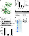
K115R RIP1 ubiquitination site mutant displays inherent kinase activity. (a) Crystal structure of RIP1 kinase domain (PDB-ID 4ITJ) illustrating the position of K115 (colored magenta) in the C-terminal domain. (b) The kinase activity of recombinant kinase domains of RIP1 WT and K115R proteins can be inhibited by necrostatin compounds as assessed by an HTRF assay. (c) Recombinant kinase domain RIP1 WT, KD and K115R proteins were incubated with Sypro orange and submitted to a temperature ramp to assess their thermal stability. Data are mean±S.E.M. values of three experiments. (d) The 293 T cells were transfected with the indicated constructs. After 24 h, cell lysates were analyzed by western blotting with the indicated antibodies. (e) Recombinant full-length RIP1 WT, D138N and K115R proteins were expressed in insect cells and purified via Flag resin (Coomassie stain, left side). WT and K115R RIP1 are phosphorylated upon purification. Equal amounts of examined full-length RIP1 proteins were investigated by western blotting with P-RIP1 antibody (right side). (f) Full-length WT and K115R RIP1 proteins display similar kinase activity. Kinase activity of equal amounts of examined full-length RIP1 proteins was measured by ADP-Glo assay
Figure 5
Figure 5
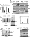
Necroptotic RIP1 ubiquitination on Lys115 regulates cell death signaling. (a) HT29 RIP1 KO cells reconstituted with the indicated constructs were treated for 24 h with TNFα 20 ng/ml (T), BV6 2 μM (B) and zVAD 20 μM (Z) as indicated. Cell viability was assessed by CellTiter-Glo. Data are mean±S.E.M. values of three experiments. (b) HT29 RIP1 KO (-/-) cells reconstituted with the indicated constructs were treated as in (a) for 0, 1, 2 or 4 h. Cell lysates were analyzed by western blotting with the indicated antibodies. (c) Reduced necroptotic ubiquitination in HT29 cells expressing K115R RIP1. WT or K115R RIP1 HT29 cells were treated with TBZ for 2 h as in (a). Cells were lysed in 6 M urea buffer, immunoprecipitated using linear or K63 linkage-specific anti-ubiquitin antibodies and analyzed by western blotting with the indicated antibodies. (d) K115 RIP1 ubiquitination is not critical for TNF-associated RIP1 ubiquitination. HT29 RIP1 KO cells reconstituted with vector, RIP1 WT or RIP1 K115R constructs were treated for 7 min with Flag-TNFα 20 ng/ml and cell lysates were immunoprecipitated with Flag-TNF. The pull-downs and cell lysates were analyzed by western blotting with the indicated antibodies. (e and f) K115 RIP1 ubiquitination is not critical for TNF-induced NF-κB and MAPK signaling in HT29 cells. (e) HT29 RIP1 KO cells reconstituted with vector, RIP1 WT or RIP1 K115R constructs were treated for indicated time points with TNFα 20 ng/ml for 4 h and IL-8 mRNA levels were examined by quantitative real-time PCR. (f) HT29 RIP1 KO cells reconstituted with vector, RIP1 WT or RIP1 K115R constructs were treated for indicated time points with TNFα 20 ng/ml and cell lysates were analyzed by western blotting with the indicated antibodies
Figure 6
Figure 6
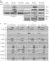
Necroptotic RIP1 ubiquitination on Lys115 regulates necrosome assembly. (a) HT29 RIP1 KO (-/-) cells reconstituted with vector, WT RIP1 or K115R RIP1 constructs were treated TNFα 20 ng/ml (T), BV6 2 μM (B) and zVAD 20 μM (Z) for indicated time points. Cell lysates were immunoprecipitated with caspase-8 antibody. The pull-downs and lysates were analyzed by western blotting with the indicated antibodies. (b) Gel filtration analysis of the necrosome. HT29 RIP1 KO cells reconstituted with vector, WT RIP1 or K115R RIP1 constructs were treated as in (a) for the indicated times. Lysates were fractionated on a Superose 6 10/300 GL column and the resulting fractions as well as total cell lysate were separated by SDS-PAGE and probed with the indicated antibodies. Asterisks denote nonspecific bands. Fraction numbers are indicated below and molecular weight markers on top of western blots

References

    1. Steller H. Mechanisms and genes of cellular suicide. Science 1995; 267: 1445–1449. - PubMed
    1. Linkermann A, Stockwell BR, Krautwald S, Anders HJ. Regulated cell death and inflammation: an auto-amplification loop causes organ failure. Nat Rev Immunol 2014; 14: 759–767. - PubMed
    1. Salvesen GS, Abrams JM. Caspase activation – stepping on the gas or releasing the brakes? Lessons from humans and flies. Oncogene 2004; 23: 2774–2784. - PubMed
    1. Linkermann A, Green DR. Necroptosis. N Engl J Med 2014; 370: 455–465. - PMC - PubMed
    1. Newton K. RIPK1 and RIPK3: critical regulators of inflammation and cell death. Trends Cell Biol 2015; 25: 347–353. - PubMed

MeSH terms