

Subfoveal choroidal thickness in patients with diabetic retinopathy and diabetic macular oedema
- PMID: 27518549
- PMCID: PMC5177755
- DOI: 10.1038/eye.2016.187
Subfoveal choroidal thickness in patients with diabetic retinopathy and diabetic macular oedema
Abstract
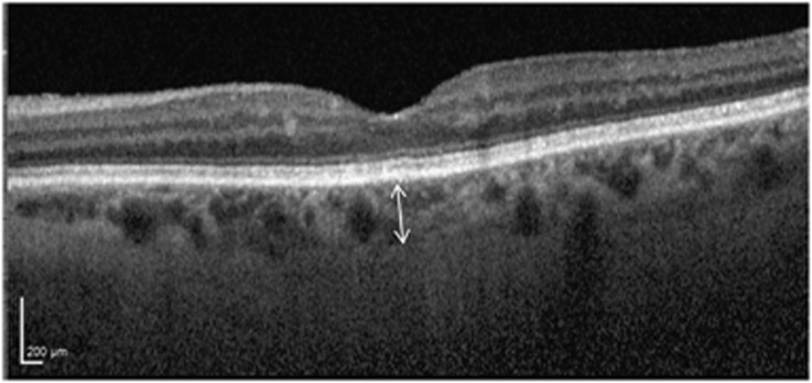
PurposeTo investigate the relationship between subfoveal choroidal thickness, severity of diabetic retinopathy (DR), and the presence of diabetic macular oedema (DMO) using enhanced depth imaging spectral domain optical coherence tomography (EDI-OCT) in patients with type 2 diabetes.MethodsA retrospective study of 145 eyes from untreated, type 2 diabetic patients who attended clinic at the Oxford Eye Hospital between January 2012 and February 2013, and underwent fundus photography and EDI-OCT imaging. Eyes were divided into two groups based on the presence or absence of foveal involving DMO and classified according to retinopathy grade: R1 (mild non-proliferative diabetic retinopathy (NPDR), R2 (moderate-severe NPDR), and R3 (proliferative diabetic retinopathy (PDR). Subfoveal choroidal thickness was measured on the EDI-OCT images and statistically analysed using Student's t-test.ResultsIn mild NPDR (n=87), the mean subfoveal choroidal thickness was 217.7 microns. In moderate-severe NPDR (n=37), the mean subfoveal choroidal thickness was 221.7 microns. In PDR (n=21), the mean subfoveal choroidal thickness was 242.1 microns. There was a statistically significant increase in choroidal thickness in PDR when compared with the mild NPDR group, P=0.027. DMO was associated with a non-statistically significant increase in choroidal thickness (225.4 microns) compared with eyes without DMO (209.3 microns), P=0.13.ConclusionSubfoveal choroidal thickness increased with the severity of diabetic retinopathy but showed no statistically significant association with the presence of DMO. This suggests that the choroidal layer is responsive to retinal vascular changes.
Conflict of interest statement
VC is a consultant of Allergan, Bayer, Novartis, and Quantel Medical and has received speaker fees from Heidelberg Engineering. The other authors declare no conflict of interest.
Figures
References
-
- Antonetti D, Klein R, Gardner T. Diabetic retinopathy. N Engl J Med 2012; 366(13): 1227–1239. - PubMed
-
- Hidayat A, Fine B. Diabetic choroidopathy: light and electron microscopic observations of seven cases. Ophthalmology 1985; 92(4): 512–522. - PubMed
-
- Cao J, McLeod D, Merges C, Lutty G. Choriocapillaris degeneration and related pathologic changes in human diabetic eyes. Arch Ophthalmol 1998; 116(5): 589–597. - PubMed
-
- Fukushima I, McLeod D, Lutty G. Intrachoroidal microvascular abnormality: a previously unrecognized form of choroidal neovascularization. Am J Ophthalmol 1997; 124(4): 473–487. - PubMed
MeSH terms
LinkOut - more resources
Full Text Sources
Other Literature Sources
Medical
