

Echocardiographic diagnosis of the different phenotypes of hypertrophic cardiomyopathy
- PMID: 27519172
- PMCID: PMC4982201
- DOI: 10.1186/s12947-016-0072-5
Echocardiographic diagnosis of the different phenotypes of hypertrophic cardiomyopathy
Abstract
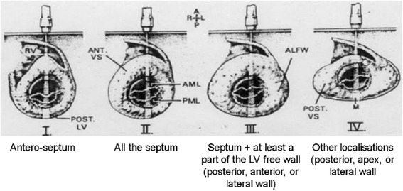
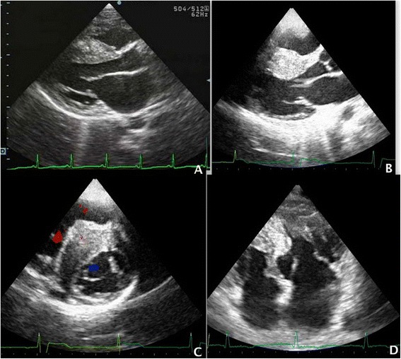
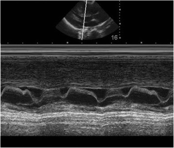
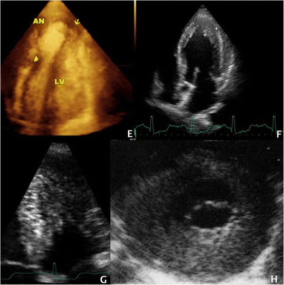
Hypertrophic Cardiomyopathy (HCM) is an inherited cardiovascular disorder of great genetic heterogeneity and has a prevalence of 0.1 - 0.2 % in the general population. Several hundred mutations in more than 27 genes, most of which encode sarcomeric structures, are associated with the HCM phenotype. Then, HCM is an extremely heterogeneous disease and several phenotypes have been described over the years. Originally only two phenotypes were considered, a more common, obstructive type (HOCM, 70 %) and a less common, non-obstructive type (HNCM, 30 %) (Maron BJ, et al. Am J Cardiol 48:418 -28, 1981). Wigle et al. (Circ 92:1680-92, 1995) considered three types of functional phenotypes: subaortic obstruction, midventricular obstruction and cavity obliteration. A leader american working group suggested that HCM should be defined genetically and not morphologically (Maron BJ, et al. Circ 113:1807-16, 2006). The European Society of Cardiology Working Group on Myocardial and Pericardial Diseases recommended otherwise a morphological classification (Elliott P, et al. Eur Heart J 29:270-6, 2008). Echocardiography is still the principal tool for the diagnosis, prognosis and clinical management of HCM. It is well known that the echocardiographic picture may have a clinical and prognostic impact. For this reason, in this article, we summarize the state of the art regarding the echocardiographic pattern of the HCM phenotypes and its impact on clinical course and prognosis.
Keywords: Cardiomiopathy; Echocardiography; Hypertrophy.
Figures
Similar articles
-
Identifying Obstructive Hypertrophic Cardiomyopathy from Nonobstructive Hypertrophic Cardiomyopathy: Development and Validation of a Model Based on Electrocardiogram Features.Glob Heart. 2023 Aug 4;18(1):40. doi: 10.5334/gh.1250. eCollection 2023. Glob Heart. 2023. PMID: 37547171 Free PMC article.
-
[Pulsed Doppler echocardiographic assessment of diastolic left ventricular hemodynamics in hypertrophic cardiomyopathy: relationship between the mode of left ventricular filling and the distribution of left ventricular hypertrophy].J Cardiogr. 1983 Sep;13(3):523-35. J Cardiogr. 1983. PMID: 6687193 Japanese.
-
Cardiac magnetic field map topology quantified by Kullback-Leibler entropy identifies patients with hypertrophic cardiomyopathy.Chaos. 2007 Mar;17(1):015118. doi: 10.1063/1.2432059. Chaos. 2007. PMID: 17411275
-
Refined echocardiographic assessment and contemporary medical treatment of obstructive hypertrophic cardiomyopathy.Cardiovasc Hematol Disord Drug Targets. 2007 Sep;7(3):174-87. doi: 10.2174/187152907781745224. Cardiovasc Hematol Disord Drug Targets. 2007. PMID: 17896958 Review.
-
The diagnosis and treatment of hypertrophic cardiomyopathy.Dtsch Arztebl Int. 2011 Apr;108(13):209-15. doi: 10.3238/arztebl.2011.0209. Epub 2011 Apr 1. Dtsch Arztebl Int. 2011. PMID: 21505608 Free PMC article. Review.
Cited by
-
Morphology of hypertrophied basal septum contributes to left ventricular outflow tract obstruction in patients with hypertrophic cardiomyopathy: a retrospective case-control study.Quant Imaging Med Surg. 2023 Jul 1;13(7):4117-4129. doi: 10.21037/qims-22-1034. Epub 2023 Apr 17. Quant Imaging Med Surg. 2023. PMID: 37456278 Free PMC article.
-
Ventricular diastolic dimension over maximal myocardial thickness is robust landmark of systolic impairment in patients with hypertrophic cardiomyopathy.Med Sci Monit. 2018 Mar 31;24:1880-1886. doi: 10.12659/msm.906111. Med Sci Monit. 2018. PMID: 29602944 Free PMC article.
-
Echocardiographic Diagnosis of Hypertrophic Cardiomyopathy by Machine Learning.Mayo Clin Proc Digit Health. 2024 Sep 3;2(4):564-573. doi: 10.1016/j.mcpdig.2024.08.009. eCollection 2024 Dec. Mayo Clin Proc Digit Health. 2024. PMID: 40206535 Free PMC article.
-
The role of echocardiography in management of hypertrophic cardiomyopathy.J Echocardiogr. 2020 Jun;18(2):77-85. doi: 10.1007/s12574-019-00454-9. Epub 2019 Dec 19. J Echocardiogr. 2020. PMID: 31858431 Free PMC article. Review.
-
Prediction of new-onset atrial fibrillation in patients with hypertrophic cardiomyopathy using machine learning.Eur J Heart Fail. 2025 Feb;27(2):275-284. doi: 10.1002/ejhf.3546. Epub 2024 Dec 18. Eur J Heart Fail. 2025. PMID: 39694602
References
-
- Elliott PM, Anastasakis A, Borger MA, et al. 2014 ESC Guidelines on diagnosis and management of hypertrophic cardiomyopathy The Task Force for the Diagnosis and Management of Hypertrophic Cardiomyopathy of the European Society of Cardiology (ESC) Eur Heart J. 2014;35(39):2733–2779. doi: 10.1093/eurheartj/ehu284. - DOI - PubMed
-
- Maron BJ, Towbin JA, Thiene G, et al. American Heart Association; Council on Clinical Cardiology, Heart Failure and Transplantation Committee; Quality of Care and Outcomes Research and Functional Genomics and Translational Biology Interdisciplinary Working Groups, Council on Epidemiology and Prevention. Circulation. 2006;113:1807–1816. doi: 10.1161/CIRCULATIONAHA.106.174287. - DOI - PubMed
-
- Rapezzi C, Arbustini E, Caforio AL, et al. Diagnostic work-up in cardiomyopathies: bridging the gap between clinical phenotypes and final diagnosis. A position statement from the ESC Working Group on Myocardial and Pericardial Diseases. Eur Heart J. 2013;34:1448–1458. doi: 10.1093/eurheartj/ehs397. - DOI - PubMed
Publication types
MeSH terms
LinkOut - more resources
Full Text Sources
Other Literature Sources
