Review
. 2016 Sep 25;428(19):3752-75.
doi: 10.1016/j.jmb.2016.08.003. Epub 2016 Aug 9.

Molecular Mechanisms of Two-Component Signal Transduction

Affiliations
Review

Molecular Mechanisms of Two-Component Signal Transduction

Christopher P Zschiedrich et al. J Mol Biol. .

Abstract

Two-component systems (TCS) comprising sensor histidine kinases and response regulator proteins are among the most important players in bacterial and archaeal signal transduction and also occur in reduced numbers in some eukaryotic organisms. Given their importance to cellular survival, virulence, and cellular development, these systems are among the most scrutinized bacterial proteins. In the recent years, a flurry of bioinformatics, genetic, biochemical, and structural studies have provided detailed insights into many molecular mechanisms that underlie the detection of signals and the generation of the appropriate response by TCS. Importantly, it has become clear that there is significant diversity in the mechanisms employed by individual systems. This review discusses the current knowledge on common themes and divergences from the paradigm of TCS signaling. An emphasis is on the information gained by a flurry of recent structural and bioinformatics studies.

Keywords: response regulator; sensor histidine kinase; signal transduction; two-component system.


Figures

Fig. 1
Fig. 1
Domain architecture of the typical two-components system. A) Schematic view of a prototypical membrane-bound sensor histidine kinase, featuring domains for signal recognition, transmission and catalysis. B) Variable extra-cytoplasmic sensor domains (SD). The most common extra-cytoplasmic SD are PAS and α-helical domains. The citrate-bound periplasmic PAS domain of CitA (PDB ID: 2J80), the sensor domain of KinD (PDB ID: 4JGO) with tandem PAS domains and the α-helical extra-cytoplasmic SD of TorS (PDB ID: 3O1H) are illustrated on the left, middle and right respectively. Ligands are shown in green and red spheres. Only the N-terminal PAS domain of KinD is responsible for ligand binding. C) Signal transduction occurs via the cytoplasmic HAMP domain. The HAMP domain of Af1503 (PDB ID: 2L7H) is shown as a representative. D) The cytoplasmic SD are responsible for intracellular signals detection and transmission. GAF (PDB ID: 4G3K) and PAS (PDB ID: 4I5S) domains are shown. E) The conserved kinase core consists of the DHp and C-terminal ATP-binding catalytic domain. The core of the kinase HK853 is illustrated (PDB ID: 3DGE). The individual dimeric structure of DHp is illustrated in green and the CA domains in red. Location of ADP and phosphorylatable histidine are shown.
Fig. 2
Fig. 2
Schematic view of the different autophosphorylation modes observed for histidine kinases. The orientation of the DHp helices determines whether phosphorylation is performed in cis (phosphorylation of the same subunit) or in trans (phosphorylation of the neighboring subunit). A) Cis (left) and trans (right) autophosphorylation for HisKA-type kinases. B) Cis autophosphorylation for the monomeric kinase EL346 is a representative for HisKA_2-type kinases. C) The sensor kinase DesK is a structural representative for HisKA_3-type kinases. Experimental evidence suggests that this kinase phosphorylates in trans, but its DHp domain is orientated as cis-phosphorylating HisKA-type SK. Examples for representative kinases of each mode are listed.
Fig. 3
Fig. 3
Complex formation and phosphotransfer of the SK DHp and RR receiver domain. A) Structural comparison of complexes of sporulation phosphorelay proteins Spo0B/Spo0F and the SK/RR pair HK853/RR468 are depicted, showing a conserved interaction mode. DHp domains, catalytic domains and RR receiver domains are illustrated in green, orange and blue, respectively. B) The interface of HK853 and RR468. Residues involved in interface formation are shown in red (HK853) and orange (RR468) spheres. The His-Asp residues involved in phosphoryl group transfer are in yellow. C) Highly correlated interface contacts identified by DCA are shown for the same protein pair. Residues involved in direct protein-protein interaction are depicted as in B. D) Close up view at the phosphotransfer active site between HK853 and RR468. Phosphotransfer is guided by the close proximity of the conserved Asp53 (yellow) and His260 (red) after complex formation. PDB IDs 3DGE and 1F51 were used.
Fig. 4
Fig. 4
Receiver domain secondary and tertiary structure. A) Secondary structure and annotation of the structural elements of a typical RR receiver domain. (blue β-strands and red α-helices.) The additional β-strand 6 and α-helix 6 of the NarL subfamily are indicated in yellow. B) Receiver domain crystal structure of PhoP, showing the alpha/beta fold [(βα)5] with 5-stranded parallel beta-sheet encompassed by two alpha-helices on one side and three on the other side. C) Superimposition of the active (red) and inactive (blue) conformation of the PhoP receiver domain. This illustrates the slight conformational changes upon phosphorylation of the receiver domain. The two conformational states of the switch residues T79 and Y98 are depicted in black. Beryllium trifluoride that mimics phosphorylation of the receiver domain is shown in cyan spheres with the conserved side of phosphorylation in yellow (Asp51). D) Close up view of the active center and switch residues of the well-studied RR PhoP, CheY and Spo0F illustrating a conserved switch mechanism for the typical receiver domain. Active conformation of the switch residues are depicted in red, the inactive state is shown in blue. The following PDB IDs were used to present the different RR receiver domains (PhoP: active [2PL1] vs. inactive [2PKX], CheY: active [1FQW] vs. inactive [2CHF] and Spo0F:active [1PUX] vs. inactive [1FSP]).
Fig. 5
Fig. 5
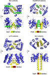
Dimerization modes of the most prevalent RR subclasses. Among the most common dimerization modes are the α1 α5 interface of DesR, α4 β5 α5 interface of PhoB, α4 loop interface of ComE and β5 α5 interface of HupR as representative for their respective RR-subfamily. Structural elements involved in interface formation are colored in red, green or yellow. The RR backbone is illustrated in blue. The same color code is used for the secondary structure of each dimerization mode. The following PDB IDs were used to present the different RR receiver domains (DesR: 4LDZ, PhoB: 1ZES, ComE: 4CBV and HupR: 2JK1).
Fig. 6
Fig. 6
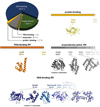
Classification of the response regulator superfamily. Pie chart presenting the percentage distribution of the 5 RR families among all classified RR. Further classification in subclasses is presented in horizontal bars. The percentage distribution of the subclasses within the respective RR family is indicated below. Structures of representative members of the most studied subclasses are illustrated in ribbon diagrams. The following PDB IDs were used to present the single RR effector domains (CheV: 3UR1, AmiR: 1S8N, CheB: 3SFT, PleD: 3IGN, RpfG: 1YOY, OmpR: 2GWR, NarL: 4HYE, NtrC: 3DZD, LytTR: 4G4K and YesN: 3LSG).
Fig. 7
Fig. 7
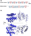
Full-length structure of ComE, a member of the LytTR subfamily of DNA-binding RR. A) Secondary structure and annotation of the structural elements of the RR ComE. β-strands are in blue and α-helices in red. B) Structure of full length ComE (dark blue: receiver domain and pale blue: effector domain). The PDB ID 4CBV was used to illustrate full-length ComE.

Similar articles

Cited by

References

    1. Ulrich LE, Zhulin IB. The MiST2 database: a comprehensive genomics resource on microbial signal transduction. Nucleic Acids Res. 2010;38:D401–D407. - PMC - PubMed
    1. Ortet P, Whitworth DE, Santaella C, Achouak W, Barakat M. P2CS: updates of the prokaryotic two-component systems database. Nucleic Acids Res. 2015;43:D536–D541. - PMC - PubMed
    1. Igo MM, Ninfa AJ, Stock JB, Silhavy TJ. Phosphorylation and dephosphorylation of a bacterial transcriptional activator by a transmembrane receptor. Genes Dev. 1989;3:1725–1734. - PubMed
    1. Szurmant H, White RA, Hoch JA. Sensor complexes regulating two-component signal transduction. Curr. Opin. Struct. Biol. 2007;17:706–715. - PMC - PubMed
    1. Szurmant H, Hoch JA. Interaction fidelity in two-component signaling. Curr. Opin. Microbiol. 2010;13:190–197. - PMC - PubMed

Publication types

MeSH terms