. 2016 Oct;186(10):2614-22.
doi: 10.1016/j.ajpath.2016.06.018. Epub 2016 Aug 9.

Activation of Liver X Receptor Attenuates Oleic Acid-Induced Acute Respiratory Distress Syndrome

Affiliations

Activation of Liver X Receptor Attenuates Oleic Acid-Induced Acute Respiratory Distress Syndrome

Zanmei Zhao et al. Am J Pathol. 2016 Oct.

Abstract

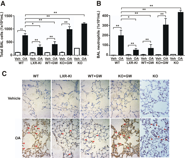
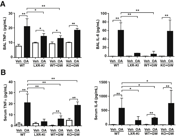
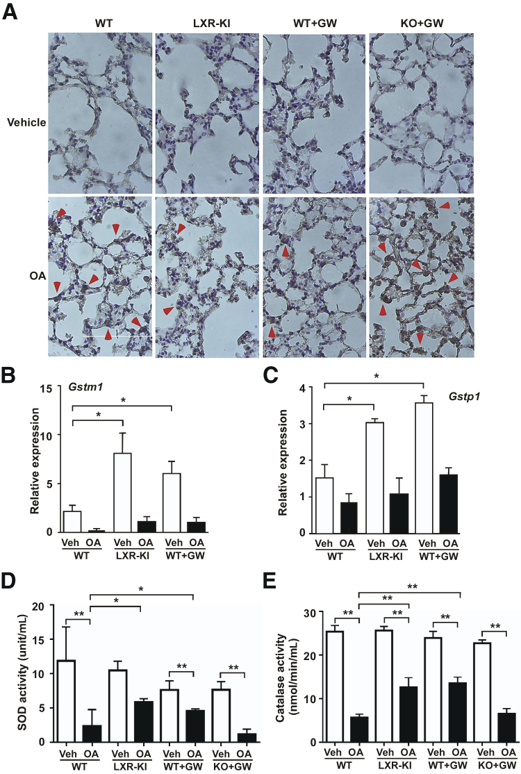
Liver X receptors (LXRs) were identified as receptors that sense oxidized cholesterol derivatives. LXRs are best known for their hepatic functions in regulating cholesterol metabolism and triglyceride synthesis, but whether and how LXRs play a role in the lung diseases is less understood. To study the function of LXRs in acute respiratory distress syndrome (ARDS), we applied the oleic acid (OA) model of ARDS to mice whose LXR was genetically or pharmacologically activated. The VP-LXRα knock-in (LXR-KI) mice, in which a constitutively activated LXRα (VP-LXRα) was inserted into the mouse LXRα locus, were used as the genetic gain-of-function model. We showed that the OA-induced lung damages, including the cytokine levels and total cell numbers and neutrophil numbers in the bronchoalveolar lavage fluid, the wet/dry weight ratio, and morphological abnormalities were reduced in the LXR-KI mice and wild-type mice treated with the LXR agonist GW3965. The pulmonoprotective effect of GW3965 was abolished in the LXR-null mice. Consistent with the pulmonoprotective effect of LXR and the induction of antioxidant enzymes by LXR, the OA-induced suppression of superoxide dismutase and catalase was attenuated in LXR-KI mice and GW3965-treated wild-type mice. Taken together, our results demonstrate that activation of LXRs can alleviate OA-induced ARDS by attenuating the inflammatory response and enhancing antioxidant capacity.


Figures

Figure 1
Figure 1
Generation of liver X receptor (LXR) α knock-in (LXR-KI) mice that bear the constitutive activation of LXRα in the lung. A: Strategy to knock-in VP-LXRα into the mouse LXRα locus. The positions and numbers of the LXRα gene exons are labeled. On gene targeting, part of exon 2, exons 3 to 7, and introns in between of the wild-type (WT) allele are replaced by the VP-LXRα-SV40-Neo cassette. B: The expression of knock-in VP-LXRα and the endogenous LXRα was determined by Northern blot analysis using a LXRα cDNA probe. Ethidium bromide staining of the agarose gel is to show the sample loading.
Figure 2
Figure 2
Activation of liver X receptor (LXR) attenuates the oleic acid (OA)–induced increases in pulmonary permeability and edema. All mice are males. Wild-type (WT), LXR knock-in (LXR-KI), and LXR-DKO [knockout (KO)] mice received a single tail vein injection of vehicle (Veh) or OA. When necessary, WT and LXR-DKO mice were treated with GW3965 (GW; daily i.p. injections at 20 mg/kg) for 7 days before being treated with Veh or OA. Mice were sacrificed 2 hours after the OA treatment and analyzed. A: The wet/dry weight ratio of the lung tissues. B: The protein concentrations in the cell-free bronchoalveolar lavage (BAL) fluid supernatants. C: Hematoxylin and eosin staining of the lung sections. n = 8 (Veh group, A and B); n = 11 (WT + OA group, A and B); n = 4 (other groups, A and B). P < 0.05, ∗∗P < 0.01. Original magnification, ×200 (C).
Figure 3
Figure 3
Activation of liver X receptor (LXR) attenuates the oleic acid (OA)–induced infiltration of neutrophils. Wild-type (WT), LXR knock-in (LXR-KI), and LXR-DKO [knockout (KO)] mice received a single tail vein injection of vehicle (Veh) or OA. When necessary, WT and LXR-DKO mice were treated with GW3965 (GW; daily i.p. injections at 20 mg/kg) for 7 days before being treated with Veh or OA. Mice were sacrificed 2 hours after the OA treatment and analyzed. Last two groups of LXR DKO mice were not treated with GW3965. A: The total bronchoalveolar lavage (BAL) cell numbers were counted using a hemocytometer. B: The polymorphonuclear neutrophils were stained with the May-Grunwald and Giemsa solution, and their cell numbers were counted. C: Immunohistochemical staining of the lung paraffin sections using the anti-myeloperoxidase (MPO) antibody. Arrowheads indicate the positive MPO staining. n = 8 (Veh group, A and B); n = 11 (WT + OA group, A and B); n = 4 (other groups, A and B). P < 0.05, ∗∗P < 0.01. Original magnification, ×400 (C).
Figure 4
Figure 4
Activation of liver X receptor (LXR) attenuates the oleic acid (OA)–induced local and systemic inflammation. Wild-type (WT), LXR knock-in (LXR-KI), and LXR-DKO [knockout (KO)] mice received a single tail vein injection of vehicle (Veh) or OA. When necessary, WT and LXR-DKO mice were treated with GW3965 (GW; daily i.p. injections at 20 mg/kg) for 7 days before being treated with Veh or OA. The mice were sacrificed before the bronchoalveolar lavage (BAL) fluid and the blood was collected. A: The concentrations of tumor necrosis factor (TNF)-α and IL-6 in the cell-free BAL fluid supernatants. B: The concentrations of TNF-α and IL-6 in the serum. n = 8 (Veh group); n = 11 (WT + OA group); n = 4 (other groups). P < 0.05, ∗∗P < 0.01.
Figure 5
Figure 5
The protective effect of liver X receptor (LXR) is associated with attenuation of oleic acid (OA)–induced oxidative stress and induction of antioxidant genes. A: Immunohistochemical staining of the lung paraffin sections using the anti–8-hydroxyguanosine antibody. Wild-type (WT), LXR knock-in (LXR-KI), and LXR-DKO [knockout (KO)] mice received a single tail vein injection of vehicle (Veh) or OA. When necessary, WT and LXR-DKO mice were treated with GW3965 (GW; daily i.p. injections at 20 mg/kg) for 7 days before being treated with Veh or OA. Mice were sacrificed 2 hours after the OA treatment and analyzed. Arrowheads indicate the positive staining. The lung expression of antioxidant genes Gstm1 (B) and Gstp1 (C) in WT and LXR-KI mice and GW3965-treated WT mice, in the absence or presence of OA treatment, was measured by real-time PCR. The activities of superoxide dismutase (SOD; D) and catalase (E) were measured in the lung homogenates. n = 5 (each group, B and C); n = 8 (WT Veh group, D and E); n = 11 (WT + OA group, D and E); n = 4 (other groups, D and E). P < 0.05, ∗∗P < 0.01. Original magnification, ×400 (A).

Similar articles

Cited by

References

    1. Dagvadorj J., Shimada K., Chen S., Jones H.D., Tumurkhuu G., Zhang W., Wawrowsky K.A., Crother T.R., Arditi M. Lipopolysaccharide induces alveolar macrophage necrosis via CD14 and the P2X7 receptor leading to interleukin-1α release. Immunity. 2015;42:640–653. - PMC - PubMed
    1. Matthay M.A., Ware L.B., Zimmerman G.A. The acute respiratory distress syndrome. J Clin Invest. 2012;122:2731–2740. - PMC - PubMed
    1. Ashbaugh D.G., Bigelow D.B., Petty T.L., Levine B.E. Acute respiratory distress in adults. Lancet. 1967;2:319–323. - PubMed
    1. Gordon D., Rubenfeld G.D., Caldwell E., Peabody E., Weaver J., Martin D.P., Neff M., Stern E.J., Hudson L.D. Incidence and outcomes of acute lung injury. N Engl J Med. 2005;353:1685–1693. - PubMed
    1. Han S., Mallampalli R.K. The acute respiratory distress syndrome: from mechanism to translation. J Immunol. 2015;194:855–860. - PMC - PubMed

MeSH terms