Review
. 2016 Oct;57(10):1789-1805.
doi: 10.1194/jlr.R070920. Epub 2016 Aug 12.

Lipid synthesis and membrane contact sites: a crossroads for cellular physiology

Affiliations
Review

Lipid synthesis and membrane contact sites: a crossroads for cellular physiology

J Pedro Fernández-Murray et al. J Lipid Res. 2016 Oct.

Abstract

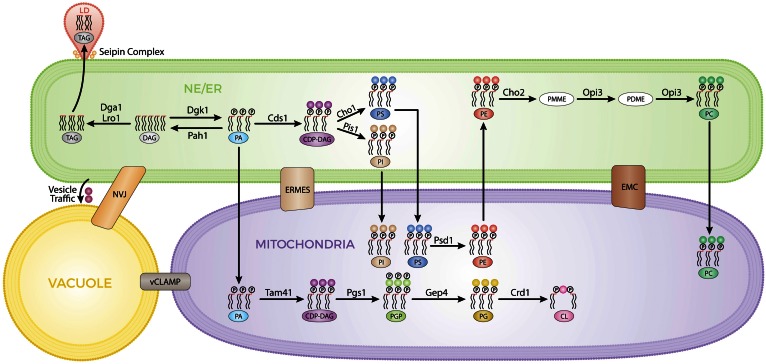
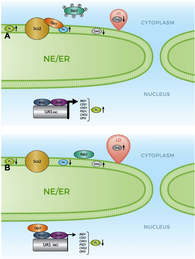
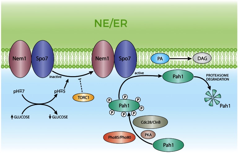
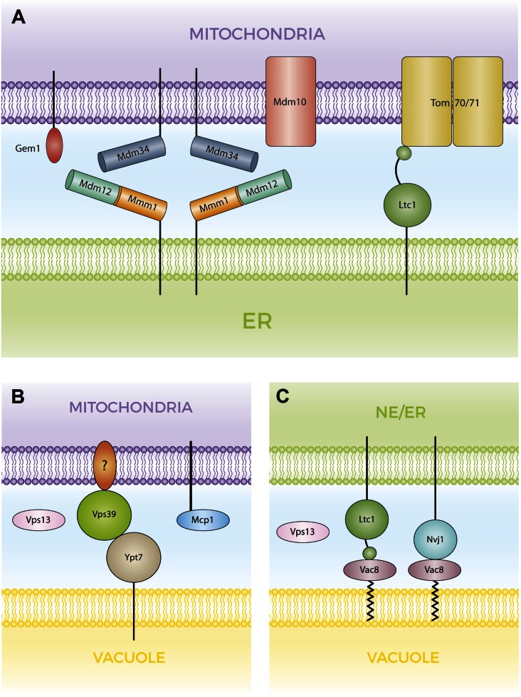
Membrane contact sites (MCSs) are regions of close apposition between different organelles that contribute to the functional integration of compartmentalized cellular processes. In recent years, we have gained insight into the molecular architecture of several contact sites, as well as into the regulatory mechanisms that underlie their roles in cell physiology. We provide an overview of two selected topics where lipid metabolism intersects with MCSs and organelle dynamics. First, the role of phosphatidic acid phosphatase, Pah1, the yeast homolog of metazoan lipin, toward the synthesis of triacylglycerol is outlined in connection with the seipin complex, Fld1/Ldb16, and lipid droplet formation. Second, we recapitulate the different contact sites connecting mitochondria and the endomembrane system and emphasize their contribution to phospholipid synthesis and their coordinated regulation. A comprehensive view is emerging where the multiplicity of contact sites connecting different cellular compartments together with lipid transfer proteins functioning at more than one MCS allow for functional redundancy and cross-regulation.

Keywords: endoplasmic reticulum; mitochondria; organelle; phosphatidic acid; triacylglycerol; vacuole.


Figures

Fig. 1.
Fig. 1.
MCSs enable metabolic flux through different compartments for PL and TAG syntheses. This figure outlines several lipid biosynthetic pathways and their locations. The MCSs bridging the gaps between the different compartments are indicated. PA is the key lipid precursor for the synthesis of TAG and PLs taking place at the nuclear envelope (NE)/ER (green). TAG is sorted into LDs (pink) connected to the ER by the seipin complex. Most of the reactions for PL synthesis take place at the ER. PS decarboxylation mediated by Psd1 and generating PE occurs in the mitochondria (purple) with transport of PS and PE indicated by arrows across these compartments. Transports of PI, PC, and PA (for the synthesis of the specific mitochondrial lipids PG and CL) into mitochondria are also indicated by arrows from the ER and into mitochondria. ER-mitochondria encounter structure (ERMES) and ER membrane protein complex (EMC) are represented as boxes connecting both compartments. An alternate route through the vacuole (yellow) bridging ER and mitochondria is shown. Vacuole and NE/ER are connected indirectly by vesicle traffic through the endomembrane system, as well as by the NVJ contact site. The vacuolar and mitochondrial patch (vCLAMP) contact site is represented as a box connecting both compartments.
Fig. 2.
Fig. 2.
Activation status of PA phosphatase, Pah1, impacts on PL and NL homeostasis. A: Pah1 in its hyperphosphorylated form is kept off the ER and unable to reach its substrate, PA. An increased ER level of PA recruits the transcriptional repressor, Opi1, to the ER membrane through the simultaneous interaction with the integral protein, Scs2, allowing for the expression of genes involved in PL synthesis. A reduced level of DAG attenuates TAG synthesis and LD formation. B: Dephosphorylated Pah1 binds to ER membranes and hydrolyzes PA generating DAG leading to TAG synthesis and LD formation. Upon reduction of the PA level at the ER, Opi1 is released and translocates to the nucleus where it represses transcription and decreasing PL synthesis.
Fig. 3.
Fig. 3.
Mechanisms of regulation of PA phosphatase, Pah1. Pah1 is phosphorylated by protein kinase A (PKA), Pho80/Pho85, and Cdc28/ClnB. In its hyperphosphorylated status, Pah1 is inactive and kept away from the membranes. The ER resident phosphatase complex, Nem1/Spo7, dephosphorylates and activates Pah1. Pah1 translocates to the ER membrane reaching its substrate PA and generates DAG. The active form of Pah1 is a substrate of the proteasome, having a reduced half-life. In turn, the phosphatase complex, Nem1/Spo7, is activated by cytosol acidification upon glucose exhaustion through an uncharacterized mechanism. Activated TORC1 prevents Nem1/Spo7 activation promoting low levels of Pah1 activity.
Fig. 4.
Fig. 4.
Molecular organization of MCSs connecting the ER, mitochondria, and vacuole. A: ERMES and Ltc1-Tom70/71 tethering complexes at ER-mitochondria contact sites. The ERMES complex is composed of four subunits. The subunits Mmm1, Mdm12, and Mdm34 carry SMP domains, physically interact forming a ternary complex and are proposed to transport lipids between the ER and mitochondria. Mdm10 is a mitochondrial β-barrel protein shared with the SAM complex. Gem1 is a substoichiometric component of ERMES, but its role is poorly characterized. Ltc1 is an ER integral protein that contains a sterol binding domain and interacts with mitochondrial Tom70 and Tom71. B: vCLAMP complexes connect mitochondria and vacuole. The vacuolar Rab GTPases, Ypt7 and Vps39, are components of this complex, whereas the mitochondrial component is not known. Vps13 is observed associated with this MCS under fermenting growth conditions, whereas the involvement of Mcp1 in this complex was deduced by genetic analysis. C: The NVJ is characterized by the interaction of Vac8 and Nvj1. Ltc1 also localizes at this MCS through its interaction with Vac8 and independent of Nvj1. Under respiratory growth conditions, Vps13 localizes at the NVJ.

References

    1. Athenstaedt K., and Daum G.. 1999. Phosphatidic acid, a key intermediate in lipid metabolism. Eur. J. Biochem. 266: 1–16. - PubMed
    1. Henry S. A., Kohlwein S. D., and Carman G. M.. 2012. Metabolism and regulation of glycerolipids in the yeast Saccharomyces cerevisiae. Genetics. 190: 317–349. - PMC - PubMed
    1. Henry S. A., and Patton-Vogt J. L.. 1998. Genetic regulation of phospholipid metabolism: yeast as a model eukaryote. Prog. Nucleic Acid Res. Mol. Biol. 61: 133–179. - PubMed
    1. Carman G. M., and Henry S. A.. 2007. Phosphatidic acid plays a central role in the transcriptional regulation of glycerophospholipid synthesis in Saccharomyces cerevisiae. J. Biol. Chem. 282: 37293–37297. - PMC - PubMed
    1. Travers K. J., Patil C. K., Wodicka L., Lockhart D. J., Weissman J. S., and Walter P.. 2000. Functional and genomic analyses reveal an essential coordination between the unfolded protein response and ER-associated degradation. Cell. 101: 249–258. - PubMed

MeSH terms

LinkOut - more resources