. 2017 May 1;72(5):595-602.
doi: 10.1093/gerona/glw151.

Sirtuin 3 Deregulation Promotes Pulmonary Fibrosis

Affiliations

Sirtuin 3 Deregulation Promotes Pulmonary Fibrosis

Meredith L Sosulski et al. J Gerontol A Biol Sci Med Sci. .

Abstract

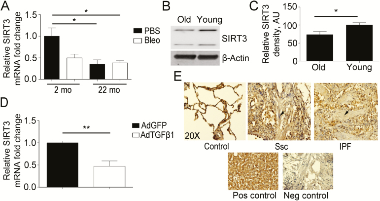
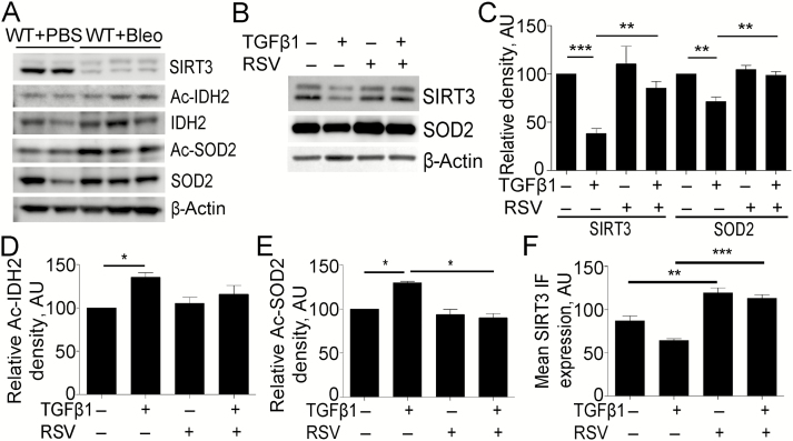
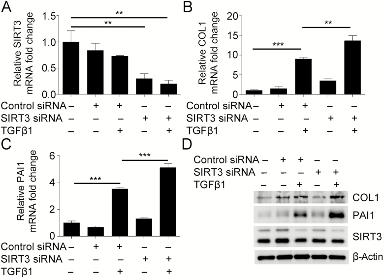
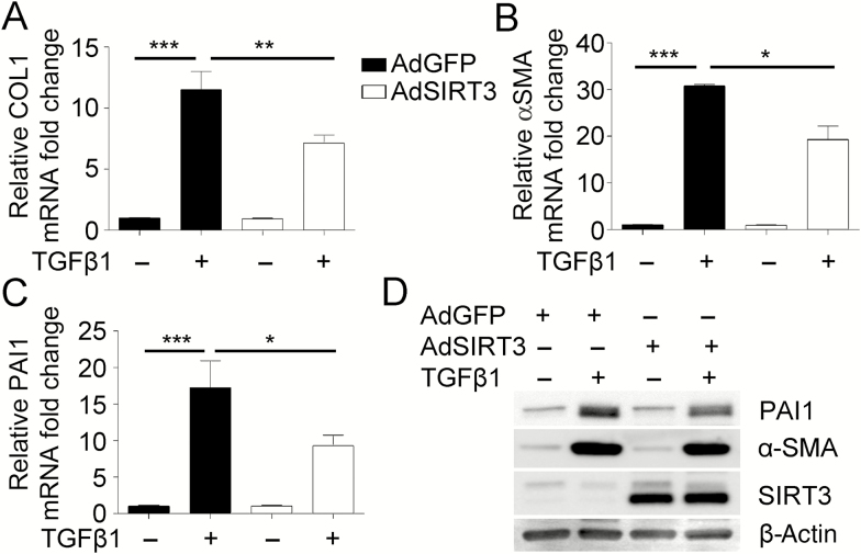
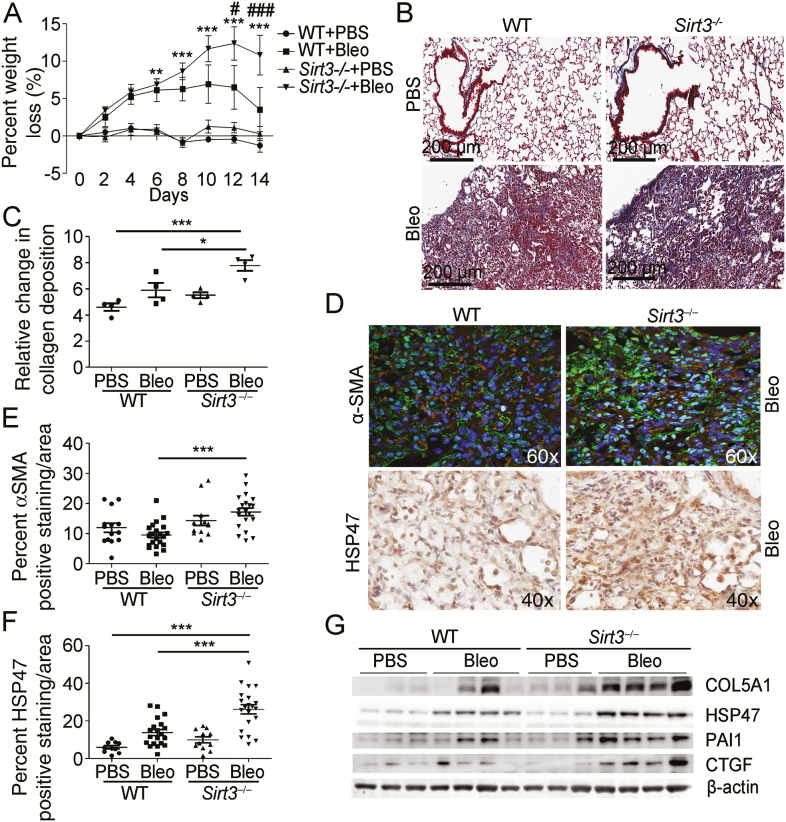
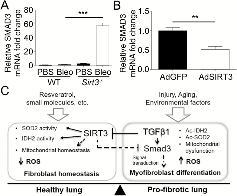
Oxidative stress leads to alveolar epithelial cell injury and fibroblast-myofibroblast differentiation (FMD), key events in the pathobiology of pulmonary fibrosis (PF). Sirtuin 3 (SIRT3) is a mitochondrial protein deacetylase regulator of antioxidant response and mitochondrial homeostasis. Here, we demonstrate reduced SIRT3 expression in the lungs of old mice compared to young mice, as well as in two murine models of PF. The analysis of the pattern of SIRT3 expression in the lungs of patients with PF revealed low SIRT3 staining within the fibrotic regions. We also demonstrated, using murine models of PF and human lung fibroblasts, that reduced SIRT3 expression in response to transforming growth factor beta 1 (TGFβ1) promotes acetylation (inactivation) of major oxidative stress response regulators, such as SOD2 and isocitrate dehydrogenase 2. Reduction of SIRT3 in human lung fibroblasts promoted FMD. By contrast, overexpression of SIRT3 attenuated TGFβ1-mediated FMD and significantly reduced the levels of SMAD family member 3 (SMAD3). Resveratrol induced SIRT3 expression and ameliorated acetylation changes induced by TGFβ1. We demonstrated that SIRT3-deficient mice are more susceptible to PF compared to control mice, and concomitantly exhibit enhanced SMAD3 expression. Collectively, these data define a SIRT3/TGFβ1 interaction during aging that may play a significant role in the pathobiology of PF.

Keywords: Age-related pathology; Lungs/pulmonary; Mitochondria; Reactive oxygen species; SIRT3.


Figures

Figure 1.
Figure 1.
Downregulation of Sirtuin 3 (SIRT3) in aging and pulmonary fibrosis (PF). (A) Real-time quantitative reverse-transcriptase polymerase chain reaction (qRT-PCR) analysis for SIRT3 mRNA expression in aging (22-month-old) and young (2-month-old) mice at 14 days after oropharyngeal aspiration of phosphate-buffered saline (PBS) or bleomycin (Bleo) (n = 5 per treatment). (B) Representative Western blot (WB) for SIRT3 expression in mouse lung fibroblasts isolated from old (22-month-old) and young (2-month-old) mice. β-Actin used as a loading control. (C) Densitometry analysis of WB for SIRT3 expression is shown in (B). (D) qRT-PCR analysis for SIRT3 mRNA expression in young mice at 14-day post-oropharyngeal aspiration of control Adenovirus-GFP (AdGFP) or Adenovirus-TGFβ1 (AdTGFβ1; n = 5 per treatment). (E) Immunohistochemistry in lung tissue samples from control (n = 10), systemic scleroderma (SSc; n = 16), and idiopathic PF (IPF; n = 13) patients show a differential expression pattern of SIRT3. Positive cells appear brown (diaminobenzidene stain). Nuclei counterstained with hematoxylin appear blue. The arrow shows a fibrotic lesion. Liver tissue was used as a positive control. Immunoglobulin G alone was used as a negative control. *p < .05, **p < .01. AU, arbitrary units.
Figure 2.
Figure 2.
Transforming growth factor beta 1 (TGFβ1) regulates Sirtuin 3 (SIRT3) concomitant with the acetylation of major oxidative stress response regulators. (A) Representative Western blot (WB) for SIRT3, acetylated isocitrate dehydrogenase 2 (Ac-IDH2), IDH2, acetylated superoxide dismutase 2, mitochondrial (SOD2) (Ac-SOD2), and SOD2 expression from wild-type (WT) mice exposed to bleomycin (Bleo) or phosphate-buffered saline (PBS) vehicle, at 14-day postexposure. β-Actin was used as a loading control. (B) Representative WB for SIRT3 and SOD2 expression from normal human lung fibroblasts (NHLF) treated or untreated, with TGFβ1 and/or resveratrol. (C) Densitometry analysis of WB for SIRT3 and SOD2 expression is shown in (B). (D) Densitometry analysis of WB for acetylated IDH2 (K413) expression from NHLF treated or untreated, with TGFβ1 and/or resveratrol shown in Supplementary Figure 2C. (E) Densitometry analysis of WB for acetylated SOD2 (K68) expression from NHLF treated or untreated, with TGFβ1 and/or resveratrol shown in Supplementary Figure 2D. (F) Quantification of representative immunofluorescence images for SIRT3 in NHLF treated, with TGFβ1 and/or resveratrol at 24 hours, shown in Supplementary Figure 2E. *p < .05, **p < .01, ***p < .005. AU, arbitrary units; RSV, resveratrol.
Figure 3.
Figure 3.
Downregulation of Sirtuin 3 (SIRT3) promotes myofibroblast differentiation mediated by transforming growth factor beta 1 (TGFβ1). (A–C) Real-time quantitative reverse-transcriptase polymerase chain reaction analysis from normal human lung fibroblasts (NHLF), transfected with siRNA targeting SIRT3, treated with or without TGFβ1, to evaluate transcriptional changes in SIRT3 and fibrotic markers collagen type I (COL1) and plasminogen activator inhibitor-1 (PAI1). (D) Representative Western blot for COL1, PAI1, and SIRT3 expression in NHLF deficient in SIRT3. β-Actin was used as a loading control. **p < .01, ***p < .005.
Figure 4.
Figure 4.
Overexpression of Sirtuin 3 (SIRT3) decreases myofibroblast differentiation potential mediated by transforming growth factor beta 1 (TGFβ1). (A–C) Real-time quantitative reverse-transcriptase polymerase chain reaction analysis from normal human lung fibroblasts (NHLF), transfected with adenovirus overexpressing SIRT3, treated with or without TGFβ1, to evaluate transcriptional changes in fibrotic markers collagen type I (COL1), α-smooth muscle actin (α-SMA), and plasminogen activator inhibitor-1 (PAI1). (D) Representative Western blot for PAI1, α-SMA, and SIRT3 expression in NHLF overexpressing SIRT3. β-Actin was used as a loading control. *p < .05, **p < .01, ***p < .005.
Figure 5.
Figure 5.
Sirtuin 3 (SIRT3)-deficient mice are susceptible to bleomycin (Bleo)-induced pulmonary fibrosis. (A) The graph depicts weight loss as a percentage in SIRT3-deficient (Sirt3−/−; n = 7 per treatment) and wild-type (WT; n = 5 per treatment) control mice after Bleo or phosphate-buffered saline (PBS) vehicle. (B and C) Representative images and quantification of Masson’s trichrome staining to evaluate collagen deposition in SIRT3-deficient and WT mice after Bleo or PBS vehicle, at 14-day postexposure. Positive collagen deposition appears blue. (D) Representative images of immunofluorescence (IF) staining for α-smooth muscle actin (α-SMA) and immunohistochemistry (IHC) staining for heat shock protein 47 (HSP47) fibrotic marker expression in SIRT3-deficient and WT control mice after Bleo, 14-day postexposure. In IF images, positive cells appear green with nuclei counterstained with 4′,6-diamidino-2-phenylindole appearing blue. In IHC images, positive cells appear brown (diaminobenzidene stain) with nuclei counterstained with hematoxylin appearing blue. (E) Quantification of IF staining for α-SMA fibrotic marker expression in SIRT3-deficient and WT control mice after Bleo or PBS vehicle, 14-day postexposure. (F) Quantification of IHC staining for HSP47 fibrotic marker expression in SIRT3-deficient and WT control mice after Bleo or PBS vehicle, 14-day postexposure. (G) Representative Western blot for fibrotic markers collagen type 5 alpha chain 1 (COL5A1), HSP47, plasminogen activator inhibitor-1 (PAI1), and connective tissue growth factor (CTGF) expression in lung tissue extracted from SIRT3-deficient mice. β-Actin was employed as a loading control. *p < .05, **p < .01, ***p < .005. In (A), **p < .01, ***p < .005 comparing Sirt3−/− Bleo-exposed group to WT PBS control group, #p < .05, ###p < .005 comparing Sirt3−/− bleomycin-exposed group to WT bleomycin-exposed group.
Figure 6.
Figure 6.
Interaction between Sirtuin 3 (SIRT3) and transforming growth factor beta 1 (TGFβ1) during fibrogenesis. (A) Real-time quantitative reverse-transcriptase polymerase chain reaction (qRT-PCR) analysis for SMAD family member 3 (SMAD3) expression in SIRT3-deficient (Sirt3−/−; n = 7 per treatment) and wild-type (WT; n = 5 per treatment) control mice after bleomycin (Bleo) or phosphate-buffered saline vehicle, at 14-day postexposure. (B) qRT-PCR confirmation for SMAD3 expression in normal human lung fibroblasts (NHLF) overexpressing SIRT3. **p < .01, ***p < .005. (C) Model for the role of SIRT3/TGFβ crosstalk in pulmonary fibrosis. The proposed model describes the role of SIRT3 in the regulation of SMAD3 and the antioxidant response during fibrogenesis. Aging, lung injury, TGFβ1 expression, genetic and environmental factors that contribute to reduction of SIRT3 favor the profibrotic effects of TGFβ1. By contrast, induction of SIRT3, by resveratrol, small molecules, or genetic approaches, enhances antifibrotic effects in the healthy lung mediated through downregulation of TGFβ1 signal transduction and an enhanced antioxidant response. Taken together, we propose that SIRT3 is critical to modulating the fibrotic response to lung injury in the elderly adults.

Similar articles

Cited by

References

    1. Roberts AB, Sporn MB, Assoian RK, et al. Transforming growth factor type beta: rapid induction of fibrosis and angiogenesis in vivo and stimulation of collagen formation in vitro. Proc Natl Acad Sci USA. 1986;83:4167–4171. doi:10.1073/pnas.83.12.4167 - PMC - PubMed
    1. Kawakami T, Ihn H, Xu W, Smith E, LeRoy C, Trojanowska M. Increased expression of TGF-beta receptors by scleroderma fibroblasts: evidence for contribution of autocrine TGF-beta signaling to scleroderma phenotype. J Invest Dermatol. 1998;110:47–51. doi:10.1046/j.1523-1747.1998.00073.x - PubMed
    1. Bhattacharyya S, Wei J, Varga J. Understanding fibrosis in systemic sclerosis: shifting paradigms, emerging opportunities. Nat Rev Rheumatol. 2012;8:42–54. doi:10.1038/nrrheum.2011.149 - PMC - PubMed
    1. Vij R, Strek ME. Diagnosis and treatment of connective tissue disease-associated interstitial lung disease. Chest. 2013;143:814–824. doi:10.1378/chest.12-0741 - PMC - PubMed
    1. Raghu G, Chen SY, Hou Q, Yeh WS, Collard HR. Incidence and prevalence of idiopathic pulmonary fibrosis in US adults 18–64 years old. Eur Respir J. 2016;48:179–186. doi:10.1183/13993003.01653-2015 - PubMed