

Longitudinal Evidence for Dissociation of Anterior and Posterior MTL Resting-State Connectivity in Aging: Links to Perfusion and Memory
- PMID: 27522073
- PMCID: PMC5028008
- DOI: 10.1093/cercor/bhw233
Longitudinal Evidence for Dissociation of Anterior and Posterior MTL Resting-State Connectivity in Aging: Links to Perfusion and Memory
Abstract
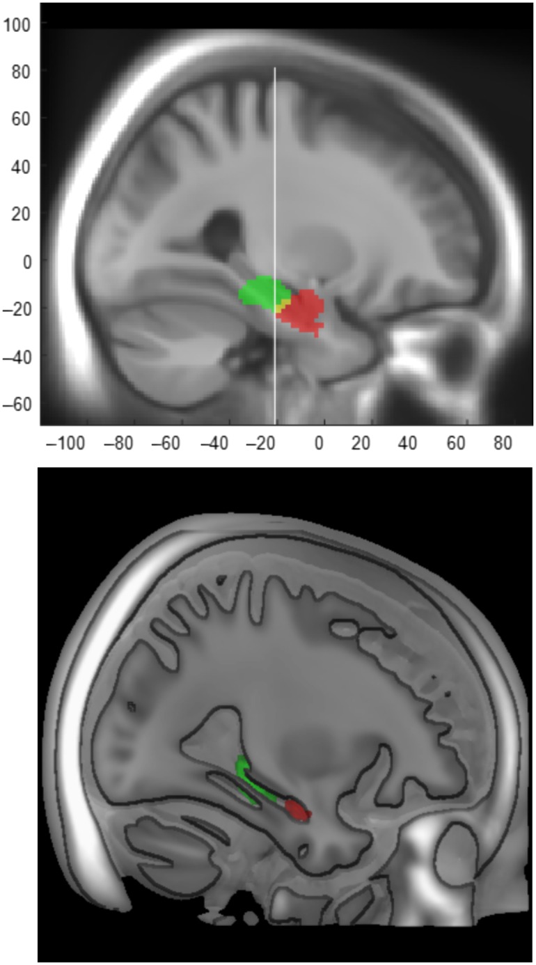
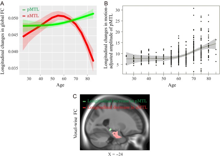
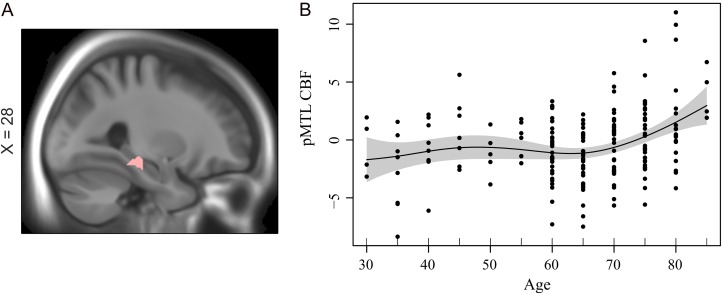
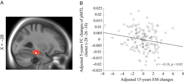
Neuroimaging studies of spontaneous signal fluctuations as measured by resting-state functional magnetic resonance imaging have revealed age-related alterations in the functional architecture of brain networks. One such network is located in the medial temporal lobe (MTL), showing structural and functional variations along the anterior-posterior axis. Past cross-sectional studies of MTL functional connectivity (FC) have yielded discrepant findings, likely reflecting the fact that specific MTL subregions are differentially affected in aging. Here, using longitudinal resting-state data from 198 participants, we investigated 5-year changes in FC of the anterior and posterior MTL. We found an opposite pattern, such that the degree of FC within the anterior MTL declined after age 60, whereas elevated FC within the posterior MTL was observed along with attenuated posterior MTL-cortical connectivity. A significant negative change-change relation was observed between episodic-memory decline and elevated FC in the posterior MTL. Additional analyses revealed age-related cerebral blood flow (CBF) increases in posterior MTL at the follow-up session, along with a positive relation of elevated FC and CBF, suggesting that elevated FC is a metabolically demanding alteration. Collectively, our findings indicate that elevated FC in posterior MTL along with increased local perfusion is a sign of brain aging that underlie episodic-memory decline.
Keywords: episodic memory, functional connectivity, longitudinal, anterior and posterior MTL, perfusion.
© The Author 2016. Published by Oxford University Press.
Figures
Similar articles
-
The retrosplenial cortex: A memory gateway between the cortical default mode network and the medial temporal lobe.Hum Brain Mapp. 2018 May;39(5):2020-2034. doi: 10.1002/hbm.23983. Epub 2018 Jan 23. Hum Brain Mapp. 2018. PMID: 29363256 Free PMC article.
-
Attenuated anticorrelation between the default and dorsal attention networks with aging: evidence from task and rest.Neurobiol Aging. 2016 Sep;45:149-160. doi: 10.1016/j.neurobiolaging.2016.05.020. Epub 2016 Jun 3. Neurobiol Aging. 2016. PMID: 27459935 Free PMC article.
-
The Longitudinal Trajectory of Default Mode Network Connectivity in Healthy Older Adults Varies As a Function of Age and Is Associated with Changes in Episodic Memory and Processing Speed.J Neurosci. 2018 Mar 14;38(11):2809-2817. doi: 10.1523/JNEUROSCI.3067-17.2018. Epub 2018 Feb 13. J Neurosci. 2018. PMID: 29440553 Free PMC article.
-
[Functional neuroimaging studies of episodic memory--functional dissociation in the medial temporal lobe structures].Brain Nerve. 2008 Jul;60(7):833-44. Brain Nerve. 2008. PMID: 18646623 Review. Japanese.
-
Medial temporal lobe activations in fMRI and PET studies of episodic encoding and retrieval.Hippocampus. 1999;9(1):7-24. doi: 10.1002/(SICI)1098-1063(1999)9:1<7::AID-HIPO2>3.0.CO;2-K. Hippocampus. 1999. PMID: 10088896 Review.
Cited by
-
Association of Cerebral Hypoperfusion and Poor Collaterals with Cognitive Impairment in Patients with Severe Vertebrobasilar Artery Stenosis.J Alzheimers Dis Rep. 2024 Jun 21;8(1):999-1007. doi: 10.3233/ADR-240007. eCollection 2024. J Alzheimers Dis Rep. 2024. PMID: 39114550 Free PMC article.
-
Medial temporal lobe connectivity and its associations with cognition in early Alzheimer's disease.Brain. 2020 Apr 1;143(4):1233-1248. doi: 10.1093/brain/awaa068. Brain. 2020. PMID: 32252068 Free PMC article.
-
Longitudinal Changes in Whole-Brain Functional Connectivity Strength Patterns and the Relationship With the Global Cognitive Decline in Older Adults.Front Aging Neurosci. 2020 Mar 17;12:71. doi: 10.3389/fnagi.2020.00071. eCollection 2020. Front Aging Neurosci. 2020. PMID: 32256339 Free PMC article.
-
Anterior-temporal network hyperconnectivity is key to Alzheimer's disease: from ageing to dementia.Brain. 2025 Jun 3;148(6):2008-2022. doi: 10.1093/brain/awaf008. Brain. 2025. PMID: 39813142 Free PMC article.
-
A Longitudinal Study of Changes in Resting-State Functional Magnetic Resonance Imaging Functional Connectivity Networks During Healthy Aging.Brain Connect. 2020 Sep;10(7):377-384. doi: 10.1089/brain.2019.0724. Epub 2020 Aug 19. Brain Connect. 2020. PMID: 32623915 Free PMC article.
References
Publication types
MeSH terms
LinkOut - more resources
Full Text Sources
Other Literature Sources
Medical
Molecular Biology Databases
Miscellaneous
