Randomized Controlled Trial
. 2017 May;58(5):600-608.
doi: 10.1177/0284185116658682. Epub 2016 Aug 20.

Spot fluoroscopy: a novel innovative approach to reduce radiation dose in neurointerventional procedures

Affiliations
Randomized Controlled Trial

Spot fluoroscopy: a novel innovative approach to reduce radiation dose in neurointerventional procedures

Ljubisa Borota et al. Acta Radiol. 2017 May.

Abstract

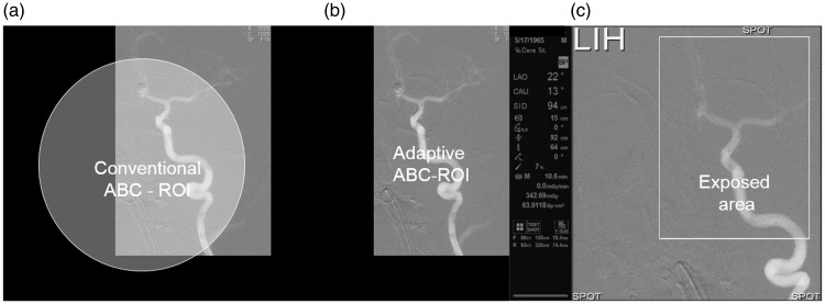
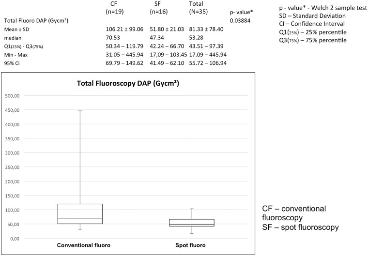
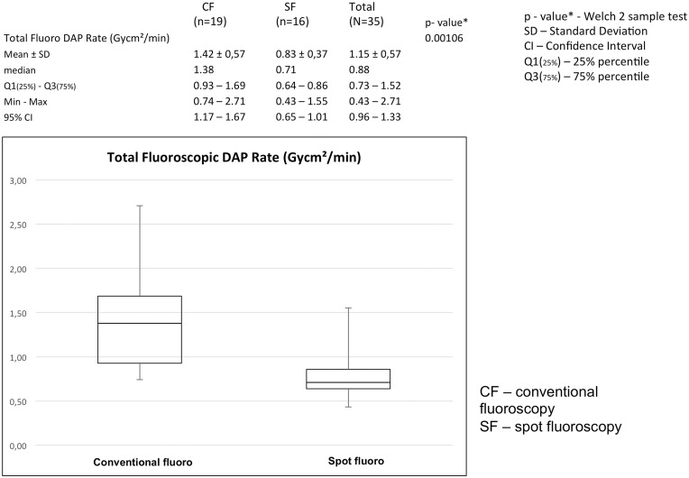
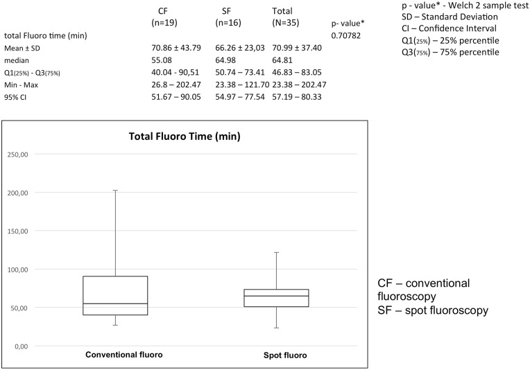
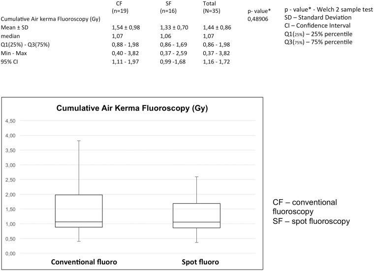
Background Increased interest in radiation dose reduction in neurointerventional procedures has led to the development of a method called "spot fluoroscopy" (SF), which enables the operator to collimate a rectangular or square region of interest anywhere within the general field of view. This has potential advantages over conventional collimation, which is limited to symmetric collimation centered over the field of view. Purpose To evaluate the effect of SF on the radiation dose. Material and Methods Thirty-five patients with intracranial aneurysms were treated with endovascular coiling. SF was used in 16 patients and conventional fluoroscopy in 19. The following parameters were analyzed: the total fluoroscopic time, the total air kerma, the total fluoroscopic dose-area product, and the fluoroscopic dose-area product rate. Statistical differences were determined using the Welch's t-test. Results The use of SF led to a reduction of 50% of the total fluoroscopic dose-area product (CF = 106.21 Gycm2, SD = 99.06 Gycm2 versus SF = 51.80 Gycm2, SD = 21.03 Gycm2, p = 0.003884) and significant reduction of the total fluoroscopic dose-area product rate (CF = 1.42 Gycm2/min, SD = 0.57 Gycm2/s versus SF = 0.83 Gycm2/min, SD = 0.37 Gycm2/min, p = 0.00106). The use of SF did not lead to an increase in fluoroscopy time or an increase in total fluoroscopic cumulative air kerma, regardless of collimation. Conclusion The SF function is a new and promising tool for reduction of the radiation dose during neurointerventional procedures.

Keywords: X-ray; collimation; digital subtraction angiography (DSA); dose saving; fluoroscopy; neurointervention.


Figures

Fig. 1.
Fig. 1.
(a) White arrow indicates the upper left corner of the ROI. (b) The upper left corner is defined, and the arrow is being moved to the lower right corner. (c, d) ROI is now defined, and only this part of the FOV, outlined by the white-lined frame, will be exposed to irradiation; the LIH (upper left corner) will be superimposed over the collimated part of the FOV. “Spot” in each corner indicates that SF will be activated.
Fig. 2.
Fig. 2.
(a) Angiography of left internal carotid artery shows an aneurysm localized proximally on the M1 segment. (b) Conventional fluoroscopic road map used for creation of last image hold. (c) LIH in the left upper corner is no longer displayed – compare with Fig. 1, definition of the FOV. “Spot” in each corner indicates that SF is now activated – only the region inside the white-lined frame is exposed to radiation. Image shows positioning of balloon in front of the aneurysm. (d) Angiography of left internal carotid artery, last run, shows occluded aneurysm and minimal flow in the neck region of the aneurysm.
Fig. 3.
Fig. 3.
(a) A situation where rectangular ROI does not correspond to a conventional circular sensing area. (b) ROI and sensing area have the same, rectangular shape and completely correspond to each other. (c) Important technical parameters are displayed to the left of the fluoroscopic image. The size and position of the rectangular ROI exposed to irradiation can be changed at any moment during the intervention depending on the size, shape, and position of the targeted vascular structure.
Fig. 4.
Fig. 4.
SF leads to a significant reduction of the fluoroscopic dose area product (Gycm2), P = 0.03884.
Fig. 5.
Fig. 5.
SF leads to a significant reduction of total fluoroscopic dose area product per minute (Gycm2/min), P = 0.00106.
Fig. 6.
Fig. 6.
SF does not lead to increase of fluoroscopy time (min). Difference between the values of fluoroscopy times is not significant, P = 0.70782.
Fig. 7.
Fig. 7.
SF does not lead to an increase of total fluoroscopic air kerma (Gy). Difference between values of total fluoroscopic air kerma is not significant, P = 0.48906.

Similar articles

Cited by

References

    1. Söderman M, Mauti M, Boon S, et al. Radiation dose in neuroradiography using image noise reduction technology: a population study based on 614 patients. Neuroradiology 2013; 55: 1365–1372. - PMC - PubMed
    1. Klein LW, Miller DL, Balter S, et al. Occupational health hazards in the interventional laboratory: Time for a safer environment. Radiology 2009; 250: 538–544. - PubMed
    1. Dose-Area Product. Conference of Radiation Control Program Directors (CRCPD) 205 Capital Avenue Frankfort, KY 4060, First published: October 2001, Reviewed/Republished: September 2008. Available at: http://www.crcpd.org/Pubs/QAC/DAP.pdf.
    1. Gallas S, Drouineau J, Gabrillargues J, et al. Feasibility, procedural morbidity and mortality, and long-term follow-up of endovascular treatment of 321 unruptured aneurysms. Am J Neuroradiol 2008; 29: 63–68. - PMC - PubMed
    1. Muroi T, Tanaka M, Shimizu Y, et al. Medical Diagnostic Imaging Apparatus. PCT/JP2013/052412. 2013.

Publication types

LinkOut - more resources