Review
. 2016 Sep;132(3):317-38.
doi: 10.1007/s00401-016-1606-5. Epub 2016 Aug 13.

Vascular, glial, and lymphatic immune gateways of the central nervous system

Affiliations
Review

Vascular, glial, and lymphatic immune gateways of the central nervous system

Britta Engelhardt et al. Acta Neuropathol. 2016 Sep.

Abstract

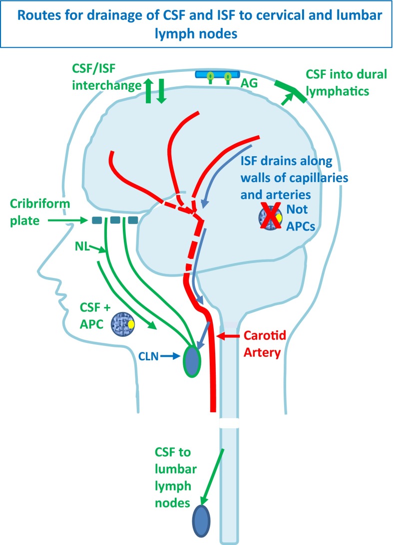
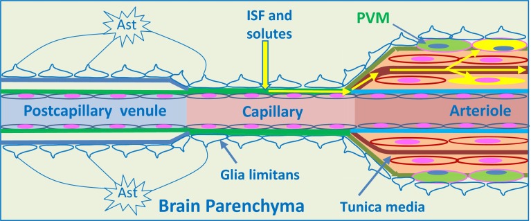
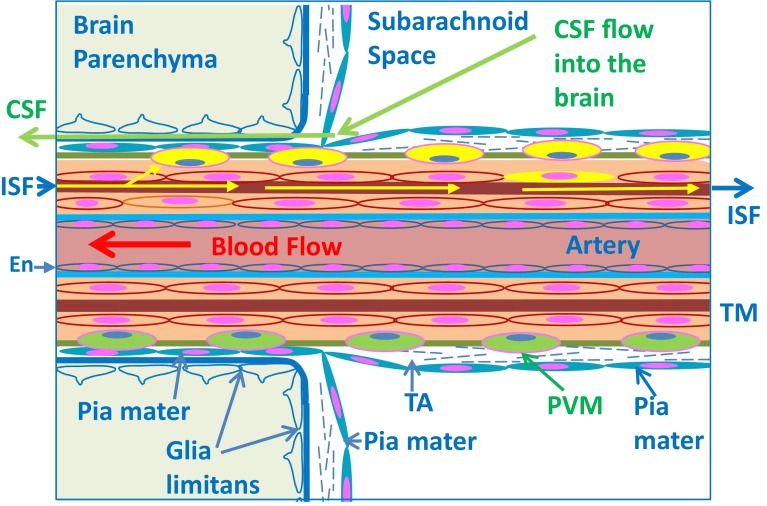
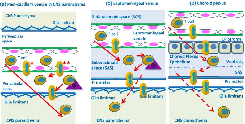
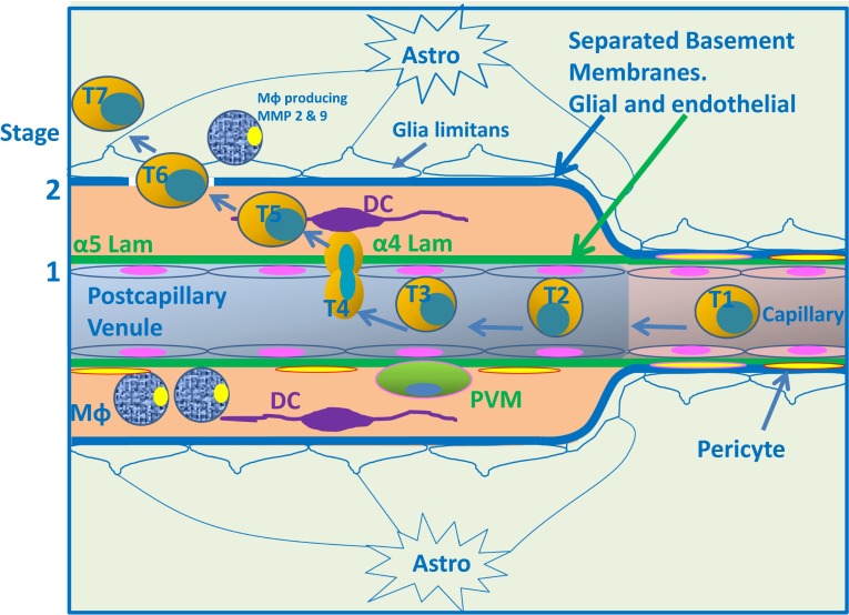
Immune privilege of the central nervous system (CNS) has been ascribed to the presence of a blood-brain barrier and the lack of lymphatic vessels within the CNS parenchyma. However, immune reactions occur within the CNS and it is clear that the CNS has a unique relationship with the immune system. Recent developments in high-resolution imaging techniques have prompted a reassessment of the relationships between the CNS and the immune system. This review will take these developments into account in describing our present understanding of the anatomical connections of the CNS fluid drainage pathways towards regional lymph nodes and our current concept of immune cell trafficking into the CNS during immunosurveillance and neuroinflammation. Cerebrospinal fluid (CSF) and interstitial fluid are the two major components that drain from the CNS to regional lymph nodes. CSF drains via lymphatic vessels and appears to carry antigen-presenting cells. Interstitial fluid from the CNS parenchyma, on the other hand, drains to lymph nodes via narrow and restricted basement membrane pathways within the walls of cerebral capillaries and arteries that do not allow traffic of antigen-presenting cells. Lymphocytes targeting the CNS enter by a two-step process entailing receptor-mediated crossing of vascular endothelium and enzyme-mediated penetration of the glia limitans that covers the CNS. The contribution of the pathways into and out of the CNS as initiators or contributors to neurological disorders, such as multiple sclerosis and Alzheimer's disease, will be discussed. Furthermore, we propose a clear nomenclature allowing improved precision when describing the CNS-specific communication pathways with the immune system.

Keywords: Alzheimer’s disease; Antigen-presenting cells; Blood–brain barrier; CNS; CSF; Dendritic cells; Glia limitans: multiple sclerosis; Immune privilege; Interstitial fluid; Lymphatic drainage.


Figures

Fig. 1
Fig. 1
Drainage pathways for CSF and interstitial fluid (ISF) to cervical lymph nodes. CSF and ISF drain to lymph nodes by different and distinct pathways. In humans, CSF drains into the blood of venous sinuses through well-developed arachnoid villi and granulations (AG). Lymphatic drainage of CSF occurs via nasal and dural lymphatics and along cranial and spinal nerve roots (outlined in green). Channels that pass from the subarachnoid space through the cribriform plate allow passage of CSF (green line) T cells and antigen-presenting cells (APC) into nasal lymphatics (NL) and cervical lymph nodes (CLN). CSF from the lumbar subarachnoid space drains to lumbar lymph nodes. ISF from the brain parenchyma drains along basement membranes in the walls of cerebral capillaries and arteries (blue arrows) to cervical lymph nodes adjacent to the internal carotid artery just below the base of the skull. This narrow intramural perivascular drainage pathway does not allow the traffic of APC. There is interchange between CSF and ISF (convective influx/glymphatic system), as CSF enters the surface of the brain alongside penetrating arteries
Fig. 2
Fig. 2
Pathways for lymphatic drainage of peripheral organs, CSF and cerebral interstitial fluid. a Tissue fluid from skin, gut, and other organs drains into blind-ended lymphatic vessels lined by endothelium. Antigen-presenting cells (APC) also drain by this route to regional lymph nodes. Valves are shown in the lymphatic vessel. b CSF and antigen-presenting cells (APC) pass from the subarachnoid space through channels in the ethmoid bone, alongside olfactory nerves (ON), to enter lymphatics in the nasal mucosa and drain to cervical lymph nodes. CSF also drains via dural lymphatics towards the deep cervical lymph nodes (see text) c Interstitial fluid (ISF) from the brain parenchyma enters basement membranes in the walls of capillaries and then basement membranes between smooth muscle cells in the tunica media of cerebral arterioles and arteries. The route of intramural perivascular drainage for ISF is indicated by blue arrows that track along the walls of intracranial arteries to cervical lymph nodes (CLN) related to the internal carotid artery at the base of the skull
Fig. 3
Fig. 3
Intramural perivascular drainage of interstitial fluid (ISF) out of the brain parenchyma. As an arteriole loses its tunica media, it becomes a capillary and then a post-capillary venule. Astrocyte end feet closely invests the surface of capillaries and form the glia limitans. Capillary walls are the site of the blood–brain barrier (BBB) for solutes; the glial and endothelial components of the basement membrane (green) are fused. The wall of the post-capillary venule is the BBB for lymphocytes and other inflammatory cells to cross from blood to CNS (see Fig. 6); the glial (blue) and endothelial (green) basement membranes are not fused. Interstitial fluid (ISF) and solutes drain from the extracellular spaces in the brain parenchyma through gaps between astrocyte end feet (yellow arrow) to enter bulk flow channels in basement membranes of cerebral capillaries. From there, ISF drains into basement membranes between smooth muscle cells in the tunica media of arterioles and arteries (yellow arrows): this is the intramural perivascular drainage pathway. Tracers following this pathway are taken up by smooth muscle cells in the tunica media and by perivascular macrophages (PVM) on the outer aspects of arterioles and arteries. Neither the endothelial basement membrane of arterioles and arteries (light blue) nor the outer basement membranes of the artery wall (green) are involved in the intramural perivascular drainage of ISF from the CNS
Fig. 4
Fig. 4
Artery at the surface of the cerebral cortex. This figure shows the relationship of the artery with the subarachnoid space, the pathway for entry of CSF into the brain, and the intramural perivascular pathway for the drainage of interstitial fluid (ISF) out of the brain. Blood flows along the lumen of the artery into the brain. The arterial endothelium (En) is separated from the tunica media (TM) by a basement membrane (light blue), that is not involved in ISF transport, and by the extracellular matrix of the tunica intima. ISF and solutes flow out of the brain along basement membranes (brown) between smooth muscle cells in the tunica media. As tracers in the ISF flow along this pathway (yellow arrows), they are taken up by perivascular macrophages (PVM—coloured yellow) and by smooth muscle cells in the tunica media. The pia mater forms a continuous layer of (blue coloured) cells; it coats the wall of the artery in the subarachnoid space, fuses with the pia mater on the surface of the brain, and extends as a layer of pia closely applied to the artery, as it enters the brain. CSF enters the brain from the subarachnoid space (the convective influx/glymphatic pathway) along the basement membrane (dark blue) that is shared by the pia mater and the astrocytes of the glia limitans. In the normal cerebral cortex, there is no perivascular (Virchow–Robin) space around arteries, as they enter the brain. Tunica adventitia (TA) coats the leptomeningeal artery in the subarachnoid space, and perivascular macrophages (PVM) are aligned along the outer parts of the artery wall
Fig. 5
Fig. 5
Three potential routes for entry of T-cells into the CNS. a Encephalitogenic T cells enter the CNS parenchyma by a first step, passing between (*) or through (**) the BBB endothelial cells of post-capillary venules into the perivascular space, where they need to see their cognate antigen on perivascular APC (triangle) before they penetrate the glia limitans as a second step (for details, see text and Fig. 6). b In EAE, T cells enter the CSF from leptomeningeal venules, where they need to see their cognate antigen on leptomeningeal macrophages (triangle) and might penetrate the pia mater and glia limitans to enter the CNS parenchyma; this route is less certain in MS (see text). c T cells pass from blood vessels into the stroma of the choroid plexus and may then penetrate the choroid plexus epithelium to enter the ventricular CSF. They then pass into the subarachnoid space (SAS) and may penetrate the CNS parenchyma by passing through the pia mater and the glia limitans in EAE; this route is less certain in MS (see text). Solid lines direct experimental evidence is available for this route; interrupted (dashed) lines indirect experimental evidence is available
Fig. 6
Fig. 6
Entry of encephalitogenic T cells directly into the CNS from the blood. A post-capillary venule in an inflamed brain showing the passage of T cells from the lumen into the CNS parenchyma in a two-step process. Step 1 as a T cell passes from the capillary (T1) to the post-capillary venule, it rolls along the surface of the endothelium (T2) and is arrested by receptors on the T cell and endothelium (T3) (see text for details). The T cell leaves the lumen by diapedesis across the endothelial barrier (T4) and passes through the endothelial basement membrane at sites containing α4 laminin. Areas of basement membrane containing α5 laminin do not allow such diapedesis. The T cell then enters the perivenular space (T5) formed by separation of elements of the glial-endothelial basement membrane (endothelial component: green; glial component: blue). At this point (T5), there is a step of “re-activation” by recognition of cognate Ag in the perivascular space. As this step also occurs in the absence of neuroinflammation, this holds true for immune surveillance. Infiltrating T cells can recognize their cognate antigen on rare perivascular dendritic cells (DC) and on monocyte-macrophages (Mφ). Step 2 of traffic from blood to CNS involves penetration of the basement membrane of the glia limitans (T6). Recognition of antigen leads to local T-cell activation and upregulation of further trafficking cues on the BBB endothelium allowing for entry of additional immune cells, including bone marrow-derived macrophages and DCs, into the perivascular space. This leads to local expression of matrixmetalloproteinases (MMP) 2 and 9 produced by macrophages (Mφ) and to cleaving of extracellular matrix receptors of astrocytic end feet that allow inflammatory cells to enter the CNS parenchyma across the glia limitans (T7)

References

    1. Abadier M, Haghayegh Jahromi N, Cardoso Alves L, Boscacci R, Vestweber D, Barnum S, Deutsch U, Engelhardt B, Lyck R. Cell surface levels of endothelial ICAM-1 influence the transcellular or paracellular T-cell diapedesis across the blood–brain barrier. Eur J Immunol. 2015;45:1043–1058. doi: 10.1002/eji.201445125. - DOI - PubMed
    1. Abbott NJ. Evidence for bulk flow of brain interstitial fluid: significance for physiology and pathology. Neurochem Int. 2004;45:545–552. doi: 10.1016/j.neuint.2003.11.006. - DOI - PubMed
    1. Abbott NJ. Blood–brain barrier structure and function and the challenges for CNS drug delivery. J Inherit Metab Dis. 2013;36:437–449. doi: 10.1007/s10545-013-9608-0. - DOI - PubMed
    1. Agrawal S, Anderson P, Durbeej M, van Rooijen N, Ivars F, Opdenakker G, Sorokin LM. Dystroglycan is selectively cleaved at the parenchymal basement membrane at sites of leukocyte extravasation in experimental autoimmune encephalomyelitis. J Exp Med. 2006;203:1007–1019. doi: 10.1084/jem.20051342. - DOI - PMC - PubMed
    1. Alcolado R, Weller RO, Parrish EP, Garrod D. The cranial arachnoid and pia mater in man: anatomical and ultrastructural observations. Neuropathol Appl Neurobiol. 1988;14:1–17. doi: 10.1111/j.1365-2990.1988.tb00862.x. - DOI - PubMed

Publication types

MeSH terms