. 2016 Sep 1;99(3):704-710.
doi: 10.1016/j.ajhg.2016.06.025. Epub 2016 Aug 11.

GNB5 Mutations Cause an Autosomal-Recessive Multisystem Syndrome with Sinus Bradycardia and Cognitive Disability

Affiliations

GNB5 Mutations Cause an Autosomal-Recessive Multisystem Syndrome with Sinus Bradycardia and Cognitive Disability

Elisabeth M Lodder et al. Am J Hum Genet. .

Erratum in

  • GNB5 Mutations Cause an Autosomal-Recessive Multisystem Syndrome with Sinus Bradycardia and Cognitive Disability.
    Lodder EM, De Nittis P, Koopman CD, Wiszniewski W, Moura de Souza CF, Lahrouchi N, Guex N, Napolioni V, Tessadori F, Beekman L, Nannenberg EA, Boualla L, Blom NA, de Graaff W, Kamermans M, Cocciadiferro D, Malerba N, Mandriani B, Coban Akdemir ZH, Fish RJ, Eldomery MK, Ratbi I, Wilde AAM, de Boer T, Simonds WF, Neerman-Arbez M, Sutton VR, Kok F, Lupski JR, Reymond A, Bezzina CR, Bakkers J, Merla G. Lodder EM, et al. Am J Hum Genet. 2016 Sep 1;99(3):786. doi: 10.1016/j.ajhg.2016.08.011. Epub 2016 Sep 1. Am J Hum Genet. 2016. PMID: 27588455 Free PMC article. No abstract available.

Abstract

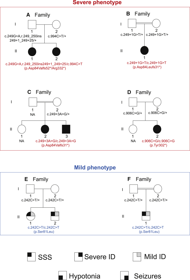
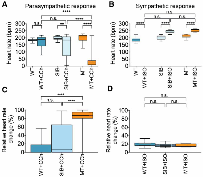
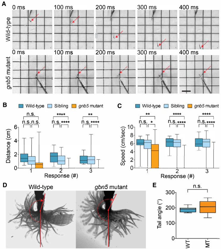
GNB5 encodes the G protein β subunit 5 and is involved in inhibitory G protein signaling. Here, we report mutations in GNB5 that are associated with heart-rate disturbance, eye disease, intellectual disability, gastric problems, hypotonia, and seizures in nine individuals from six families. We observed an association between the nature of the variants and clinical severity; individuals with loss-of-function alleles had more severe symptoms, including substantial developmental delay, speech defects, severe hypotonia, pathological gastro-esophageal reflux, retinal disease, and sinus-node dysfunction, whereas related heterozygotes harboring missense variants presented with a clinically milder phenotype. Zebrafish gnb5 knockouts recapitulated the phenotypic spectrum of affected individuals, including cardiac, neurological, and ophthalmological abnormalities, supporting a direct role of GNB5 in the control of heart rate, hypotonia, and vision.

Keywords: G-protein signaling; heart rate; hypotonia; intellectual disability; parasympathetic system; whole-exome sequencing.


Figures

Figure 1
Figure 1
Pedigrees from the Six Families Investigated in this Study Affected members of families A to D (upper red-lined panel) and E to F (lower blue-lined panel) show severe and mild manifestation of the core symptoms of the syndrome defined in this study. Filled symbols represent individuals with severe sinus sick syndrome (SSS; top left quarter), intellectual disability (ID; top right quarter), hypotonia (bottom left quarter), and seizures (bottom right quarter), whereas a light-gray top left quarter indicates the presence of mild ID. Genotypes are specified according to GenBank: NM_006578.3.
Figure 2
Figure 2
Cardiac Function in gnb5 Mutant Zebrafish (A–D) Box-whisker plots demonstrate the heart rate response and the relative heart rate change of 5 dpf wild-type (WT), sibling (SIB), and gnb5 mutant (MT) larvae. Embryos at 5 dpf were embedded in 0.3% agarose prepared in E3 medium containing 16 mg/ml Tricaine. Basal heart rates were recorded first. Then, (A and C) 400 μM of the parasympathetic agonist carbachol (CCh; Sigma-Aldrich C4382) (WT n = 10, SIB n = 39, MT n = 14) or (B and D) 100 μM of the sympathetic agonist isoproterenol hydrochloride (ISO; Sigma-Aldrich 1351005) (WT n = 12, SIB n = 22, MT n = 9) was added and incubated for 30 min and heart rates were measured. Recordings were performed at 150 frames per second and were analyzed with ImageJ. The relative heart rate change is the percentage change between the basal heart rate measured and the heart rate after addition of CCh or ISO. n denotes the number of fish used per dataset. Differences between two groups were analyzed via the Student’s t test. Differences between more than two groups were analyzed via one-way ANOVA with Tukey’s post-hoc test. Data are shown as mean ± SEM, and p < 0.05 was considered significant. p < 0.05, ∗∗p ≤ 0.01, ∗∗∗p ≤ 0.001, ∗∗∗∗p ≤ 0.0001. p > 0.05 was considered not significant (n.s.). bpm, beats per minute.
Figure 3
Figure 3
Neurologic Function in gnb5 Mutant Zebrafish (A–C) Touch-evoked escape response assay in which three consecutive tactile stimuli were applied. Embryos at 3 dpf were placed in the middle of a standard 88 mm petri dish containing E3 medium. Three consecutive tactile stimuli were applied by touching the tail of the embryo with an insect pin. Stimuli were only applied when the embryo was still. Behavior was recorded with a standard camera (30 fps) and analyzed with ImageJ (NIH) and the plugin MTrackJ. (A) shows representative responses of 3 dpf wild-type and gnb5 mutant embryos to a touch stimulus. Scale bar, 0.5 cm. Box-whisker plots show quantification of the (B) swimming distance and (C) swimming speed in TL wild-types (n = 19), siblings (n = 46), and gnb5 mutants (n = 27). (D and E) Analysis of maximum tail movement at 5 dpf. Larvae at 5 dpf were sedated in E3 containing 16 mg/ml Tricaine and embedded in 0.5% UltraPureTM agarose (Invitrogen 16500-500) in a 35 mm glass bottom dish. After setting, the agarose was cut away caudal to the swimming bladder, leaving the tail free to move. The dish was filled with E3 medium and embryos were left to recover from the sedation for 10 min at 28°C. Next, a maximal escape response was elicited by repeatedly touching the head of the embryo with an insect pin. Recordings were performed at 280 fps, for 30 s, with a high-speed CCD camera (Hamamatsu Photonics K.K., C9300-221) and analyzed with ImageJ (angle tool). (D) shows representative minimum projection images of tail movement in wild-type and gnb5 mutant embryos, including tail angle analysis. The tail angle represents the angle between the head-tail midline axis in resting state and a line that was drawn from just caudal of the swimbladder to the tip of the tail at maximal tail movement. (E) Tail angle quantification is displayed in box-whisker plots (wild-type n = 10, gnb5 mutants n = 10). fps, frames per second. n denotes the number of fish used per dataset. Differences between two groups were analyzed via the Student’s t test. Differences between more than two groups were analyzed via one-way ANOVA with Tukey’s post-hoc test. Data are shown as mean ± SEM, and p < 0.05 was considered significant. p < 0.05, ∗∗p ≤ 0.01, ∗∗∗p ≤ 0.001, ∗∗∗∗p ≤ 0.0001. p > 0.05 was considered not significant (n.s.).

References

    1. Daly A.F., Lysy P.A., Desfilles C., Rostomyan L., Mohamed A., Caberg J.H., Raverot V., Castermans E., Marbaix E., Maiter D. GHRH excess and blockade in X-LAG syndrome. Endocr. Relat. Cancer. 2016;23:161–170. - PMC - PubMed
    1. Daly A.F., Yuan B., Fina F., Caberg J.H., Trivellin G., Rostomyan L., de Herder W.W., Naves L.A., Metzger D., Cuny T. Somatic mosaicism underlies X-linked acrogigantism syndrome in sporadic male subjects. Endocr. Relat. Cancer. 2016;23:221–233. - PMC - PubMed
    1. Trivellin G., Daly A.F., Faucz F.R., Yuan B., Rostomyan L., Larco D.O., Schernthaner-Reiter M.H., Szarek E., Leal L.F., Caberg J.H. Gigantism and acromegaly due to Xq26 microduplications and GPR101 mutation. N. Engl. J. Med. 2014;371:2363–2374. - PMC - PubMed
    1. Krishnan A., Mustafa A., Almén M.S., Fredriksson R., Williams M.J., Schiöth H.B. Evolutionary hierarchy of vertebrate-like heterotrimeric G protein families. Mol. Phylogenet. Evol. 2015;91:27–40. - PubMed
    1. Alfaiz A.A., Micale L., Mandriani B., Augello B., Pellico M.T., Chrast J., Xenarios I., Zelante L., Merla G., Reymond A. TBC1D7 mutations are associated with intellectual disability, macrocrania, patellar dislocation, and celiac disease. Hum. Mutat. 2014;35:447–451. - PubMed

MeSH terms