. 2016 Sep:11:239-248.
doi: 10.1016/j.ebiom.2016.08.006. Epub 2016 Aug 4.

Sickle Cell Trait Increases Red Blood Cell Storage Hemolysis and Post-Transfusion Clearance in Mice

Affiliations

Sickle Cell Trait Increases Red Blood Cell Storage Hemolysis and Post-Transfusion Clearance in Mice

David O Osei-Hwedieh et al. EBioMedicine. 2016 Sep.

Abstract

Background: Transfusion of blood at the limits of approved storage time is associated with lower red blood cell (RBC) post-transfusion recovery and hemolysis, which increases plasma cell-free hemoglobin and iron, proposed to induce endothelial dysfunction and impair host defense. There is noted variability among donors in the intrinsic rate of storage changes and RBC post-transfusion recovery, yet genetic determinants that modulate this process are unclear.

Methods: We explore RBC storage stability and post-transfusion recovery in murine models of allogeneic and xenogeneic transfusion using blood from humanized transgenic sickle cell hemizygous mice (Hbatm1PazHbbtm1TowTg(HBA-HBBs)41Paz/J) and human donors with a common genetic mutation sickle cell trait (HbAS).

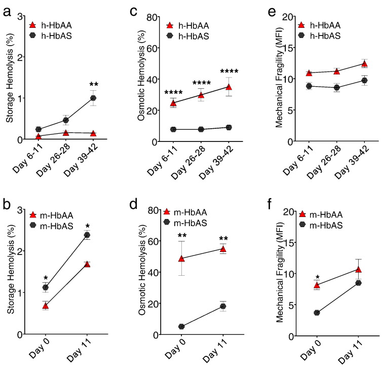
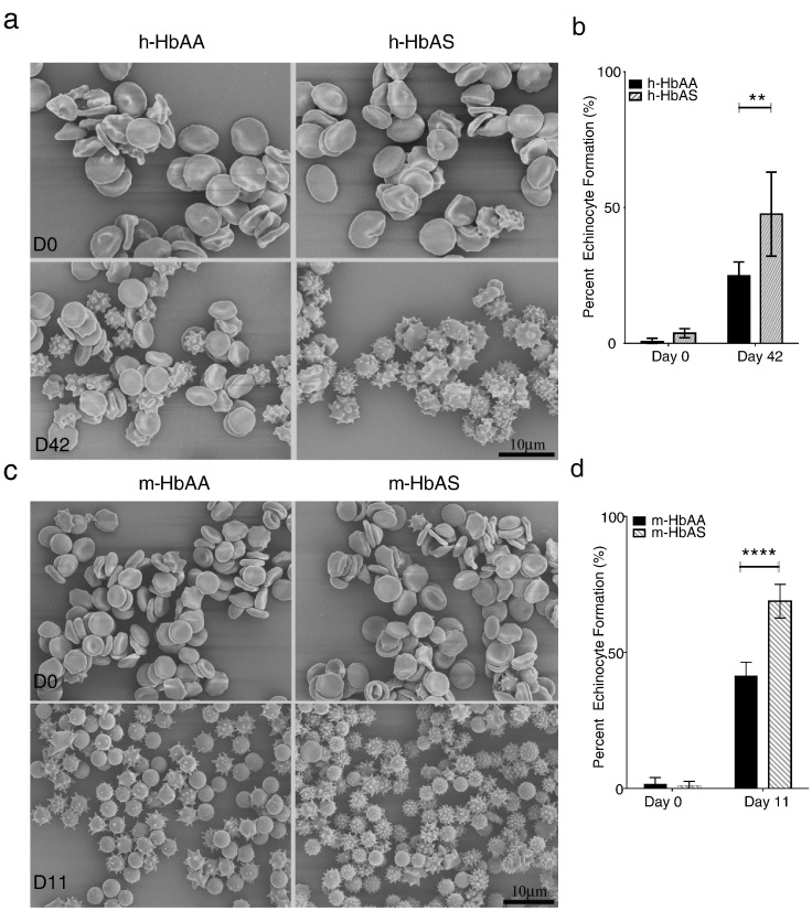
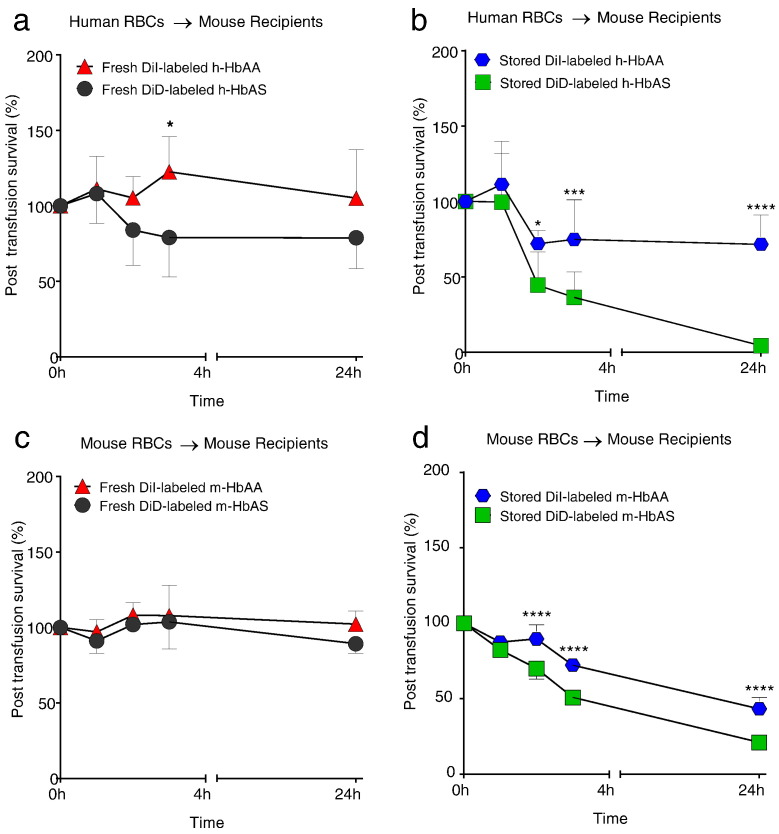
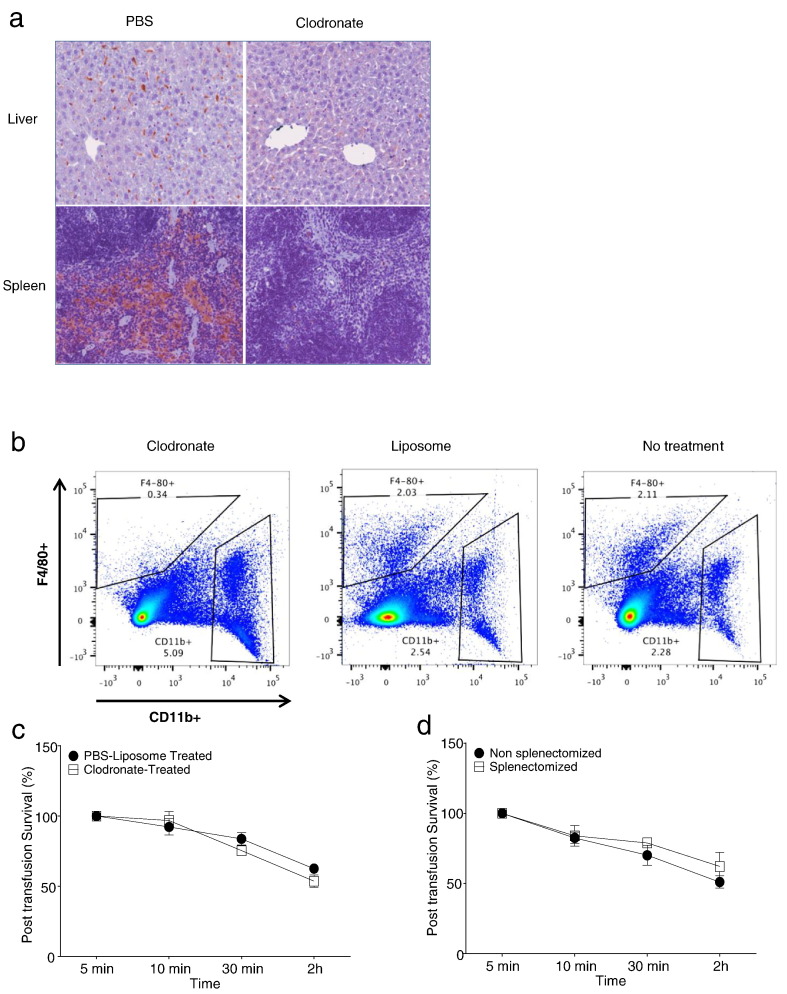
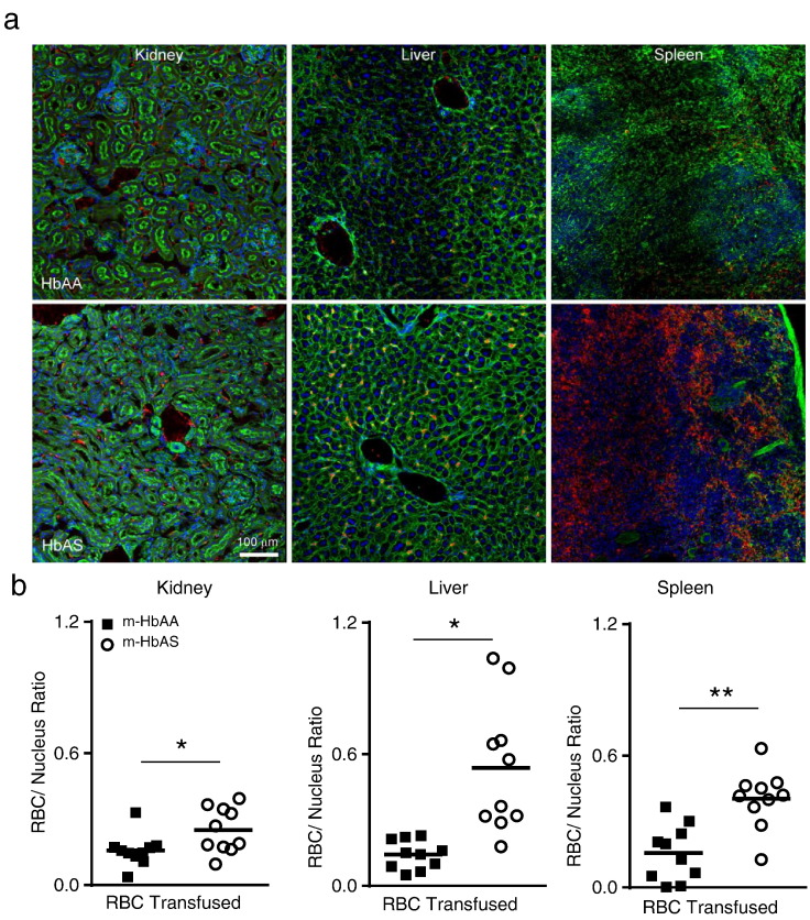
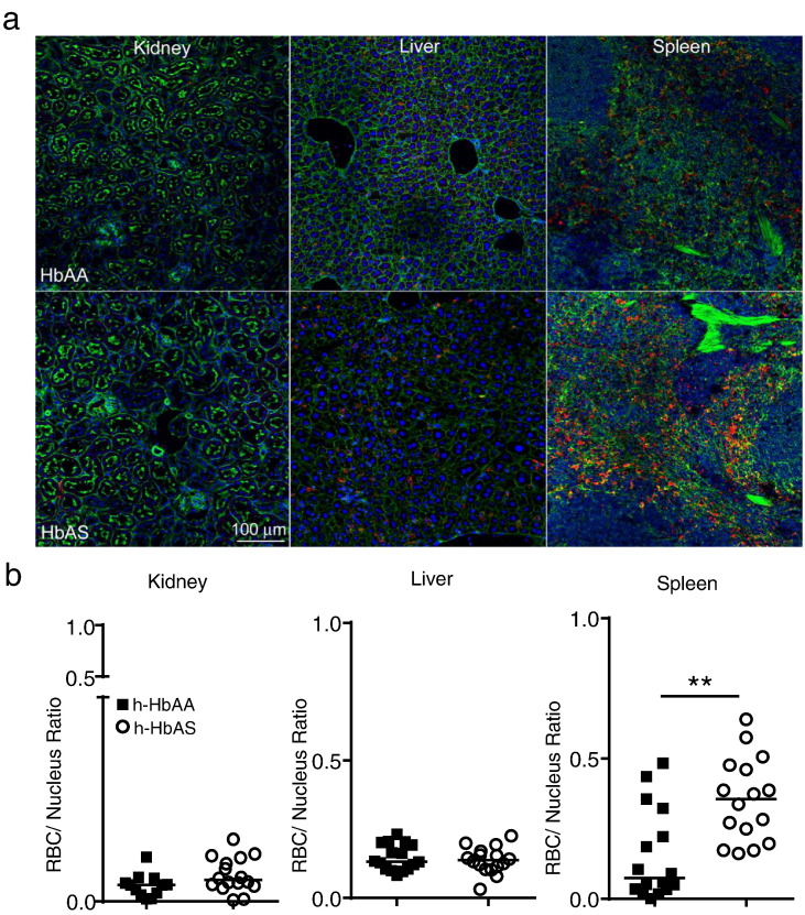
Findings: Human and transgenic HbAS RBCs demonstrate accelerated storage time-dependent hemolysis and reduced post-transfusion recovery in mice. The rapid post-transfusion clearance of stored HbAS RBC is unrelated to macrophage-mediated uptake or intravascular hemolysis, but by enhanced sequestration in the spleen, kidney and liver. HbAS RBCs are intrinsically different from HbAA RBCs, with reduced membrane deformability as cells age in cold storage, leading to accelerated clearance of transfused HbAS RBCs by entrapment in organ microcirculation.

Interpretation: The common genetic variant HbAS enhances RBC storage dysfunction and raises provocative questions about the use of HbAS RBCs at the limits of approved storage.

Keywords: Blood; Post-transfusion survival; RBC hemolysis; Red cell storage; Sickle cell trait; Transfusion practice.


Figures

Fig. 1
Fig. 1
HbAS is associated with higher storage hemolysis in RBCs. Fresh leukoreduced human RBC units (n = 3 HbAA and n = 9 HbAS donors) processed and stored under standard blood banking conditions and murine RBC units (n = 3 HbAA RBCs and n = 3 HbAS RBCs pooled samples, where each pooled sample contained RBCs from n = 11 mice) were stored at 4 °C, sampled and tested at various times during a 42-day or 11-day storage period, respectively. (a,b) At the end of storage, there was a higher concentration of free hemoglobin in HbAS RBC samples compared to HbAA RBCs. (c,d) HbAS RBCs exhibited increased resilience under hypotonic-induced osmotic shock compared to HbAA RBCs throughout storage, as measured by the fraction of lysed RBCs in a hypotonic solution. (e,f) Human and murine RBCs showed no difference at the end of storage under mechanical stress induced by agitation with one metal bead (3/32″) for 180 min in a 96-well plate. HbAA RBCs are indicated as red triangle and HbAS RBCs are indicated as black circle. Human HbAA (h-HbAA), Human HbAS (h-HbAS), Mouse HbAA (m-HbAA), Mouse HbAS (m-HbAS). The results are presented as mean ± SEM. *p < 0.05; **p < 0.01; ****p < 0.0001 analyzed by 2Way ANOVA, GraphPad Prism 6.0.
Fig. 2
Fig. 2
Echinocyte formation is increased in HbAS RBCs compared to HbAA RBCs during storage. RBC images were taken with a JEOL JSM- 6335F scanning electron microscope. (a) Upper panel shows representative fields from fresh and stored human HbAA and HbAS RBCs at the beginning and the end of 39–42 day storage. (b) Five fields per sample were counted and analyzed from one healthy and one SCT representative donors at the beginning and end of storage. Human and mouse RBC SEM analysis was performed twice. (c) Lower panel shows representative fields from fresh and 11-day stored murine RBCs obtained from WT C57BL/6 and Berkeley hemizygous mice (One pooled sample each where n = 11 mice donor RBCs/pooled sample). (d) Six fields were counted and analyzed for echinocyte formation from each pooled sample. The results are presented as mean ± SEM, where echinocyte formation represents % echinocyte per total number of cells in fields counted. **p < 0.01; ****p < 0.0001 analyzed by 2Way ANOVA, GraphPad Prism 6.0.
Fig. 3
Fig. 3
Stored human and murine HbAS RBCs show accelerated post-transfusion clearance compared to stored HbAA RBCs. (a) WT C57BL/6 recipient mice (n = 6) were transfused with a 50:50 mixture of fresh DiI-labeled h-HbAA RBCs (indicated as red triangle) and fresh DiD-labeled h-HbAS RBCs (indicated as black circle). (b) N = 9 WT C57BL/6 recipients were transfused with a 50:50 mixture of 39-day stored DiI-labeled h-HbAA RBCs (indicated as blue circle) and stored DiD-labeled h-HbAS RBCs (indicated as green square) both re-suspended to a 55% hematocrit following labeling. (c) WT C57BL/6 mice recipients (n = 6) were transfused with a 50:50 mixture of fresh DiI-labeled m-HbAA RBCs (indicated as red triangle) and fresh DiD-labeled m-HbAS RBCs (indicated as black circle). (d) WT recipient mice (n = 6) were transfused with 11-day stored DiI-labeled m-HbAA RBCs (indicated as blue circle) and DiD-labeled stored m-HbAS RBCs (indicated as green square). All mice were 8–12 weeks of age and received a total volume of 200 μl of leukoreduced HbAA and HbAS RBCs (100 μl each). To mimic similar conditions, human and murine RBCs were stored in glass. Post-transfusion recovery was measured by dual-label cell tracking by flow cytometry unless stated otherwise. The results are presented as mean ± SD. *p < 0.05; ***p < 0.001; ****p < 0.0001 analyzed by 2Way ANOVA, GraphPad Prism 6.0.
Fig. 4
Fig. 4
Clodronate treatment or splenectomy does not alter stored m-HbAS RBC post-transfusion survival. (a) WT recipient mice were treated with clodronate or PBS liposomes 24 h prior to liver and spleen harvest for immunohistochemistry. Representative images are shown indicating staining for F4/80 + macrophages. (b) WT mice received either clodronate, PBS liposomes or no treatment. Mice were euthanized after 24 h. Spleens were homogenized and stained with F4/80 and CD11b antibodies. Samples were analyzed using flow cytometry to quantify the F4/80 + population. (c) WT recipient mice were transfused with 11-day stored m-HbAS RBCs 24 h following clodronate (n = 3) or PBS liposome (n = 3) injection (2 mg i.p.). (d) WT recipient mice were transfused with 11-day stored m-HbAS RBCs 5 d following splenectomy (n = 4) or sham procedure (n = 6) Results presented are mean ± SD.
Fig. 5
Fig. 5
Splenectomy does not increase kidney and liver sequestration of stored m-HbAS RBCs following transfusion. Splenectomized WT C57BL/6 recipient mice (n = 7) or sham operated (n = 5) were transfused with stored m-HbAS RBCs. Kidney and liver organs were harvested 2 h post-transfusion, fixed in 2% PFA and processed for confocal imaging. (a) Confocal images showing sequestered m-HbAS RBCs in recipient mouse tissues (Magnification 20 ×, Green: phalloidin, Blue: DAPI, Red: Cy3-labeled m-HbAS RBCs). (b) Individual points represent sequestered m-HbAS RBCs normalized to nuclei number per section quantified by fluorescence intensity using NIS Elements. Two sections from each recipient mouse organ were analyzed.
Fig. 6
Fig. 6
Increased tissue sequestration of stored m-HbAS RBCs within kidney, liver and spleen organs following transfusion when compared with stored m-HbAA RBCs. WT C57BL/6 recipient mice (n = 5 per group) were transfused with stored cy3 labeled m-HbAA or m-HbAS RBCs. Kidney, liver and spleen organs were harvested 2 h post-transfusion for confocal imaging. (a) Confocal images showing sequestered m-HbAA and m-HbAS RBCs in recipient mouse tissues (Magnification 20 ×, Green: phalloidin, Blue: DAPI, Red: cy3 RBCs). (b) Individual points represent sequestered cy3-labeled RBCs normalized to nuclei number per section quantified by fluorescence intensity using NIS Elements. Two sections from each recipient mouse organ were analyzed (n = 10 sections per group). Statistical Analysis by two-tailed Mann Whitney U, *p < 0.05, **p < 0.01, lines indicate the median.
Fig. 7
Fig. 7
Increased sequestration of stored h-HbAS RBCs in the spleen following transfusion when compared with stored h-HbAA RBCs. WT C57BL/6 recipient mice (n = 8 per group) were transfused with conventionally stored h-HbAA or h-HbAS RBCs. Kidney, liver and spleen organs were harvested 2 h post-transfusion for confocal imaging. (a) Confocal images showing sequestered h-HbAA and h-HbAS RBCs in recipient mouse tissues (Magnification 20 ×, Green: phalloidin, Blue: DAPI, Red: Glycophorin A: FITC antibody for human RBCs). (b) Individual points represent sequestered h-RBCs normalized to nuclei number per section quantified by fluorescence intensity using NIS Elements. Two sections from each recipient mouse organ were analyzed (n = 16 sections per group). Statistical Analysis by two-tailed Mann Whitney U, **p < 0.01, lines indicate the median.

References

    1. Awwad H.K., Moussa L., Sheraki A.S. The effect of red cell aging on chromium-51 binding and in vitro elution. J. Nucl. Med. 1966;7:687–695. - PubMed
    1. Bazin R., Aubin E., Boyer L., St-AMOUR I., Roberge C., Lemieux R. Functional in vivo characterization of human monoclonal anti-D in NOD-scid mice. Blood. 2002;99:1267–1272. - PubMed
    1. Brittenham G.M., Schechter A.N., Noguchi C.T. Hemoglobin S polymerization: primary determinant of the hemolytic and clinical severity of the sickling syndromes. Blood. 1985;65:183–189. - PubMed
    1. Callender S.T., Nickel J.F. Sickle cell disease; studied by measuring the survival of transfused red blood cells. J. Lab. Clin. Med. 1949;34:90–104. - PubMed
    1. Connor J., Pak C.C., Schroit A.J. Exposure of phosphatidylserine in the outer leaflet of human red blood cells. Relationship to cell density, cell age, and clearance by mononuclear cells. J. Biol. Chem. 1994;269:2399–2404. - PubMed

MeSH terms