

Hydrothermally processed 1D hydroxyapatite: Mechanism of formation and biocompatibility studies
- PMID: 27524076
- PMCID: PMC4987716
- DOI: 10.1016/j.msec.2016.06.047
Hydrothermally processed 1D hydroxyapatite: Mechanism of formation and biocompatibility studies
Abstract
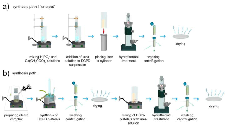
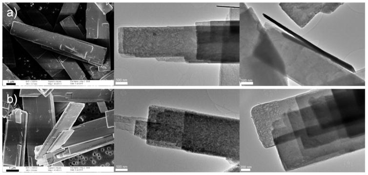
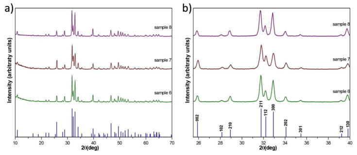
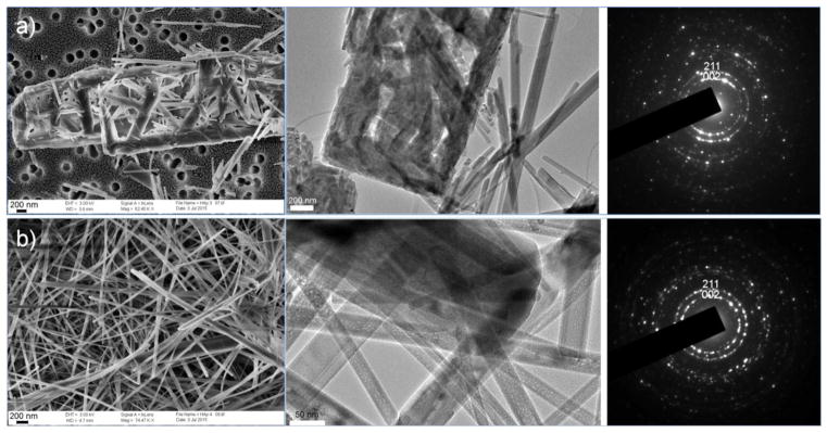
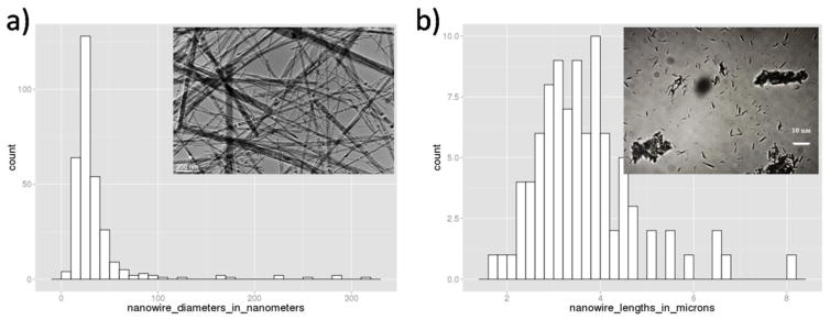
Recent developments in bone tissue engineering have led to an increased interest in one-dimensional (1D) hydroxyapatite (HA) nano- and micro-structures such as wires, ribbons and tubes. They have been proposed for use as cell substrates, reinforcing phases in composites and carriers for biologically active substances. Here we demonstrate the synthesis of 1D HA structures using an optimized, urea-assisted, high-yield hydrothermal batch process. The one-pot process, yielding HA structures composed of bundles of ribbons and wires, was typified by the simultaneous occurrence of a multitude of intermediate reactions, failing to meet the uniformity criteria over particle morphology and size. To overcome these issues, the preparation procedure was divided to two stages: dicalcium phosphate platelets synthesized in the first step were used as a precursor for the synthesis of 1D HA in the second stage. Despite the elongated particle morphologies, both the precursor and the final product exhibited excellent biocompatibility and caused no reduction of viability when tested against osteoblastic MC3T3-E1 cells in 2D culture up to the concentration of 2.6mg/cm(2). X-ray powder diffraction combined with a range of electron microscopies and laser diffraction analyses was used to elucidate the formation mechanism and the microstructure of the final particles. The two-step synthesis involved a more direct transformation of DCP to 1D HA with the average diameter of 37nm and the aspect ratio exceeding 100:1. The comparison of crystalline domain sizes along different crystallographic directions showed no signs of significant anisotropy, while indicating that individual nanowires are ordered in bundles in the b crystallographic direction of the P63/m space group of HA. Intermediate processes, e.g., dehydration of dicalcium phosphate, are critical for the formation of 1D HA alongside other key aspects of this phase transformation, it must be investigated in more detail in the continuous design of smart HA micro- and nano-structures with advanced therapeutic potentials.
Keywords: Biomedical; Hydrothermal; Hydroxyapatite; Nanowires; Particle size distribution.
Copyright © 2016 Elsevier B.V. All rights reserved.
Figures
Similar articles
-
One- and three-dimensional growth of hydroxyapatite nanowires during sol-gel-hydrothermal synthesis.ACS Appl Mater Interfaces. 2012 Mar;4(3):1490-9. doi: 10.1021/am201735k. Epub 2012 Feb 15. ACS Appl Mater Interfaces. 2012. PMID: 22296410
-
Preparation and properties of in-situ growth of carbon nanotubes reinforced hydroxyapatite coating for carbon/carbon composites.Mater Sci Eng C Mater Biol Appl. 2017 Jan 1;70(Pt 1):805-811. doi: 10.1016/j.msec.2016.09.060. Epub 2016 Sep 28. Mater Sci Eng C Mater Biol Appl. 2017. PMID: 27770958
-
Fabrication and characterization of needle-like nano-HA and HA/MWNT composites.J Mater Sci Mater Med. 2008 Jan;19(1):75-81. doi: 10.1007/s10856-007-3107-5. Epub 2007 Jun 19. J Mater Sci Mater Med. 2008. PMID: 17577639
-
Hydroxyapatite-titanium bulk composites for bone tissue engineering applications.J Biomed Mater Res A. 2015 Feb;103(2):791-806. doi: 10.1002/jbm.a.35198. Epub 2014 Apr 25. J Biomed Mater Res A. 2015. PMID: 24737723 Review.
-
High-aspect-ratio nanostructured hydroxyapatite: towards new functionalities for a classical material.Chem Sci. 2023 Dec 1;15(1):55-76. doi: 10.1039/d3sc05344j. eCollection 2023 Dec 20. Chem Sci. 2023. PMID: 38131070 Free PMC article. Review.
Cited by
-
Hydroxyapatite and Titanium Dioxide Nanoparticles: Radiolabelling and In Vitro Stability of Prospective Theranostic Nanocarriers for 223Ra and 99mTc.Nanomaterials (Basel). 2020 Aug 20;10(9):1632. doi: 10.3390/nano10091632. Nanomaterials (Basel). 2020. PMID: 32825280 Free PMC article.
-
Preparation of Micro/Nano-Structure Copper-Substituted Hydroxyapatite Scaffolds with Improved Angiogenesis Capacity for Bone Regeneration.Materials (Basel). 2018 Aug 23;11(9):1516. doi: 10.3390/ma11091516. Materials (Basel). 2018. PMID: 30142939 Free PMC article.
-
Enhancing bioactivity and stability of polymer-based material-tissue interface through coupling multiscale interfacial interactions with atomic-thin TiO2 nanosheets.Nano Res. 2023;16(4):5247-5255. doi: 10.1007/s12274-022-5153-1. Epub 2022 Dec 5. Nano Res. 2023. PMID: 36532602 Free PMC article.
-
Hydrothermal synthesis of nanocrystalline hydroxyapatite-graphene nanosheet on Ti-6Al-7Nb: mechanical and in vitro corrosion performance.J Mater Sci Mater Med. 2021 Apr 1;32(4):40. doi: 10.1007/s10856-021-06514-w. J Mater Sci Mater Med. 2021. PMID: 33792780 Free PMC article.
-
Microwave-Heating-Assisted Synthesis of Ultrathin and Ultralong Hydroxyapatite Nanowires Using Biogenic Creatine Phosphate and Their Derived Flexible Bio-Paper with Drug Delivery Function.Molecules. 2025 Feb 21;30(5):996. doi: 10.3390/molecules30050996. Molecules. 2025. PMID: 40076221 Free PMC article.
References
-
- Roeder RK, Converse GL, Kane RJ, Yue W. Hydroxyapatite-reinforced polymer biocomposites for synthetic bone substitutes. JOM. 2008;60:38–45.
-
- Aston DE, Bow JR, Gangadean DN. Mechanical properties of selected nanostructured materials and complex bio-nano, hybrid and hierarchical systems. Int Mater Rev. 2013;58:167–202. doi: 10.1179/1743280412Y.0000000012. - DOI
MeSH terms
Substances
Grants and funding
LinkOut - more resources
Full Text Sources
Other Literature Sources
Miscellaneous
