Review
. 2016 Aug 15;20(1):228.
doi: 10.1186/s13054-016-1400-8.

Critical care ultrasonography in acute respiratory failure

Affiliations
Review

Critical care ultrasonography in acute respiratory failure

Philippe Vignon et al. Crit Care. .

Abstract

Acute respiratory failure (ARF) is a leading indication for performing critical care ultrasonography (CCUS) which, in these patients, combines critical care echocardiography (CCE) and chest ultrasonography. CCE is ideally suited to guide the diagnostic work-up in patients presenting with ARF since it allows the assessment of left ventricular filling pressure and pulmonary artery pressure, and the identification of a potential underlying cardiopathy. In addition, CCE precisely depicts the consequences of pulmonary vascular lesions on right ventricular function and helps in adjusting the ventilator settings in patients sustaining moderate-to-severe acute respiratory distress syndrome. Similarly, CCE helps in identifying patients at high risk of ventilator weaning failure, depicts the mechanisms of weaning pulmonary edema in those patients who fail a spontaneous breathing trial, and guides tailored therapeutic strategy. In all these clinical settings, CCE provides unparalleled information on both the efficacy and tolerance of therapeutic changes. Chest ultrasonography provides further insights into pleural and lung abnormalities associated with ARF, irrespective of its origin. It also allows the assessment of the effects of treatment on lung aeration or pleural effusions. The major limitation of lung ultrasonography is that it is currently based on a qualitative approach in the absence of standardized quantification parameters. CCE combined with chest ultrasonography rapidly provides highly relevant information in patients sustaining ARF. A pragmatic strategy based on the serial use of CCUS for the management of patients presenting with ARF of various origins is detailed in the present manuscript.

Keywords: Echocardiography; Echocardiography Doppler; Pulmonary edema; Respiratory distress syndrome; Respiratory insufficiency; Ultrasonography.


Figures
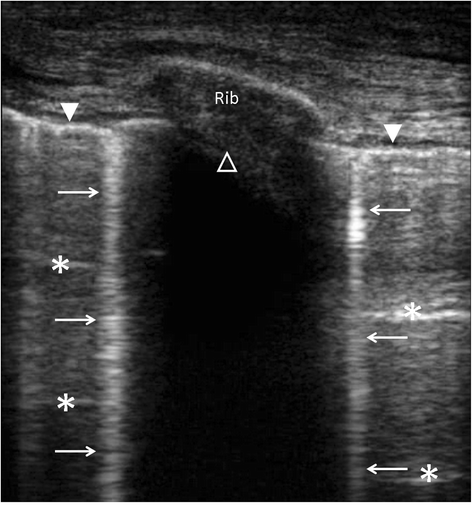
Fig. 1
Fig. 1
Longitudinal view of an intercostal space using chest ultrasonography disclosing the pleural line (closed white arrow head), A-lines (white asterisks), B-lines (white arrows), and shadowing related to the rib (open white arrow head)
Fig. 2
Fig. 2
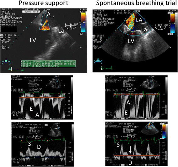
Transesophageal echocardiographic hemodynamic monitoring in a patient with a known ischemic cardiomyopathy who failed ventilator weaning. Under pressure support, a trivial mitral regurgitation was disclosed by color Doppler mapping in the two-chamber view (upper left), and left cardiac filling pressures were low, as reflected by an inverted mitral E/A Doppler pattern (middle left) and a predominant pulmonary vein S wave (lower left). During the spontaneous breathing trial, a severe mitral regurgitation occurred (upper right), mitral Doppler pattern was restrictive (middle right), and pulmonary vein Doppler disclosed a reversed D wave consistent with massive mitral insufficiency (lower right, arrow). The acute mitral regurgitation was attributed to a papillary muscle dysfunction secondary to a transient myocardial ischemic event. LA left atrium, La left auricle, LV left ventricle. Adapted from [7]

Comment in

  • Pulmonary vein signal in mitral regurgitation.
    Balthazar T, Jacobs B, Voigt JU. Balthazar T, et al. Crit Care. 2018 May 11;22(1):123. doi: 10.1186/s13054-018-2031-z. Crit Care. 2018. PMID: 29747648 Free PMC article. No abstract available.

References

    1. Mayo P, Beaulieu Y, Doelken P, Feller-Kopman D, Harrod C, Kaplan A, et al. American College of Chest Physicians/La Société de Réanimation de Langue Française statement on competence in critical care ultrasonography. Chest. 2009;135:1050–60. doi: 10.1378/chest.08-2305. - DOI - PubMed
    1. Vignon P, Mentec H, Terre S, Gastinne H, Guéret P, Lemaire F. Diagnostic accuracy and therapeutic impact of transthoracic and transesophageal echocardiography in mechanically ventilated patients in the ICU. Chest. 1994;106:1829–34. doi: 10.1378/chest.106.6.1829. - DOI - PubMed
    1. Bouhemad B, Mongodi S, Via G, Rouquette I. Ultrasound for “lung monitoring” of ventilated patients. Anesthesiology. 2015;122:437–47. doi: 10.1097/ALN.0000000000000558. - DOI - PubMed
    1. Silva S, Biendel C, Ruiz J, Olivier M, Bataille B, Geeraerts T, et al. Usefulness of cardiothoracic chest ultrasound in the management of acute respiratory failure in critical care practice. Chest. 2013;144:859–65. doi: 10.1378/chest.13-0167. - DOI - PubMed
    1. Gandhi SK, Powers JC, Nomeir AM, Fowle K, Kitzman DW, Rankin KM, et al. The pathogenesis of acute pulmonary edema associated with hypertension. N Engl J Med. 2001;344:17–22. doi: 10.1056/NEJM200101043440103. - DOI - PubMed

MeSH terms