. 2016 Aug 23;16(8):2219-2230.
doi: 10.1016/j.celrep.2016.07.039. Epub 2016 Aug 11.

Necroptosis Promotes Staphylococcus aureus Clearance by Inhibiting Excessive Inflammatory Signaling

Affiliations

Necroptosis Promotes Staphylococcus aureus Clearance by Inhibiting Excessive Inflammatory Signaling

Kipyegon Kitur et al. Cell Rep. .

Abstract

Staphylococcus aureus triggers inflammation through inflammasome activation and recruitment of neutrophils, responses that are critical for pathogen clearance but are associated with substantial tissue damage. We postulated that necroptosis, cell death mediated by the RIPK1/RIPK3/MLKL pathway, would function to limit pathological inflammation. In models of skin infection or sepsis, Mlkl-/- mice had high bacterial loads, an inability to limit interleukin-1b (IL-1b) production, and excessive inflammation. Similarly, mice treated with RIPK1 or RIPK3 inhibitors had increased bacterial loads in a model of sepsis. Ripk3-/- mice exhibited increased staphylococcal clearance and decreased inflammation in skin and systemic infection, due to direct effects of RIPK3 on IL-1b activation and apoptosis. In contrast to Casp1/4-/- mice with defective S. aureus killing, the poor outcomes of Mlkl-/- mice could not be attributed to impaired phagocytic function. We conclude that necroptotic cell death limits the pathological inflammation induced by S. aureus.


Figures

Figure 1
Figure 1. MLKL contributes to the clearance of S. aureus from infected skin
(A–I) Mlkl−/− and wild type (WT) mice were infected subcutaneously with 2 × 10⁶ CFU/mouse of S. aureus (SA) for 5 days and the area of infection biopsied for analysis. (A) S. aureus colony forming units (CFU) recovered (B) Representative images of mice showing skin lesions caused by S. aureus infection. (C) Quantification of lesion sizes from (B). (D and E) Cytotoxicity in HaCaT (C) or primary keratinocytes (HEKn) (D) pretreated with 10 μM necrosulfonamide (NSA) and infected with WT S. aureus. (F) Cytotoxicity in MLKL-deficient HaCaT infected with S. aureus for 4 h. (G) Immunoblot of HaCaT with MLKL knocked down by siRNA or scrambled control. (H, I and J) Number of neutrophils (PMNs) (H), macrophages (Macs) (I), and gamma-delta (γδ) T cells (J) on Mlkl−/− and WT mice. (K) Cytokines from S. aureus infected WT and Mlkl−/− mice (“nd” means “not detected”). (L) Representative images of trichrome stain on skin biopsies of PBS or S. aureus-infected mice. Scale bars, 100 μm. (M) Western blot showing cleaved caspase-1 (p20), full-length caspase-1, actin and ponceau on skin homogenate of infected WT and Mlkl−/− mice. Each point represents a mouse and lines show median values. Data are represented as bar graphs with mean ± SEM. Results shown are pooled from at least two independent experiments. *p<0.05, **p<0.01. See also Figures S1 and S2; and Tables S1 and S2.
Figure 2
Figure 2. Lack of caspases-1/4 is detrimental in S. aureus skin infection
(A–G) Casp1/4−/− and wild type (WT) mice were infected subcutaneously with 2 × 10⁶ CFU/mouse of S. aureus (SA) for 5 days and the area of infection biopsied for analysis. (A) S. aureus colony forming units (CFU) recovered (B) Representative images of mice showing skin lesions caused by S. aureus infection. (C) Quantification of lesion sizes from (B). (D, E and F) Number of neutrophils (PMNs) (D), macrophages (Macs) (E), and gamma-delta (γδ) T cells (F) on Mlkl−/− and WT mice. (G) Cytokines from S. aureus infected WT and Mlkl−/− mice (“nd” means “not detected”). Each point represents a mouse and lines show median values. Data are represented as bar graphs with mean ± SEM. Results shown are pooled from at least two independent experiments. *p<0.05, **p<0.01. See also Table S3.
Figure 3
Figure 3. Pharmacological blockade of necroptosis leads to worse outcome during S. aureus skin infection
(A) Cytotoxicity in keratinocyte cell line (HaCaT) pretreated with necrostatin-1 stable (Nec-1s) and infected with S. aureus MOI10 for 4 h. (B–I) Mice were infected subcutaneously with S. aureus (SA) and treated with 10mg/kg necrostatin-1 stable (Nec) or DMSO control for 5 days and their skin tissues biopsied for analysis. (B) S. aureus CFU recovered on day 5 from Nec-1s (N) or DMSO-treated mice. (C) Representative images of mice showing lesions on day 5 of skin infection. (D) Quantification of lesion sizes on day 5. (E–G) Number of neutrophils (PMNs) (E), gamma-delta (γδ) T cells (F), and macrophages (Macs) (G) on infected area. (H) Cytokines from S. aureus-infected mice (“nd” means “not detected”). (I) Representative images of trichrome stain on skin biopsies of PBS or S. aureus-infected mice. Scale bars, 100 μm. Each point represents a mouse and lines show median values. Data are represented as bar graphs with mean ± SEM. Results shown are pooled from at least two independent experiments. *p<0.05, **p<0.01. See also Figure S3 and Table S4.
Figure 4
Figure 4. Ripk3−/− mice have improved outcome in S. aureus skin infection
(A) Cytotoxicity in HaCaT pretreated with 10 μM GSK‘872 and infected with S. aureus. (B–J) Ripk3−/− and wild type (WT) mice were infected subcutaneously with S. aureus (SA) for 5 days and skin tissues biopsied for analysis. (B) S. aureus CFU recovered on day 5 on day of infection. (C) Representative images of mice showing lesions on day 5 of skin infection (D) Quantification of lesion sizes on day 5. (E–G) Number of neutrophils (PMNs) (E), gamma-delta (γδ) T cells (F), and macrophages (Macs) (G) on infected area. (H) Cytokines after 5 days of infection. (I) Representative images of trichrome stain on skin biopsies of S. aureus-infected mice. Scale bars, 100 μm. (J) Number of total Annexin V+ cells and number of Annexin V+ immune cells (CD54+) after 5 days of infection. Each point represents a mouse and lines show median values. Data are represented as bar graphs with mean ± SEM. Results shown are pooled from at least two independent experiments. *p<0.05, **p<0.01. See also Table S5.
Figure 5
Figure 5. Activation of caspases-8, -3 and -7 by S. aureus
(A) Western blot showing caspase-8 activation in THP-1 cells after S. aureus infection. (B) Caspases-3/7 activation in HaCaT cells pretreated with 20 μM caspase 3/7 inhibitor or DMSO for 1 h and stimulated with WT or agr S. aureus strains for 4 h. (C) Caspases-3/7 activation in THP-1 cells pretreated with 20 μM caspase 3/7 inhibitor or DMSO for 1 h and stimulated with S. aureus for 2 h. (D) Cytotoxicity in WT, Casp8−/−Ripk3−/− and Mlkl−/− BMDMs after S. aureus infection. (E) Cytotoxicity in THP-1 cells treated with DMSO or caspases-3/7 inhibitor and infected with S. aureus for 2 h. (F) Confocal images of skin biopsies obtained from S. aureus-infected WT and Ripk3−/− mice and stained for pan cytokeratin (Red) and cleaved caspase-3 (Green, arrow). Magnification of 200x; Scale bars, 100 μm. Each point represents a mouse and lines show median values. Data are represented as bar graphs with mean ± SEM. Results shown are pooled from at least two independent experiments. *p<0.05, **p<0.01.
Figure 6
Figure 6. Caspases-1/4 are necessary for S. aureus killing by host cells
(A) S. aureus killing by primary keratinocytes (HEKn cells) pretreated with 10 μM NSA or 50 μM ZVAD, infected with S. aureus for 2 h and treated with gentamicin for 4 h. (B) S. aureus uptake by mouse peritoneal exudate cells (PECs) infected with S. aureus for 1 h. (C) S. aureus killing by PECs infected with S. aureus for 24 h. (D) S. aureus uptake by BMDMs from mice after infection for 1 h. (E) Gentamicin protection assay showing S. aureus killing by BMDMs from WT, Ripk3−/−, Mlkl−/− and Casp1/4−/− mice. (F) IL-1β released by BMDMs from WT, Ripk3−/−, Mlkl−/− and Casp1/4−/− mice after stimulation for 4h with S. aureus. (G) S. aureus uptake by Casp8−/−Ripk3−/− (C8−/−R3−/−) BMDMs from mice after infection for 1 h. (H) Gentamicin protection assay showing S. aureus killing by BMDMs from WT and Casp8−/−Ripk3−/− mice. (I) IL-1β released by BMDMs from WT and Casp8−/−Ripk3−/− mice after stimulation for 4h with S. aureus. Data are represented as bar graphs with mean ± SEM. Results shown are pooled from at least two independent experiments. *p<0.05.
Figure 7
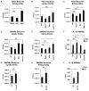
Figure 7. Necroptosis and caspase-1/4 are necessary for bacterial clearance and survival in a murine model of S. aureus sepsis
(A) S. aureus CFU recovered from the blood of mice treated with Nec-1s and infected with 1 × 10⁸ CFU/mouse of S. aureus via retro-orbital route. Mice were sacrificed 24 hours or 48 hours after infection. (B) S. aureus CFU recovered from the blood of mice treated with GSK3002963 and infected with 1 × 10⁸ CFU/mouse of S. aureus via retro-orbital route. Mice were sacrificed 24 hours after infection. (C) Kaplan-Meier survival curves of WT (n=19), Ripk3−/− (n=8), Mlkl−/− (n=9) and Casp1/4−/− (n=5) mice after retro-orbital injection with 1 × 10⁸ CFU/mouse of S. aureus. Each point represents a mouse and lines show median values. Results shown are pooled from at least two independent experiments. *p<0.05, **p<0.01.

Similar articles

Cited by

References

    1. Abtin A, Jain R, Mitchell AJ, Roediger B, Brzoska AJ, Tikoo S, Cheng Q, Ng LG, Cavanagh LL, von Andrian UH, et al. Perivascular macrophages mediate neutrophil recruitment during bacterial skin infection. Nat Immunol. 2014;15:45–53. - PMC - PubMed
    1. Aziz M, Jacob A, Wang P. Revisiting caspases in sepsis. Cell Death Dis. 2014;5:e1526. - PMC - PubMed
    1. Berger SB, Bertin J, Gough PJ. Drilling into RIP1 biology: what compounds are in your toolkit? Cell Death Dis. 2015;6:e1889. - PMC - PubMed
    1. Cai Z, Jitkaew S, Zhao J, Chiang HC, Choksi S, Liu J, Ward Y, Wu LG, Liu ZG. Plasma membrane translocation of trimerized MLKL protein is required for TNF-induced necroptosis. Nat Cell Biol. 2014;16:55–65. - PMC - PubMed
    1. Chan FK, Luz NF, Moriwaki K. Programmed necrosis in the cross talk of cell death and inflammation. Annu Rev Immunol. 2015;33:79–106. - PMC - PubMed

MeSH terms

LinkOut - more resources