

Magnetite nanoparticles for cancer diagnosis, treatment, and treatment monitoring: recent advances
- PMID: 27524934
- PMCID: PMC4981486
- DOI: 10.1016/j.mattod.2015.08.022
Magnetite nanoparticles for cancer diagnosis, treatment, and treatment monitoring: recent advances
Abstract
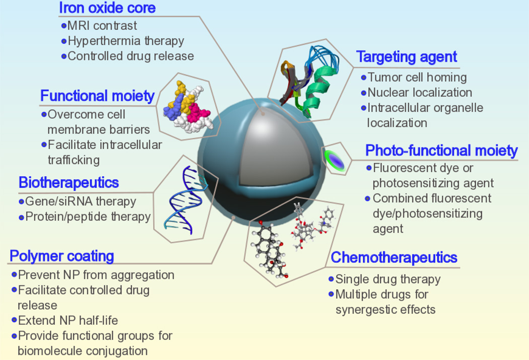
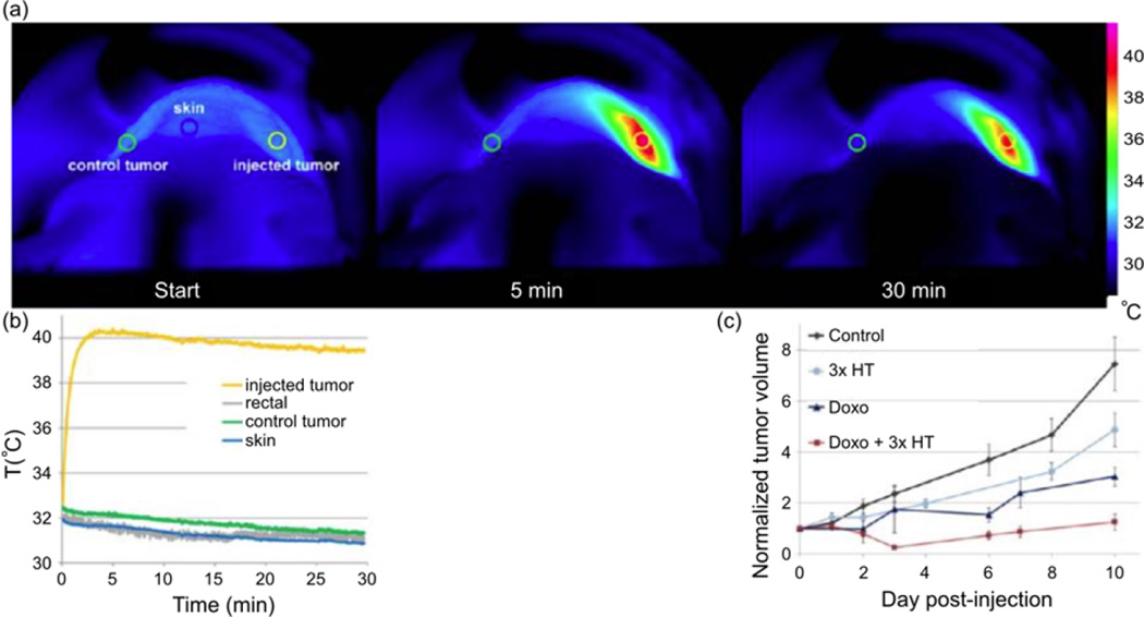
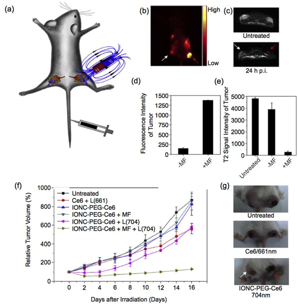
The development of nanoparticles (NPs) for use in all facets of oncological disease detection and therapy has shown great progress over the past two decades. NPs have been tailored for use as contrast enhancement agents for imaging, drug delivery vehicles, and most recently as a therapeutic component in initiating tumor cell death in magnetic and photonic ablation therapies. Of the many possible core constituents of NPs, such as gold, silver, carbon nanotubes, fullerenes, manganese oxide, lipids, micelles, etc., iron oxide (or magnetite) based NPs have been extensively investigated due to their excellent superparamagnetic, biocompatible, and biodegradable properties. This review addresses recent applications of magnetite NPs in diagnosis, treatment, and treatment monitoring of cancer. Finally, some views will be discussed concerning the toxicity and clinical translation of iron oxide NPs and the future outlook of NP development to facilitate multiple therapies in a single formulation for cancer theranostics.
Figures
Similar articles
-
Theranostic Nanoparticles for RNA-Based Cancer Treatment.Acc Chem Res. 2019 Jun 18;52(6):1496-1506. doi: 10.1021/acs.accounts.9b00101. Epub 2019 May 28. Acc Chem Res. 2019. PMID: 31135134 Free PMC article. Review.
-
A review of small molecules and drug delivery applications using gold and iron nanoparticles.Int J Nanomedicine. 2019 Mar 11;14:1633-1657. doi: 10.2147/IJN.S184723. eCollection 2019. Int J Nanomedicine. 2019. PMID: 30880970 Free PMC article. Review.
-
Inorganic Nanoparticles for Cancer Therapy: A Transition from Lab to Clinic.Curr Med Chem. 2018;25(34):4269-4303. doi: 10.2174/0929867325666171229141156. Curr Med Chem. 2018. PMID: 29284391 Review.
-
Polymer coated gold-ferric oxide superparamagnetic nanoparticles for theranostic applications.J Nanobiotechnology. 2018 Oct 13;16(1):80. doi: 10.1186/s12951-018-0405-7. J Nanobiotechnology. 2018. PMID: 30316298 Free PMC article.
-
Long-term live cells observation of internalized fluorescent Fe@C nanoparticles in constant magnetic field.J Nanobiotechnology. 2019 Feb 6;17(1):27. doi: 10.1186/s12951-019-0463-5. J Nanobiotechnology. 2019. PMID: 30728022 Free PMC article.
Cited by
-
Biocompatible Iron Oxide Nanoparticles for Targeted Cancer Gene Therapy: A Review.Nanomaterials (Basel). 2022 Sep 24;12(19):3323. doi: 10.3390/nano12193323. Nanomaterials (Basel). 2022. PMID: 36234452 Free PMC article. Review.
-
The Potential of Magnetic Nanoparticles for Diagnosis and Treatment of Cancer Based on Body Magnetic Field and Organ-on-the-Chip.Adv Pharm Bull. 2019 Aug;9(3):360-373. doi: 10.15171/apb.2019.043. Epub 2019 Aug 1. Adv Pharm Bull. 2019. PMID: 31592054 Free PMC article. Review.
-
Iron oxide nanoparticles as multimodal imaging tools.RSC Adv. 2019 Dec 6;9(69):40577-40587. doi: 10.1039/c9ra08612a. eCollection 2019 Dec 3. RSC Adv. 2019. PMID: 35542631 Free PMC article. Review.
-
Fortification of Iron Oxide as Sustainable Nanoparticles: An Amalgamation with Magnetic/Photo Responsive Cancer Therapies.Int J Nanomedicine. 2023 Oct 4;18:5607-5623. doi: 10.2147/IJN.S404394. eCollection 2023. Int J Nanomedicine. 2023. PMID: 37814664 Free PMC article. Review.
-
Magnetic particle mapping using magnetoelectric sensors as an imaging modality.Sci Rep. 2019 Feb 14;9(1):2086. doi: 10.1038/s41598-018-38451-0. Sci Rep. 2019. PMID: 30765847 Free PMC article.
References
-
- Ryu JH, et al. Advanced Drug Delivery Reviews. 2012;64(13):1447. - PubMed
Grants and funding
LinkOut - more resources
Full Text Sources
Other Literature Sources
Molecular Biology Databases
Miscellaneous