

Adrenal Mitochondria and Steroidogenesis: From Individual Proteins to Functional Protein Assemblies
- PMID: 27524977
- PMCID: PMC4965458
- DOI: 10.3389/fendo.2016.00106
Adrenal Mitochondria and Steroidogenesis: From Individual Proteins to Functional Protein Assemblies
Abstract
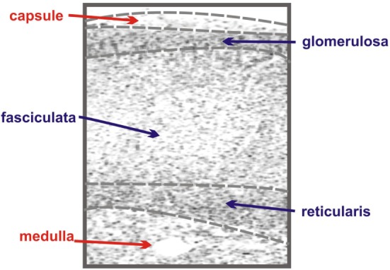
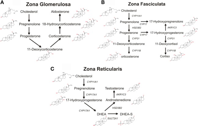
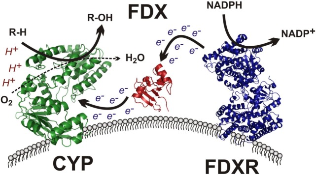
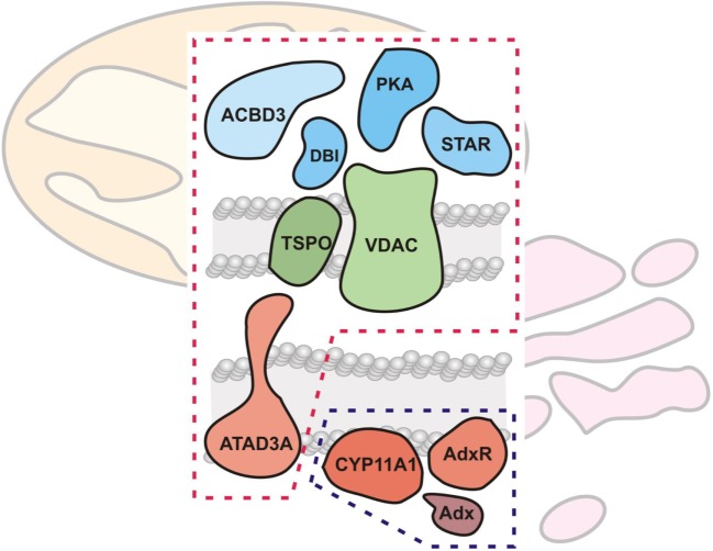
The adrenal cortex is critical for physiological function as the central site of glucocorticoid and mineralocorticoid synthesis. It possesses a great degree of specialized compartmentalization at multiple hierarchical levels, ranging from the tissue down to the molecular levels. In this paper, we discuss this functionalization, beginning with the tissue zonation of the adrenal cortex and how this impacts steroidogenic output. We then discuss the cellular biology of steroidogenesis, placing special emphasis on the mitochondria. Mitochondria are classically known as the "powerhouses of the cell" for their central role in respiratory adenosine triphosphate synthesis, and attention is given to mitochondrial electron transport, in both the context of mitochondrial respiration and mitochondrial steroid metabolism. Building on work demonstrating functional assembly of large protein complexes in respiration, we further review research demonstrating a role for multimeric protein complexes in mitochondrial cholesterol transport, steroidogenesis, and mitochondria-endoplasmic reticulum contact. We aim to highlight with this review the shift in steroidogenic cell biology from a focus on the actions of individual proteins in isolation to the actions of protein assemblies working together to execute cellular functions.
Keywords: cholesterol transport; cytochrome P450 enzyme system; endoplasmic reticulum; mitochondria; steroidogenic acute regulatory protein; translocator protein; voltage-dependent anion channels.
Figures
Similar articles
-
Steroid hormone synthesis in mitochondria.Mol Cell Endocrinol. 2013 Oct 15;379(1-2):62-73. doi: 10.1016/j.mce.2013.04.014. Epub 2013 Apr 28. Mol Cell Endocrinol. 2013. PMID: 23628605
-
Role of mitochondria in steroidogenesis.Best Pract Res Clin Endocrinol Metab. 2012 Dec;26(6):771-90. doi: 10.1016/j.beem.2012.05.002. Epub 2012 Jun 16. Best Pract Res Clin Endocrinol Metab. 2012. PMID: 23168279 Review.
-
Reactive oxygen disrupts mitochondria in MA-10 tumor Leydig cells and inhibits steroidogenic acute regulatory (StAR) protein and steroidogenesis.Endocrinology. 2003 Jul;144(7):2882-91. doi: 10.1210/en.2002-0090. Endocrinology. 2003. PMID: 12810543
-
Mitochondria-associated membrane formation in hormone-stimulated Leydig cell steroidogenesis: role of ATAD3.Endocrinology. 2015 Jan;156(1):334-45. doi: 10.1210/en.2014-1503. Endocrinology. 2015. PMID: 25375035
-
Roles of microfilaments and intermediate filaments in adrenal steroidogenesis.Microsc Res Tech. 1997 Mar 15;36(6):463-79. doi: 10.1002/(SICI)1097-0029(19970315)36:6<463::AID-JEMT4>3.0.CO;2-J. Microsc Res Tech. 1997. PMID: 9142693 Review.
Cited by
-
CRISPR-Cas9 Mediated TSPO Gene Knockout alters Respiration and Cellular Metabolism in Human Primary Microglia Cells.Int J Mol Sci. 2019 Jul 9;20(13):3359. doi: 10.3390/ijms20133359. Int J Mol Sci. 2019. PMID: 31323920 Free PMC article.
-
A molecular framework for autistic experiences: Mitochondrial allostatic load as a mediator between autism and psychopathology.Front Psychiatry. 2022 Nov 25;13:985713. doi: 10.3389/fpsyt.2022.985713. eCollection 2022. Front Psychiatry. 2022. PMID: 36506457 Free PMC article.
-
Transgenerational hypocortisolism and behavioral disruption are induced by the antidepressant fluoxetine in male zebrafish Danio rerio.Proc Natl Acad Sci U S A. 2018 Dec 26;115(52):E12435-E12442. doi: 10.1073/pnas.1811695115. Epub 2018 Dec 10. Proc Natl Acad Sci U S A. 2018. PMID: 30530669 Free PMC article.
-
Estrogenic control of mitochondrial function.Redox Biol. 2020 Apr;31:101435. doi: 10.1016/j.redox.2020.101435. Epub 2020 Jan 23. Redox Biol. 2020. PMID: 32001259 Free PMC article. Review.
-
A Novel Intronic Pathogenic Variant in STAR With a Dominant Negative Mechanism Causes Attenuated Lipoid Congenital Adrenal Hyperplasia.J Investig Med High Impact Case Rep. 2021 Jan-Dec;9:23247096211014685. doi: 10.1177/23247096211014685. J Investig Med High Impact Case Rep. 2021. PMID: 33966472 Free PMC article.
References
Publication types
LinkOut - more resources
Full Text Sources
Other Literature Sources