. 2016 Jul 1;5(7):e88.
doi: 10.1038/cti.2016.30. eCollection 2016 Jul.

Characterizing the inflammatory response in esophageal mucosal biopsies in children with eosinophilic esophagitis

Affiliations

Characterizing the inflammatory response in esophageal mucosal biopsies in children with eosinophilic esophagitis

Wael N Sayej et al. Clin Transl Immunology. .

Abstract

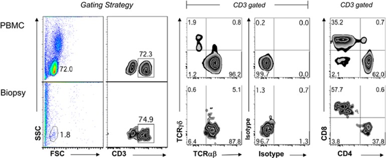
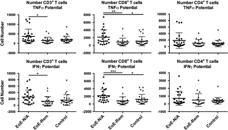
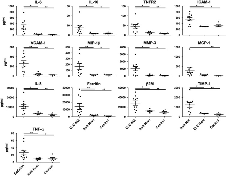
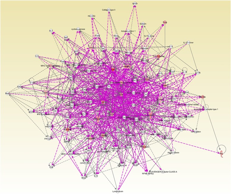
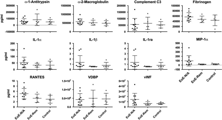
Eosinophilic esophagitis (EoE) is an emerging allergic, IgE- and non-IgE (Th2 cell)-mediated disease. There are major gaps in the understanding of the basic mechanisms that drive the persistence of EoE. We investigated whether esophageal biopsies from children with EoE demonstrate an inflammatory response that is distinct from normal controls. We prospectively enrolled 84 patients, of whom 77 were included in our analysis, aged 4-17 years (12.8±3.8 years; 81% males). Five esophageal biopsies were collected from each patient at the time of endoscopy. Intramucosal lymphocytes were isolated, phenotyped and stimulated with phorbol 12-myristate 13-acetate/ionomycin to measure their potential to produce cytokines via flow cytometry. We also performed cytokine arrays on 72-h biopsy culture supernatants. CD8(+) T cells, compared with CD4(+) T cells, synthesized more TNF-α and interferon (IFN)-γ after mitogen stimulation in the EoE-New/Active vs EoE-Remission group (P=0.0098; P=0.02) and controls (P=0.0008; P=0.03). Culture supernatants taken from explant esophageal tissue contained 13 analytes that distinguished EoE-New/Active from EoE-Remission and Controls. Principal component analysis and cluster analysis based on these analytes distinctly separated EoE-New/Active from EoE-Remission and Controls. In summary, we have identified a previously unappreciated role for CD8(+) T lymphocytes with potential to produce TNF-α and IFN-γ in EoE. Our results suggest that CD8(+) T cells have a role in the persistence or progression of EoE. We have also identified a panel of analytes produced by intact esophageal biopsies that differentiates EoE-New/Active from EoE-Remission and controls. Our results suggest that esophageal epithelial cells may have specific immune effector functions in EoE that control the type and amplitude of inflammation.


Figures

Figure 1
Figure 1
Flow cytometry gating strategy of cells isolated from esophageal biopsies: peripheral blood mononuclear cells were utilized to establish the lymphocyte gate (top panels). Flow cytometry plots representative of peripheral blood mononuclear cells and cells extracted from the biopsies are shown (bottom panels). Cells were identified as lymphocytes based on their forward (FSC) and size scatter (SSC) and analyzed for their expression of TCRαβ, TCRγδ, CD3, CD4 and CD8.
Figure 2
Figure 2
The potential of esophageal mucosal CD3+, CD3+CD8+ and CD3+CD4+ T lymphocytes to produce TNF-α or IFN-γ after phorbol 12-myristate 13-acetate/ionomycin stimulation was measured using flow cytometry. Graphic representation of the absolute number of CD3+, CD8+ and CD4+ T cells with potential to produce TNF-α and IFN-γ. There was no difference in TNF-α and IFN-γ potential in CD3+CD4+ cells (Table 3). *P<0.05, **P<0.01.
Figure 3
Figure 3
Esophageal explant culture supernatant analytes with differences among study groups. Supernatants were analyzed via multiplex cytokine analysis (Myriad RBM). Analytes with significant differences among the groups are shown. Kruskal–Wallis test followed by Dunn's multiple comparisons test was used to identify if there was a significant difference among the groups. *P<0.05, **P<0.01.
Figure 4
Figure 4
Patient group separation based on the cytokine network. (a) Unsupervised PCA of EoE-New/Active, EoE-Remission and Controls based on multiplex cytokine analysis from 72 h culture of esophageal biopsies. Two-dimensional PCA mapping represented 83% of variance (PC1=76% and PC2=7%). Each number represents a patient and patient groups are color-coded. (b) Unsupervised cluster analysis using cytokine levels between patient groups. Individual squares represent the cytokine concentration for the given cytokine (column) in a patient (row), with orange indicating higher cytokine levels and yellow indicating lower cytokine levels.
Figure 5
Figure 5
Pathway analysis using IPA based on the concentration of analytes in esophageal mucosal biopsy culture supernatants. We input the log2 ratio and P value of the difference between means of 13 factors that were different between EoE-New/Active and Normal controls into IPA software. The figure demonstrated the vast and complex interaction of the cytokines identified and how these cytokines revolve around TNF-α.
Figure 6
Figure 6
Esophageal explant culture supernatant analytes with no significant differences among study groups. Supernatants were analyzed via multiplex cytokine analysis (Myriad RBM). Analytes without significant differences among the groups are shown. Kruskal–Wallis test followed by Dunn's multiple comparisons test was used to identify if there was a significant difference among the groups. *P<0.05, **P<0.01.

References

    1. Blanchard C, Wang N, Stringer KF, Mishra A, Fulkerson PC, Abonia JP et al. Eotaxin-3 and a uniquely conserved gene-expression profile in eosinophilic esophagitis. J Clin Invest 2006; 116: 536–547. - PMC - PubMed
    1. Konikoff MR, Blanchard C, Kirby C, Buckmeier BK, Cohen MB, Heubi JE et al. Potential of blood eosinophils, eosinophil-derived neurotoxin, and eotaxin-3 as biomarkers of eosinophilic esophagitis. Clin Gastroenterol Hepatol 2006; 4: 1328–1336. - PubMed
    1. Blanchard C, Stucke EM, Rodriguez-Jimenez B, Burwinkel K, Collins MH, Ahrens A et al. A striking local esophageal cytokine expression profile in eosinophilic esophagitis. J Allergy Clin Immunol 2011; 127: 208–217. - PMC - PubMed
    1. Dellon ES, Rusin S, Gebhart JH, Covey S, Higgins LL, Beitia R et al. Utility of a Noninvasive Serum Biomarker Panel for Diagnosis and Monitoring of Eosinophilic Esophagitis: A Prospective Study. Am J Gastroenterol 2015; 110: 821–827. - PMC - PubMed
    1. Liacouras CA, Furuta GT, Hirano I, Atkins D, Attwood SE, Bonis PA et al. Eosinophilic esophagitis: updated consensus recommendations for children and adults. J Allergy Clin Immunol 2011; 128: 3–20. - PubMed

LinkOut - more resources