

A Rare Case of Tuberculosis with Sacrococcygeal Involvement Miming a Neoplasm
- PMID: 27525144
- PMCID: PMC4971299
- DOI: 10.1155/2016/7286806
A Rare Case of Tuberculosis with Sacrococcygeal Involvement Miming a Neoplasm
Abstract
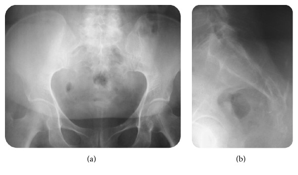
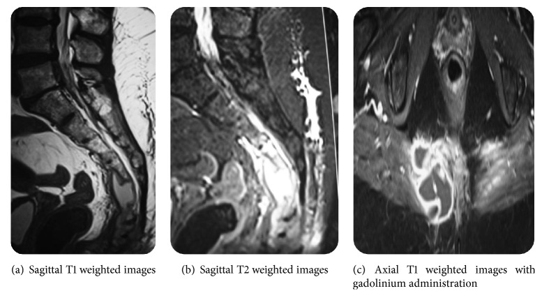
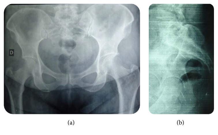
Infection of the lumbosacral junction by tuberculosis is quite rare and occurs in only 1 to 2% of all cases of spinal tuberculosis; moreover, isolated sacrococcygeal or coccygeal tuberculosis is much rarer. Failure to identify and treat these areas of involvement at an early stage may lead to serious complications such as vertebral collapse, spinal compression, and spinal deformity. In the present paper, we report an uncommon case of spinal tuberculosis with sacrococcygeal location revealed by a chronic low back pain that was successfully managed. Computed tomography scan and magnetic resonance imaging of the pelvis revealed a lytic lesion affecting both of sacrum and coccyx causing osseous destruction and suggesting a malignant process. A surgical biopsy was performed to establish the tissue diagnosis. Histopathological report confirmed the diagnosis of skeletal tuberculosis. The patient was treated with antibacillary chemotherapy for a period of 9 months. The follow-up period was of 36 months. There was a full recovery and the patient was asymptomatic.
Figures


Similar articles
-
Rare case of sacrococcygeal tuberculosis mimicking as an anal fistula.Int J Surg Case Rep. 2018;49:74-77. doi: 10.1016/j.ijscr.2018.05.033. Epub 2018 Jun 28. Int J Surg Case Rep. 2018. PMID: 29966953 Free PMC article.
-
Global Reconstruction for Extensive Destruction in Tuberculosis of the Lumbar Spine and Lumbosacral Junction: A Case Report.Global Spine J. 2015 Aug;5(4):e17-21. doi: 10.1055/s-0034-1395780. Epub 2014 Nov 10. Global Spine J. 2015. PMID: 26225288 Free PMC article.
-
A rare location of sacral tuberculosis: A report of three cases.Eur J Rheumatol. 2014 Jun;1(2):78-80. doi: 10.5152/eurjrheumatol.2014.014. Epub 2014 Jun 1. Eur J Rheumatol. 2014. PMID: 27708880 Free PMC article.
-
Sacrococcygeal chordoma: clinicoradiological study.Urol Int. 1990;45(6):372-5. doi: 10.1159/000281746. Urol Int. 1990. PMID: 2288057 Review.
-
Sacral tuberculosis: a case report and review of the literature.Surg Neurol. 2004 Feb;61(2):136-9; discussion 139-41. doi: 10.1016/s0090-3019(03)00265-9. Surg Neurol. 2004. PMID: 14751619 Review.
Cited by
-
Mycobacterium xenopi related spine infections: A case report and systematic literature review.One Health. 2023 Feb 11;16:100502. doi: 10.1016/j.onehlt.2023.100502. eCollection 2023 Jun. One Health. 2023. PMID: 36817979 Free PMC article.
-
Bone and Joint Tuberculosis: The Experience From a Tuberculosis Department in Northern Greece.Case Rep Infect Dis. 2025 May 22;2025:1632733. doi: 10.1155/crdi/1632733. eCollection 2025. Case Rep Infect Dis. 2025. PMID: 40443738 Free PMC article.
-
Rare case of sacrococcygeal tuberculosis mimicking as an anal fistula.Int J Surg Case Rep. 2018;49:74-77. doi: 10.1016/j.ijscr.2018.05.033. Epub 2018 Jun 28. Int J Surg Case Rep. 2018. PMID: 29966953 Free PMC article.
References
-
- Oniankitan O., Fianyo E., Kakpovi K., Agoda-Koussema L. K., Mijiyawa M. Sacrum Pott's disease: a rare location of spine tuberculosis. The Egyptian Rheumatologist. 2014;36(4):209–211. doi: 10.1016/j.ejr.2014.02.001. - DOI
-
- Agrawal A., Shanthi V., Mohan K. M., Reddy G. V. Lumbosacral spinal tuberculosis: a case report and review of literature. The Egyptian Orthopaedic Journal. 2015;50(1):73–75. doi: 10.4103/1110-1148.163163. - DOI
LinkOut - more resources
Full Text Sources
Other Literature Sources
