. 2016:2016:6127340.
doi: 10.1155/2016/6127340. Epub 2016 Jul 20.

Cerebral Blood Flow, Heart Rate, and Blood Pressure Patterns during the Tilt Test in Common Orthostatic Syndromes

Affiliations

Cerebral Blood Flow, Heart Rate, and Blood Pressure Patterns during the Tilt Test in Common Orthostatic Syndromes

Peter Novak. Neurosci J. 2016.

Abstract

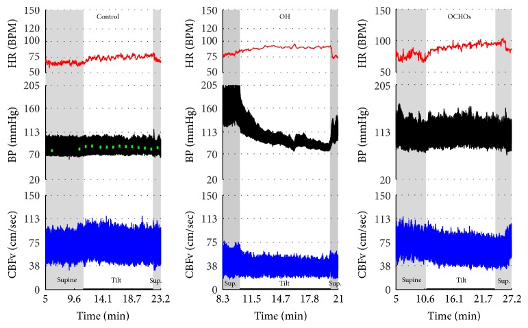
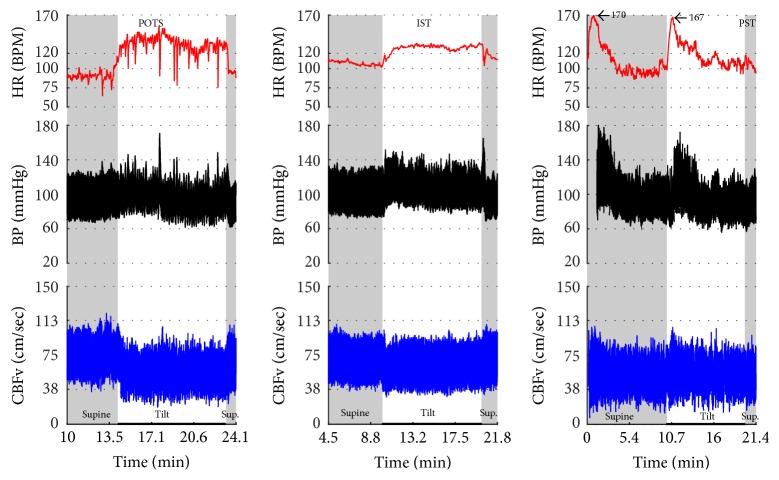
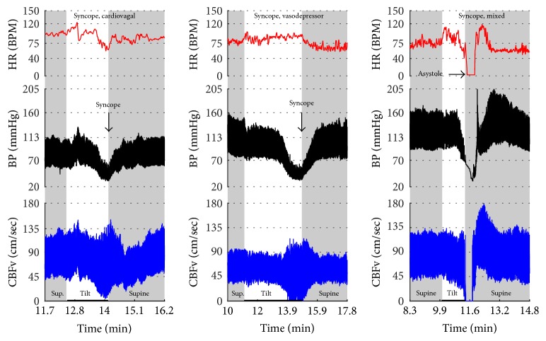
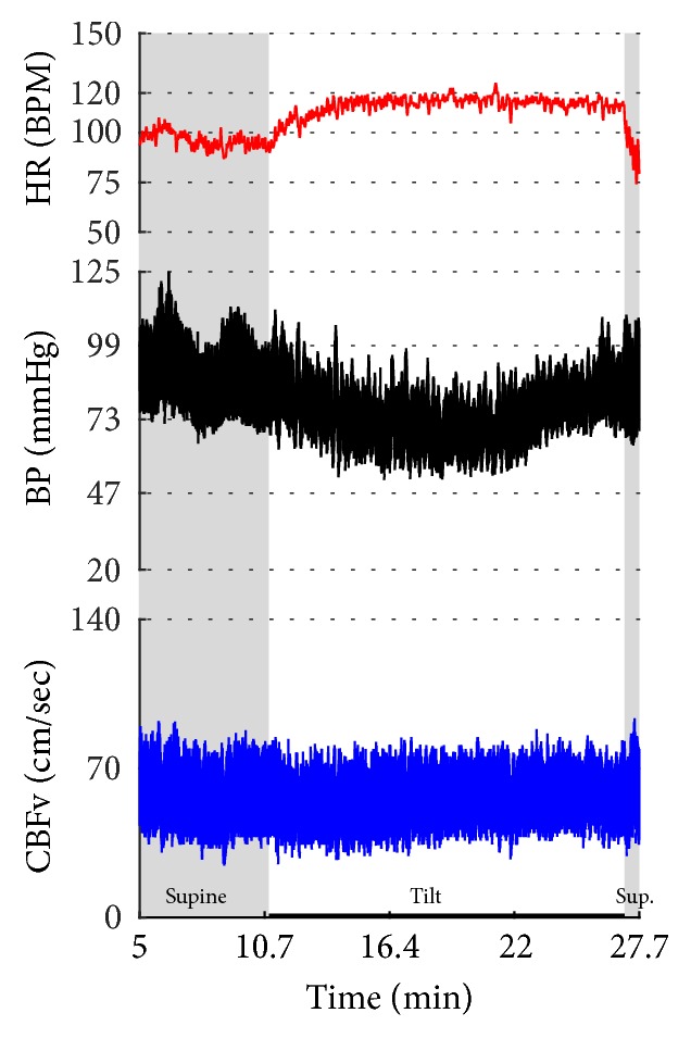
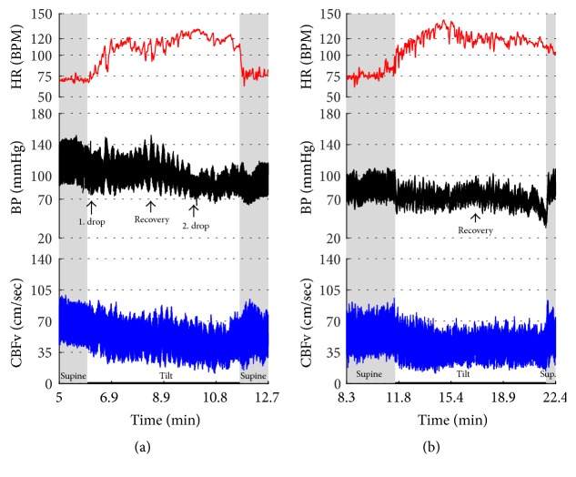
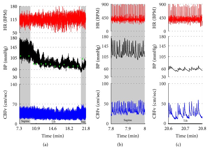
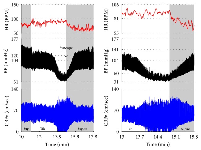
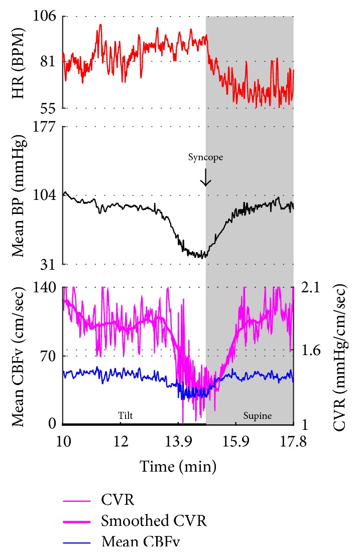
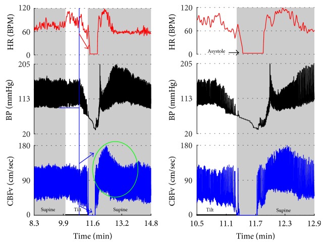
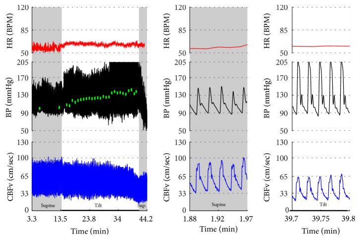
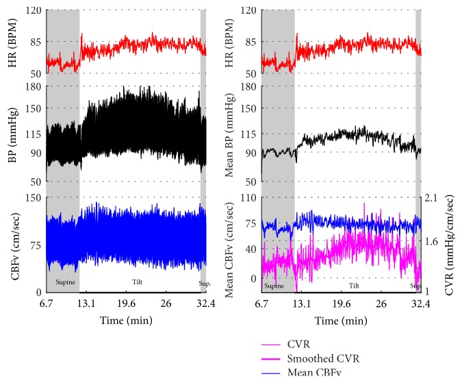
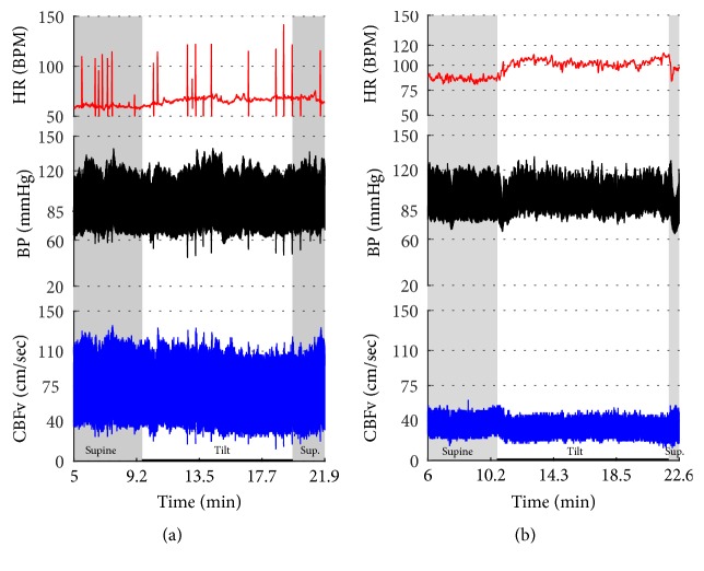
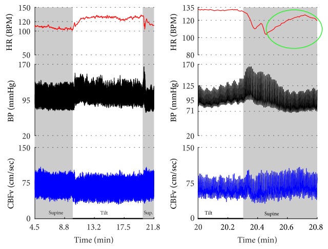
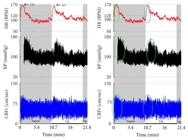
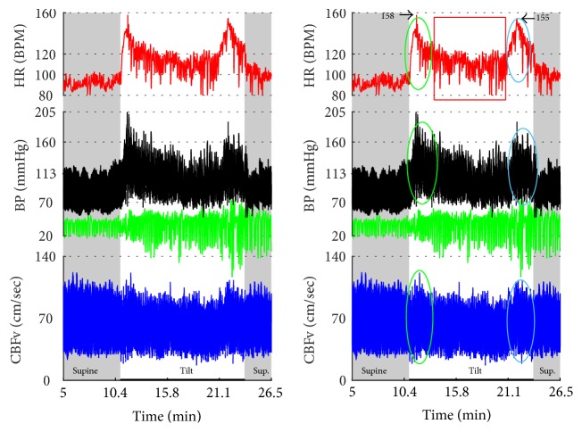
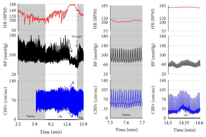
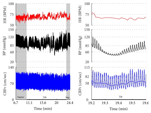
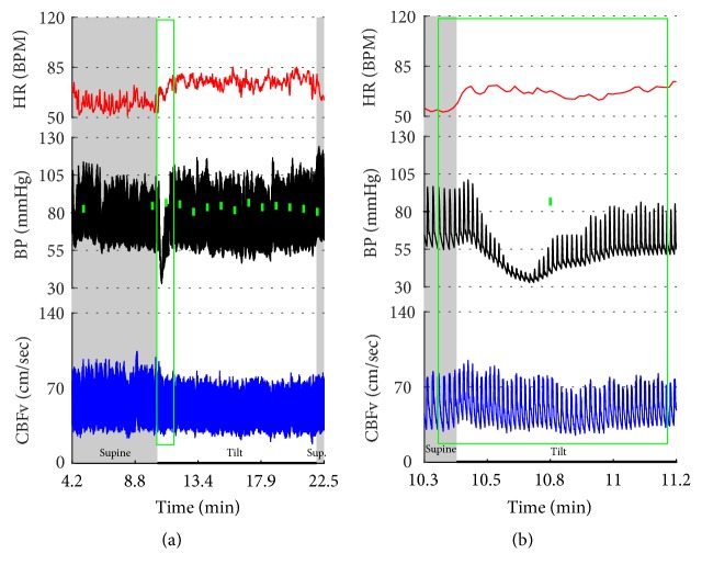
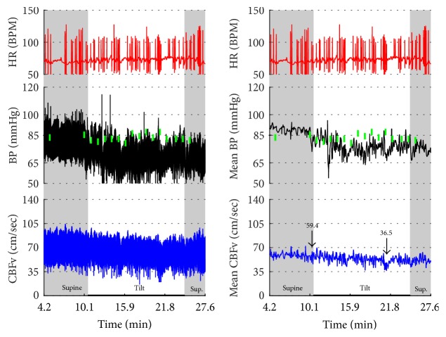
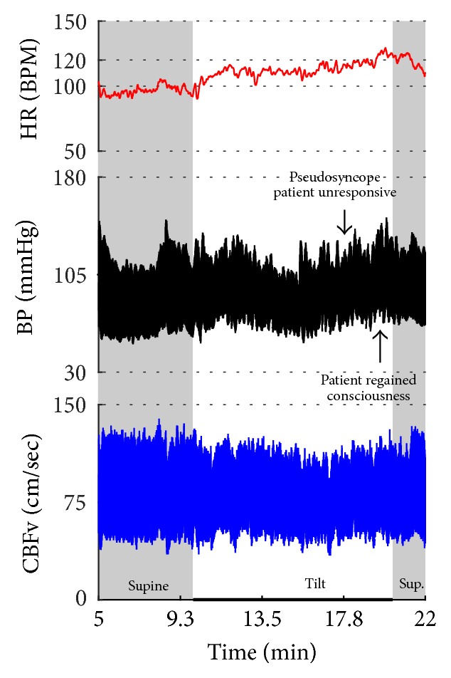
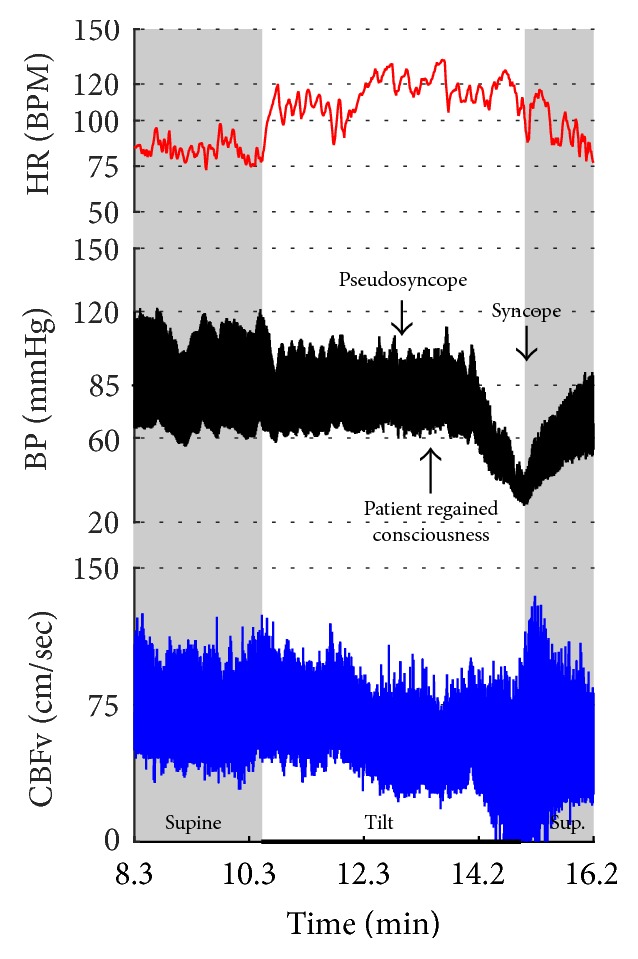
Objective. The head-up tilt test is widely used for evaluation of orthostatic intolerance. Although orthostatic symptoms usually reflect cerebral hypoperfusion, the cerebral blood flow velocity (CBFv) profile in orthostatic syndromes is not well described. This study evaluated CBFv and cardiovascular patterns associated with the tilt test in common orthostatic syndromes. Methods. This retrospective study analyzed the tilt test of patients with history of orthostatic intolerance. The following signals were recorded: ECG, blood pressure, CBFv using transcranial Doppler, respiratory signals, and end tidal CO2. Results. Data from 744 patients were analyzed. Characteristic pattern associated with a particular orthostatic syndrome can be grouped into abnormalities predominantly affecting blood pressure (orthostatic hypotension, orthostatic hypertension syndrome, vasomotor oscillations, and neurally mediated syncope-cardioinhibitory, vasodepressor, and mixed), cerebral blood flow (orthostatic hypoperfusion syndrome, primary cerebral autoregulatory failure), and heart rate (tachycardia syndromes: postural tachycardia syndrome, paroxysmal sinus tachycardia, and inappropriate sinus tachycardia). Psychogenic pseudosyncope is associated with stable CBFv. Conclusions. The tilt test is useful add-on in diagnosis of several orthostatic syndromes. However diagnostic criteria for several syndromes had to be modified to allow unambiguous pattern classification. CBFv monitoring in addition to blood pressure and heart rate may increase diagnostic yield of the tilt test.


Figures

Figure 1
Figure 1
Comparisons of normal responses to the tilt (control), orthostatic hypotension (OH), and orthostatic cerebral hypoperfusion syndrome (OCHOs). Normal heart rate increment (≥10 and <30 BPM) is seen in all examples. Orthostatic blood pressure is stable in a healthy control subject and OCHOs while it is reduced in OH. Orthostatic cerebral blood flow velocity is stable in a control subject and reduced in OH and OCHOs. CBFv can be normal or reduced in OH, depending on functioning of cerebral autoregulation and severity of OH. Green boxes represent mean blood pressure obtained from the upper arm. HR = heart rate, BP = blood pressure, and CBFv = cerebral blood flow velocity.
Figure 2
Figure 2
Comparisons of tachycardia syndromes including postural tachycardia syndrome (POTS), inappropriate sinus tachycardia (IST), and paroxysmal sinus tachycardia (PST). During supine position, the heart rate is normal (<100) in POTS, consistently elevated in IST, and transiently elevated in PST. The excessive tachycardia during the tilt is seen in all shown syndromes, being continuous in POTS and IST, and intermittent in PST. Blood pressure is stable in all examples. CBFv is reduced in POTS, normal in IST, and intermittently elevated in PST.
Figure 3
Figure 3
Comparisons of three main types of neurally mediated syncope. Syncope is associated with profound decline in BP and in diastolic CBFv. HR and BP responses differentiate each type of syncope while CBFv responses are similar among all syncope types. HR declines before BP in cardiovagal syncope. HR decline is absent in the vasodepressor syncope. HR and BP decline simultaneously in mixed syncope. CBFv shows typical vasodilation pattern in all types of syncope that is characterized by a decline in diastolic and increase in systolic CBFv. The diastolic CBFv is equal or close to zero during syncope.
Figure 4
Figure 4
Orthostatic hypotension with stable orthostatic CBFv. There is immediate decline in BP at the onset of tilt and the decline further progressed towards the end of the tilt. The HR increment was normal and CBFv was stable during the tilt. Note details of signals in (b) and (c). Data from 60-year-old man.
Figure 5
Figure 5
Transient orthostatic hypotension. This example shows a progressive decline in BP during the first 5 minutes of the tilt followed by the recovery of BP towards the end of the tilt. CBFv was normal and stable during the tilt. Data from 42-year-old woman.
Figure 6
Figure 6
Orthostatic hypotension with reduced orthostatic CBFv. (a) shows the drop of BP (1. drop) at the beginning of the tilt with a recovery (recovery) followed by a further decline in BP at the second half of the tilt (2. drop). The HR increment was preserved during the tilt. CBFv was progressively declining during the tilt. Patient was very dizzy and anxious and she requested to terminate the tilt at the 6th minute. (b) shows data from the same person a year later. The second tilt test was remarkably similar to the first one showing orthostatic hypotension with similar recovery and final decline of the blood pressure. CBFv was reduced throughout the tilt. Data from 26-year-old woman.
Figure 7
Figure 7
This example shows combination of (1) primary cerebral autoregulatory failure (pCAF); (2) severe orthostatic hypotension with reduced orthostatic CBFv; and (3) atrial fibrillation. Supine BP is elevated while supine CBFv is reduced. The pattern of elevated BP and reduced CBFv during supine position is due to abnormal cerebral vasoconstriction consistent with pCAF. There was severe OH with a progressive decrease in BP during the tilt. Diastolic CBFv was reduced during the tilt but less than systolic CBFv that can be seen in mild cerebral vasodilatation that compensates for reduced orthostatic BP. HR responses to the tilt were absent. Note random, noise-like pattern of HR due atrial fibrillation. The HR fluctuated wildly (50–180 BPM) and not all electrical systoles were transmitted in the mechanical systoles (c) resulting in marked variations of BP and CBFv. Supine hypertension and orthostatic hypotension are a marker of severe autonomic adrenergic failure. Green boxes represent mean blood pressure obtained from the arm. Data from 69-year-old man with multiple system atrophy.
Figure 8
Figure 8
Mixed syncope. In this subject, initial slowing of HR from 123 to 81 BPM (thin red arrow) is not cardioinhibitory response but reflects a functioning baroreflex since it is associated with elevation of BP (thin black arrow) and CBFv (thin blue arrow). The onset of the cardioinhibitory reaction is marked by the vertical red line with HR 108 BPM (thick red arrow) and coincides with a BP drop (thick black arrow) and patient became quickly unconscious. The pulsatility index increased during syncope (systolic CBFv increased and diastolic CFBv decreased) which is consistent with cerebral vasodilatation. The vasodilatation started early (thick blue arrows), and the changes were discernible in the doppler audio signal before noticeable changes in heart rate or BP. The cardiac slowing followed with delayed BP decrease being characteristic of cardioinhibitory syncope. Patient lost consciousness when the systolic BP declined below 60 mmHg, as expected. Data from 20 y/o woman.
Figure 9
Figure 9
Vasodepressor syncope. There is a progressive drop in BP without bradycardia till syncope occurred. HR started to slow down only after a subject was tilted back to supine position. The syncope is associated with a characteristic cerebral vasodilatation pattern. Data from 27-year-old woman.
Figure 10
Figure 10
Cerebral vasodilation during syncope. The cerebral vascular resistance is reduced during syncope which is consistent with cerebral vasodilation. The same subject as in Figure 9.
Figure 11
Figure 11
Cardioinhibitory syncope. In this subject, slowing of HR, BP drop, and cerebral vasodilation began almost simultaneously (marked by blue line) and progressed rapidly into asystole of 27 seconds. Note the vasodilatory pattern in CBFv early in the evolution of syncope (blue arrows) followed by reactive hyperemia (green circle) with elevated systolic and diastolic CBFv. Data from 39-year-old man.
Figure 12
Figure 12
Orthostatic cerebral hypoperfusion syndrome (OCHOs). The tilt provoked a progressive increase in BP that was seen in both finger (black trace) and arm (green boxes) BP. The finger systolic BP exceeded the upper range of our device, 205 mmHg. The CBFv was progressively declining during the tilt that was consistent with cerebral vasoconstriction. Patient was very dizzy and agitated during the tilt. Data from 66-year-old woman.
Figure 13
Figure 13
Orthostatic hypertension syndrome. BP was elevated during the tilt while CBFv was stable. HR responses were normal during the tilt. Note increased cerebral vascular resistance (CRV) that is consistent with cerebral vasoconstriction. Data from 39-year-old man.
Figure 14
Figure 14
Primary cerebral autoregulatory failure (pCAF, (b)). HR and BP were normal at supine and during the tilt. CBFv was markedly reduced at the supine position and was further reduced during the tilt. Data from 47-year-old man. Patient was referred for evaluation of chronic fatigue, difficulites with attention, and chronic dizziness that was both supine and postural. For comparison, a healthy 80-year-old woman has normal CBFv (a).
Figure 15
Figure 15
Inappropriate sinus tachycardia (IST). Note elevated resting heart rate (>100 BMP) which is further increased during the tilt. This response satisfies criteria for POTS (last baseline HR = 101.3, last HR during the tilt = 132.6, and increment > 30 BPM and BPM > 120) except that the continuous resting supine tachycardia is inconsistent with POTS. Second clue that this is not POTS is an episode of HR increment exceeding 120 BPM (green oval) at supine position after completing the tilt. Mean supine HR = 106.6 ± 2.8, range 97.1–114.3 BPM, mean orthostatic HR 128 ± 2.4, and range 121.8–134.0 BPM.
Figure 16
Figure 16
Paroxysmal sinus tachycardia (PST) due to anxiety. The patient, a 34 y/o woman, was referred for evaluation of postural tachycardia syndrome (POTS). During montage in supine position before placing the blood pressure sensor (i.e., why the initial portion of recording is missing), she became very anxious after she was informed that she will be tilted in several minutes. The anxiety was associated with transient tachycardia 170 BPM, elevated BP, and CBFv (green ovals). Similar pattern (transient tachycardia, elevated BP, and CBFv) was observed at the onset of the tilt (blue oval). The supine tachycardia of similar character to that of the tilt confirms that this is not POTS but IST. Furthermore, the tachycardia is usually sustained and/or it is progressively increased during the tilt in POTS. CBFv is usually unchanged or decreased in POTS during the tilt.
Figure 17
Figure 17
Postural tachycardia syndrome (POTS) or paroxysmal sinus tachycardia (PST) due to the anxiety reaction? The supine variables were normal. The tilt test induced initial tachycardia associated with elevation of BP and CBFv (green ovals). This reaction is due to anxiety since BP and CBFv are elevated. In POTS, CBFv is usually unchanged or reduced. Similar reaction occurred at the end of the tilt (blue ovals). Note that patient was hyperventilating during the tilt (green tracing). The tilt test was done without any medication. Trial of beta blockers failed to improve orthostatic intolerance. Final diagnosis was PST. Mean supine HR was 91.5 ± 4.9 BPM; mean orthostatic HR during more steady HR (demarcated by a red box) was 109.3 ± 8.5 BPM. The mean HR during the whole tilting was 118.5 ± 13.8 BPM. Data from 25-year-old woman.
Figure 18
Figure 18
Inappropriate sinus tachycardia complicated by vasodepressor syncope. The average HR was above 100 BPM with the maximal heart rate 149 BPM during the supine position. The tilt provoked HR increase and marked oscillations in all signals including HR, BP, and CBFv. At the 5th minute of the tilt, BP suddenly declined (black arrow) that was accompanied by tachycardia 179 BPM culminated in a syncope. The decline in BP without bradycardia is characteristic of vasodepressor syncope due to reduced peripheral resistance. CBFv declined and the pulsatility index (systolic CBFv – diastolic CBFv) increased (blue arrows) that indicates cerebral vasodilation. Data from 26-year-old woman.
Figure 19
Figure 19
Vasomotor oscillations. Note marked fluctuations of BP occurring approximately every minute. CBFv also fluctuates in synchrony with BP. Patient was mildly anxious during the tilt test but she was not dizzy. Data from 47-year-old woman.
Figure 20
Figure 20
The transient drop in BP at the beginning of the tilt (designated by a green box) is artificial. The oscillometric device showed much higher BP (the small filled green box in (b)). Another clue that this is a technical artifact is the fact that there is no corresponding HR reaction and also CBFv is unchanged. The tilt test was normal. Data from 38-year-old woman.
Figure 21
Figure 21
This example shows arteficial drift in the plethysmographic finger BP that may be misinterpreted as orthostatic hypotension. The spurious decline in BP continued in spite of multiple repositionings of a sensor. Note that oscillometric BP from the upper arm (green boxes) shows absent orthostatic hypotension. There are a number of extrasystoles in the HR. BP was interpreted as normal. CBFv responses to the tilt were abnormal as there was a drop in CBFv from the supine 59.4 cm/sec to 36.5 cm/sec at the end of the tilt. Patient was dizzy during the tilt. The final diagnosis was orthostatic cerebral hypoperfusion syndrome. Data from 43-year-old man.
Figure 22
Figure 22
Psychogenic unresponsiveness. A patient became progressively obtunded and confused, experienced eyelid flutter, became aphasic, and finally did lose the consciousness during the tilt. The patient responded to reassurance and she regained consciousness while in the upright position. All monitoring variables were normal during the spell; patient became slightly tachycardiac after the spell. Data from 37-year-old woman.
Figure 23
Figure 23
POTS + psychogenic unresponsiveness + syncope. The tilt provoked symptomatic excessive heart rate increment associated with a drop in CBFv without orthostatic hypotension which satisfies criteria for POTS. Then the patient became progressively less responsive which was consistent with pseudosyncope (psychogenic unresponsiveness) since CBFv was stable. The patient responded to reassurance and she regained consciousness later during the tilting. However, subsequently the patient experienced true mixed syncope at the 5th minute of the tilt (the tilt starts at minute 10.6 of the recording) and the tilt was terminated. Final diagnoses: (1) POTS; (2) psychogenic unresponsiveness; and (3) mixed syncope. Data from 21-year-old woman.

References

    1. Smith J. J., Porth C. M., Erickson M. Hemodynamic response to the upright posture. Journal of Clinical Pharmacology. 1994;34(5):375–386. doi: 10.1002/j.1552-4604.1994.tb04977.x. - DOI - PubMed
    1. Kenny R. A., Ingram A., Bayliss J., Sutton R. Head-up tilt: a useful test for investigating unexplained syncope. The Lancet. 1986;1(8494):1352–1355. - PubMed
    1. Moya A., Sutton R., Ammirati F., et al. Guidelines for the diagnosis and management of syncope (version 2009) European Heart Journal. 2009;30(21):2631–2671. doi: 10.1093/eurheartj/ehp298. - DOI - PMC - PubMed
    1. Novak V., Spies J. M., Novak P., McPhee B. R., Rummans T. A., Low P. A. Hypocapnia and cerebral hypoperfusion in orthostatic intolerance. Stroke. 1998;29(9):1876–1881. doi: 10.1161/01.str.29.9.1876. - DOI - PubMed
    1. Novak P. Quantitative autonomic testing. Journal of Visualized Experiments. 2011;(53) doi: 10.3791/2502.e2502 - DOI - PMC - PubMed

LinkOut - more resources