Review
. 2016 Aug 4:11:20.
doi: 10.1186/s13013-016-0076-9. eCollection 2016.

Physiotherapy scoliosis-specific exercises - a comprehensive review of seven major schools

Affiliations
Review

Physiotherapy scoliosis-specific exercises - a comprehensive review of seven major schools

Hagit Berdishevsky et al. Scoliosis Spinal Disord. .

Abstract

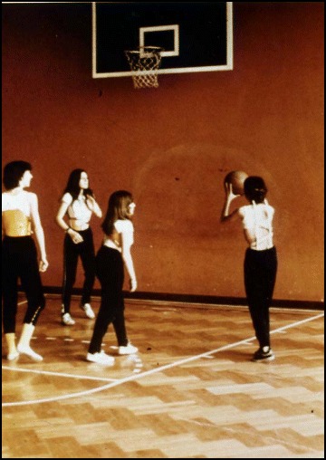
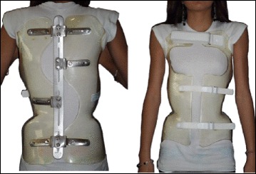
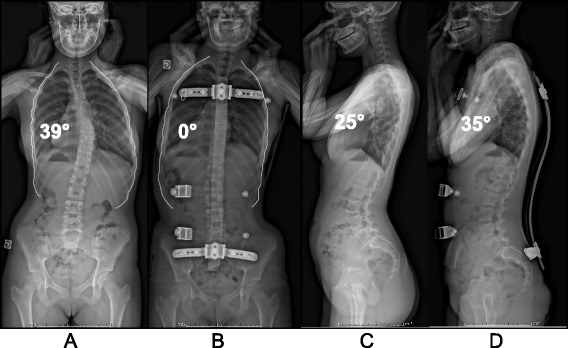
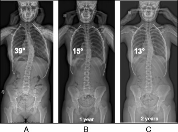
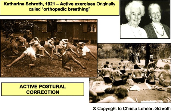
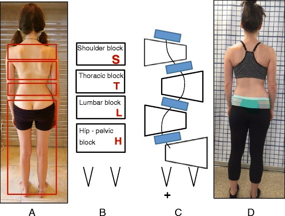
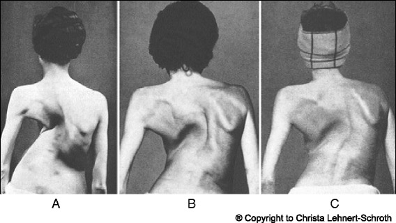
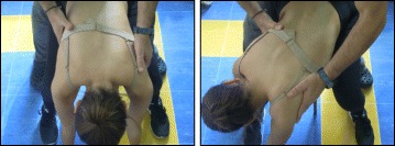
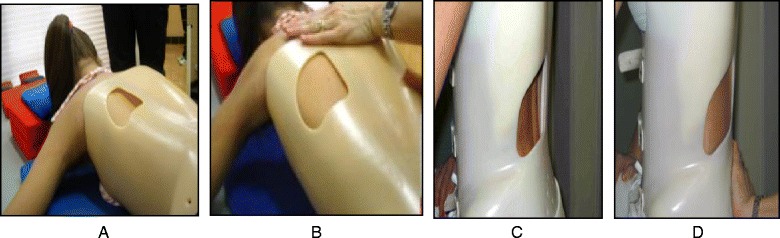
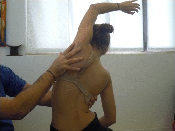
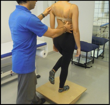
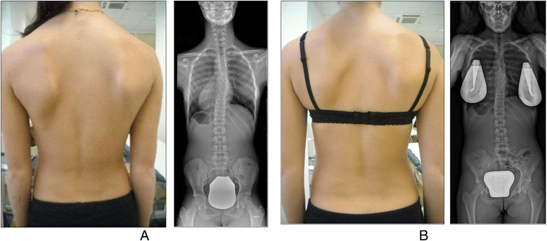
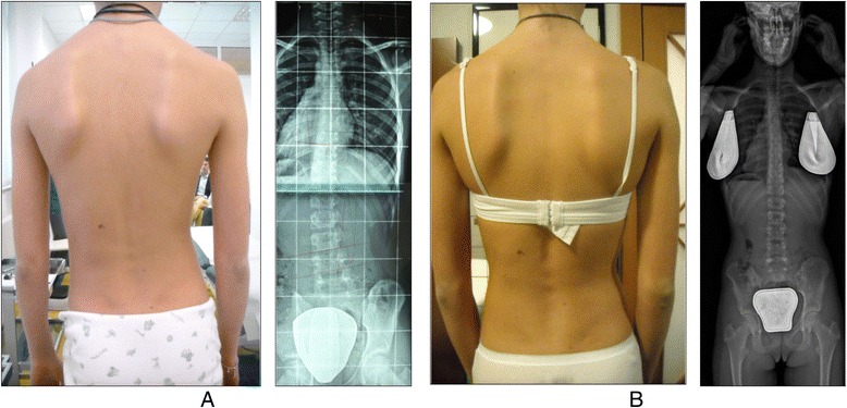
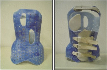
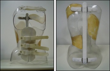
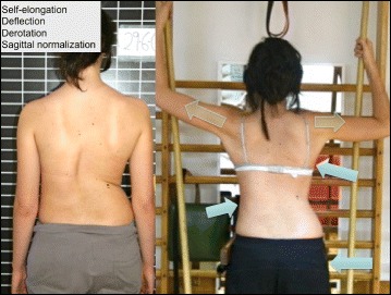
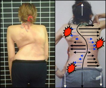
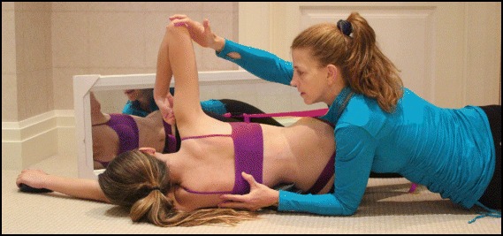
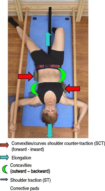
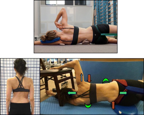
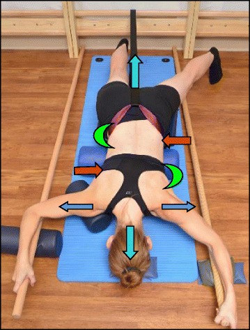
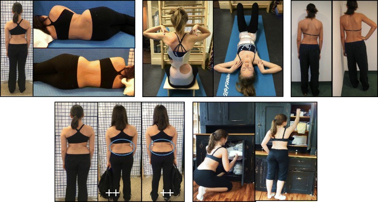
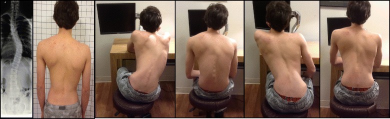
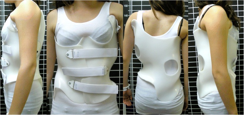
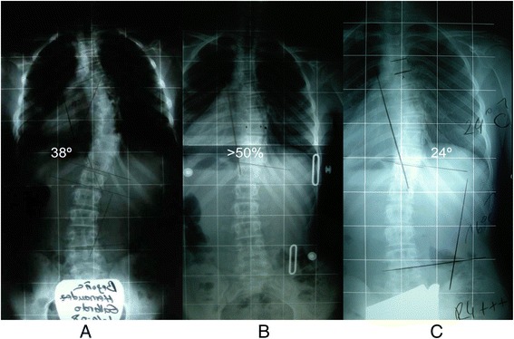
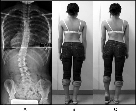
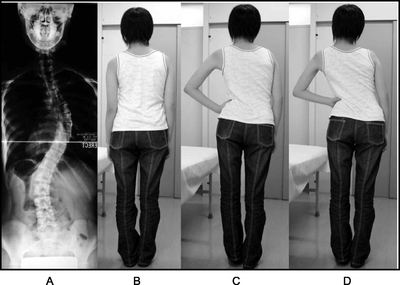
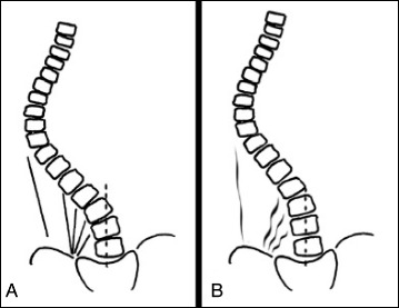
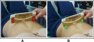
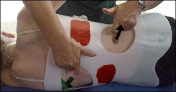
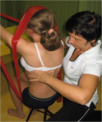
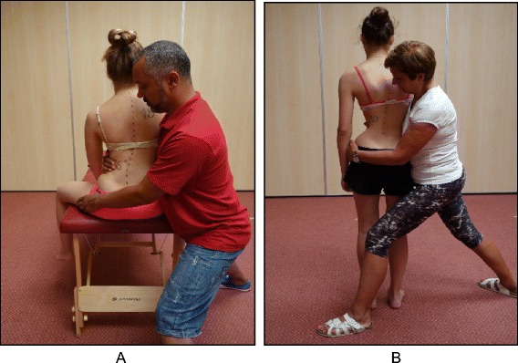
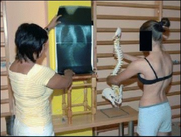
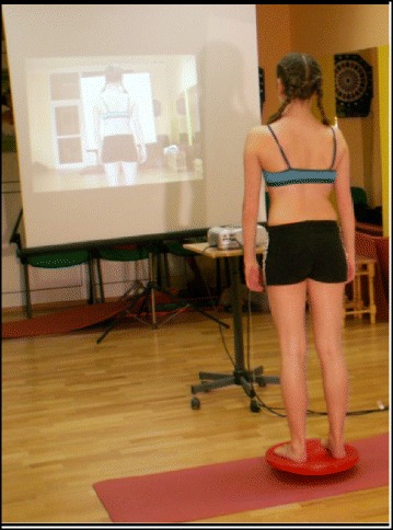
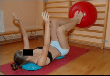
In recent decades, there has been a call for change among all stakeholders involved in scoliosis management. Parents of children with scoliosis have complained about the so-called "wait and see" approach that far too many doctors use when evaluating children's scoliosis curves between 10° and 25°. Observation, Physiotherapy Scoliosis Specific Exercises (PSSE) and bracing for idiopathic scoliosis during growth are all therapeutic interventions accepted by the 2011 International Society on Scoliosis Orthopaedic and Rehabilitation Treatment (SOSORT). The standard features of these interventions are: 1) 3-dimension self-correction; 2) Training activities of daily living (ADL); and 3) Stabilization of the corrected posture. PSSE is part of a scoliosis care model that includes scoliosis specific education, scoliosis specific physical therapy exercises, observation or surveillance, psychological support and intervention, bracing and surgery. The model is oriented to the patient. Diagnosis and patient evaluation is essential in this model looking at a patient-oriented decision according to clinical experience, scientific evidence and patient's preference. Thus, specific exercises are not considered as an alternative to bracing or surgery but as a therapeutic intervention, which can be used alone or in combination with bracing or surgery according to individual indication. In the PSSE model it is recommended that the physical therapist work as part of a multidisciplinary team including the orthopeadic doctor, the orthotist, and the mental health care provider - all are according to the SOSORT guidelines and Scoliosis Research Society (SRS) philosophy. From clinical experiences, PSSE can temporarily stabilize progressive scoliosis curves during the secondary period of progression, more than a year after passing the peak of growth. In non-progressive scoliosis, the regular practice of PSSE could produce a temporary and significant reduction of the Cobb angle. PSSE can also produce benefits in subjects with scoliosis other than reducing the Cobb angle, like improving back asymmetry, based on 3D self-correction and stabilization of a stable 3D corrected posture, as well as the secondary muscle imbalance and related pain. In more severe cases of thoracic scoliosis, it can also improve breathing function. This paper will discuss in detail seven major scoliosis schools and their approaches to PSSE, including their bracing techniques and scientific evidence. The aim of this paper is to understand and learn about the different international treatment methods so that physical therapists can incorporate the best from each into their own practices, and in that way attempt to improve the conservative management of patients with idiopathic scoliosis. These schools are presented in the historical order in which they were developed. They include the Lyon approach from France, the Katharina Schroth Asklepios approach from Germany, the Scientific Exercise Approach to Scoliosis (SEAS) from Italy, the Barcelona Scoliosis Physical Therapy School approach (BSPTS) from Spain, the Dobomed approach from Poland, the Side Shift approach from the United Kingdom, and the Functional Individual Therapy of Scoliosis approach (FITS) from Poland.


Figures

Fig. 1
Fig. 1
Dr. Jean Claude de Mauroy, co-inventor of the new Lyon ARTbrace (Asymmetrical Rigid Torsion Brace)
Fig. 2
Fig. 2
Image on the left: The Vicious cycle. Dr. Stokes and Burwell hypothesized that the vicious cycle of scoliosis curve progression begins with a triggering event which leads to the formation of wedged vertebrae. Wedged vertebrae cause the spine to curve which results in continuous asymmetric loading on the spine. This in turn, can potentially promote asymmetric growth of the spine and progression of scoliosis curves as asymmetric growth increases the wedging of the vertebrae and perpetuates the cycle to continue. The image on the right shows a scoliosis patient sitting with increased asymmetric loading of the spine as described in Stokes’ ‘Vicious Cycle”. The large red “X” on the image indicates that this is not the desired posture. The image on the right shows the same scoliosis patient sitting with improved asymmetric loading of the spine as she performs scoliosis specific physiotherapy exercises in accordance with the Lyon approach
Fig. 3
Fig. 3
Scoliosis patient developing self-awareness of postural defects with the help of a video recorder and real-time video feedback
Fig. 4
Fig. 4
Active thoracic mobilization, promoting kyphosis, using the Lyon method
Fig. 5
Fig. 5
Active lumbar correction, promoting lordosis, using the Lyon method
Fig. 6
Fig. 6
(a, b): Active thoracic shift exercise with a dowel (a) and a Swiss-ball (b) using the Lyon method
Fig. 7
Fig. 7
Active thoracic shift and derotation exercise using the Lyon method. Arrows in the radiograph and the diagram show the direction of the thoracic shift and the derotation of the ribcage as the exercise is performed using the Lyon method
Fig. 8
Fig. 8
(a, b): Balance and proprioception exercises on a Swiss-ball (a) and on a balance board (b) using the Lyon method
Fig. 9
Fig. 9
Spinal stabilization exercises using the Lyon method
Fig. 10
Fig. 10
Several standard Lyon exercises in a Lyon plaster cast promoting core strength (top left), breathing and thoracic shift (bottom), and elongation
Fig. 11
Fig. 11
(a, b): Several standard Lyon exercises in a Lyon plaster cast promoting postural correction (a) and core strengthening (b)
Fig. 12
Fig. 12
Activity level recommendations by age per the Lyon School in accordance with the Lyon treatment principles
Fig. 13
Fig. 13
Lyon method breathing exercises using a breathing machine, performed while wearing a Lyon plaster cast, increases lung capacity
Fig. 14
Fig. 14
Active thoracic mobilization using the Lyon method. Arrows in the diagram on the right show the direction of thoracic mobilization of the ribcage
Fig. 15
Fig. 15
Active lumbar mobilization using the Lyon method. The diagram on the right shows lumbar scoliosis
Fig. 16
Fig. 16
Mobilization of the costovertebral joints using the Lyon method
Fig. 17
Fig. 17
The Lyon method encourages athletic activities while wearing the Lyon brace. This group of scoliosis patients are playing basketball, which aids vertical stretching and spinal flexibility
Fig. 18
Fig. 18
Anterior (left) and posterior (right) views of the new asymmetric rigid torsion brace (ARTbrace) made from 4 mm polycarbonate. The main biomechanical concepts are based on elongation along the vertical axis, lateral inflexion in the frontal plane, and derotation of the spine in order to obtain a correction of the scoliotic curve
Fig. 19
Fig. 19
(a, b, c, d): Radiographic series of a female patient with progressive scoliosis (patient ‘S’). Initial PA (a) and lateral (c) radiographs at the time of diagnosis at age 13 show a thoracic T5-T12 scoliosis of 39° Cobb angle (a) and a 25° hypokyphosis (c). Repeat radiographs taken in the ARTbrace show a decreased Cobb angle on PA radiograph (b) and an increased kyphosis on lateral radiograph (d)
Fig. 20
Fig. 20
(a, b, c): Radiographic series of patient ‘S’ at one year (b) and two years (c) after brace weaning. Compared to the initial radiograph (a), the final radiograph (c) shows a reduction in curve Cobb angle of over 50 %
Fig. 21
Fig. 21
Asklepios Katharina Schroth Spinal Deformities Rehabilitation Centre in Bad Sobernheim, Germany. Formerly called the Katharina Schroth Klinik
Fig. 22
Fig. 22
Axel Hennes, head of the physical therapy department at the Asklepios Katharina Schroth Spinal Deformities Rehabilitation Centre in Bad Sobernheim, Germany
Fig. 23
Fig. 23
History of the Schroth method. Katharina Schroth with her daughter, Christa Lehnert-Schroth (top right). Patients with scoliosis exercising outdoors at the Katharina Schroth Klinik (bottom right; left)
Fig. 24
Fig. 24
(a, b, c, d): Schroth Body Blocks. The Schroth system of scoliosis curve classification is derived from the Schroth principle of dividing the body into “body blocks” as pictured anatomically (a) and schematically (b). Scoliosis causes the body blocks to become deformed, changing their geometric shape from a rectangle (b) to a trapezium (c). A patient with a major lumbar scoliosis left convex curve has a lumbar block shifted to the left and a hip-pelvic block shifted to the right (d)
Fig. 25
Fig. 25
(a, b, c): Severe scoliosis in a 24-year-old female patient. Initial photograph (a) before beginning scoliosis treatment shows total left trunk atrophy with a prominent right thoracic rib hump. Photographs of the same patient 9 months (b) and 12 months (c) after intensive scoliosis therapy with rotational angular breathing (RAB) exercises (also called orthopaedic breathing exercises) according to the Schroth method show a visible improvement of the scoliosis
Fig. 26
Fig. 26
Schroth method lumbar mobilization (a) and curve flexibility (b) exercises
Fig. 27
Fig. 27
The Schroth “50 x Pezziball” exercise where the patient sits on a Swiss-ball in front of a mirror (a) and performs active 3D auto self-correction using the wall bar (b)
Fig. 28
Fig. 28
The Schroth prone exercise with activation of the iliopsoas muscle (right hip flexion). Blue arrows represent trunk elongation with caudal and cranial forces. Red arrows represent areas of muscle activation around the convexities towards the midline. Green half-moons represent areas of expansion of the concavities. Red circles represent additional corrective forces: red circles around the right lower extremity and the right upper extremity represent iliopsoas activation and shoulder traction/counter-traction, respectively, resulting in correction of the lumbar and thoracic curves
Fig. 29
Fig. 29
The Schroth “Sail” exercise where the patient stands on a half foam-roll with two poles and performs active stabilization. The red circle represents the concavity (weak side according to Schroth). During active stabilization, the patient is consciously expanding the left rib cage with right directional breathing, opening the collapsed left lung, while maintaining 3D postural correction
Fig. 30
Fig. 30
The “Muscle-cylinder” exercise (also known as the “Side-lying” exercise), focusing mainly on the correction of the lumbar scoliosis curve. During this exercise, the patient lies on the lumbar convex side. The lumbar convexity is supported by a rice bag to help align the spine in the horizontal plane. The patient’s right leg is supported by a stool (in case of 4C/major lumbar scoliosis) and the patient’s right arm is supported on a chair during the exercise. Light blue arrows represent trunk elongation with cranial and caudal forces. Green half-moons represent areas of expansion of the concavities. Red arrows represent areas of muscle activation, approximating the convexities towards midline, and the direction of the correction. The dark blue arrow pointing upwards from the right elbow represents the shoulder traction, which is an isometric tension from the shoulder in a lateral/outward direction with a fixed scapula as a continuation of the transversal expansion in the proximal thoracic region
Fig. 31
Fig. 31
Patients performing Schroth 3D postural corrections in sitting and standing positions. These postural corrections are practiced during activities of daily living in order to change habitual default postures and improve alignment, pain, and curve progression
Fig. 32
Fig. 32
Scientific Exercise Approach to Scoliosis (SEAS) school leaders Antonio Negrini (a), Michele Romano (b), and Alessandra Negrini (c)
Fig. 33
Fig. 33
Right thoracic curve mobilization in preparation for bracing is aimed at increasing the range of motion of the spine according to the SEAS method
Fig. 34
Fig. 34
SEAS exercises in brace. The patient is in a relaxed position lying prone (a) and then lifts the trunk away from the sternal part of the brace to increase the thoracic kyphosis (b). Similarly, the patient is in a relaxed standing position (c) and moves the abdomen posteriorly away from the abdominal part of the brace to increase the force on the lumbar pressure pad (d)
Fig. 35
Fig. 35
SEAS mobilization and flexibility exercises of the spine to improve joint mobility for better posture correction
Fig. 36
Fig. 36
Assistive devices like balance boards are used at the beginning of learning the SEAS method
Fig. 37
Fig. 37
SEAS exercises aimed to improve balance while maintaining active self-correction either by standing on one leg on a balance board (a) or by performing a knee-bending exercise on the balance board (b)
Fig. 38
Fig. 38
SEAS principles of maintaining self-correction during activities of daily living such as sitting (a), sitting leaning forward in preparation for standing and sit-to-stand (b, c), standing (d), and landing on a wall (e, f)
Fig. 39
Fig. 39
SEAS encourages patients to participate in sports and athletic activities
Fig. 40
Fig. 40
SEAS encourages patients to live a normal life
Fig. 41
Fig. 41
Photograph and radiograph of a patient with scoliosis before SEAS (a) and 24 months later after 2 years of SEAS without bracing (b)
Fig. 42
Fig. 42
Photograph and radiograph of a patient with scoliosis before SEAS (a) and 43 months later after 3.5 years of SEAS without bracing (b)
Fig. 43
Fig. 43
The Sibilla brace. Designed for mild progressive AIS, the Sibilla brace is prescribed for scoliosis curves up to 30° Cobb angle
Fig. 44
Fig. 44
The Sforzesco brace. Designed for severe scoliosis in adolescents, the Sforzesco brace is prescribed for scoliosis curves up to 45°–50° Cobb angle or more if surgery is not an option
Fig. 45
Fig. 45
(a, b, c): The BSPTS founders Elena Salvá (a), Dr. Gloria Quera-Salvá (b), and Dr. Manuel Rigo (c)
Fig. 46
Fig. 46
(a, b, c, d): The BSPTS system of scoliosis curve classification illustrated with photographs and body block diagrams. The four scoliosis curve types in this classification system are 3C (a), 4C (b), N3N4 (c), and single lumbar or thoracolumbar (d). The 3C curve is a major thoracic scoliosis curve with a compensatory lumbar and pelvic shift (a). The 4C curve is a major lumbar scoliosis curve with a thoracic and lumbar shift (b). The N3N4 curve is a major thoracic scoliosis with or without a lumbar curve but with the pelvis in a neutral position (c). The single lumbar or thoracolumbar curve is a single curve scoliosis with an uncoupled pelvic shift and no thoracic curvature (d)
Fig. 47
Fig. 47
Rigo classification for BSPTS bracing and physical therapy
Fig. 48
Fig. 48
(a, b): Active 3D self-correction exercises. During active 3D self-correction, patients expand the collapsed areas and open the concavities by performing rotational angular breathing (RAB) and specific arm positions (a). During the Schroth-derotation sitting exercise (b), the patient sits on a chair, with a pole in either hand planted on the ground, while performs corrections 1–5, while stabilizing her curve specific corrections. (48b provided with the permission of Andrea Lebel, RPT, MCPA, Ottawa, Canada)
Fig. 49
Fig. 49
Patient with a major left lumbar-thoracolumbar scoliosis curve with a right pelvic shift performs a two-pole standing exercise applying the BSPTS principles of correction 1–5. Light brown arrows represent bilateral shoulder traction, which is required for stabilization during active self-correction. Light blue arrows represent bilateral shoulder counter-traction, which is required for midline spinal alignment. The light blue arrow pointing to the patient’s pelvis represents pelvic correction from the right to the midline, which is required exercise when the patient has a major lumbar or thoracolumbar scoliosis curve
Fig. 50
Fig. 50
Patient using postural correction and the corrective expansion/contention technique to achieve the best possible correction. The blue and black arrows represent trunk expansion during the first principle of correction. Later, the blue arrows are converted into forces represented by the red shapes, which work around the prominences to move the prominences forward and inward
Fig. 51
Fig. 51
(a, b): Before (a) and during (b) rotation angular breathing (RAB). The arrows represent directional breathing used to fill the collapsed lungs with air and reshape the thorax (b)
Fig. 52
Fig. 52
Diagrammatic depiction of the activation of the iliopsoas muscle in a lumbar scoliosis curve. The arrows show the direction of activation from the origin to the insertion points of the iliopsoas, promoting curve de-flexion and derotation towards the right
Fig. 53
Fig. 53
Physical therapist, Dr. Hagit Berdishevsky, assisting a patient in mobilizing the collapsed ribs on the left concave side and expanding the ribcage in an outwards and backwards direction
Fig. 54
Fig. 54
Schroth supine exercise for patients with a major lumbar curve. Turquoise arrows represent the cranial elongation and caudal traction force. The Green half-moon represents the area of expansion of the concavity. Light blue arrows on the patient’s arms represent bilateral shoulder traction, which is an isometric tension from the shoulder in a lateral/outward direction with a fixed scapula as a continuation of the transversal expansion in the proximal thoracic region. The arm position and muscle activation during bilateral shoulder traction can assist in active self-elongation and in preventing postural collapse. Red arrows represent counter traction forces – contraction around the convexities/the curves in a forward and inward direction towards neutral spine
Fig. 55
Fig. 55
The Schroth “Side-lying” exercise for major lumbar curves (top) and major thoracic curves (bottom). During this exercise, the patient lies on the lumbar convex side. Light blue arrows represent trunk elongation with cranial and caudal forces. Green half-moons represent areas of expansion of the concavities. Red arrows represent areas of muscle activation, approximating the convexities towards midline, and the direction of the correction to correct the convexities. The dark blue arrow pointing upwards from the right elbow represents the shoulder traction
Fig. 56
Fig. 56
The Schroth prone exercise. The leg of the lumbar convex side (left leg) is abducted and the pelvis is supported and elevated by a footstool. The lower abdomen is supported by a roll, as is the right shoulder, to facilitate trunk stabilization during the exercise. Blue arrows on the shoulders represent bilateral shoulder traction. Turquoise arrows represent trunk elongation with cranial elongation and caudal traction forces. Red arrows represent areas of muscle activation around the convexities towards the midline. Green half-moons represent areas of expansion of the concavities
Fig. 57
Fig. 57
The “Muscle-Cylinder” exercise for major lumbar curves in patients with moderate-severe scoliosis (as seen in the radiograph) helps patients achieve spinal alignment and a corrected posture
Fig. 58
Fig. 58
Patients with scoliosis demonstrate how they perform activities of daily living (ADLs) with a proper posture such sleeping, standing, carrying a bag, bending, lifting, and reaching, as well as sleeping and sitting in a brace
Fig. 59
Fig. 59
Education and multi-step training is required to achieve the correct sitting posture
Fig. 60
Fig. 60
Different views of the Rigo-Chêneau brace. From left to right: later view of the thoracic concave side, frontal, posterior, and lateral view of the thoracic convex side
Fig. 61
Fig. 61
(a, b, c): Series of radiographs of a patient with progressive scoliosis treated with the Rigo-Chêneau brace. a The initial radiograph shows a thoracic scoliosis curve of 38° Cobb. b In-brace radiograph shows a greater than 50 % scoliosis curve correction. c Out-of-brace radiograph at Risser 4 (end of growth) shows that the thoracic curve has been decreased significantly to 24° Cobb angle, a curve reduction of >35 % compared to the initial radiograph
Fig. 62
Fig. 62
Dr. Krystyna Dobosiewicz (1931–2007), founder of the DoboMed treatment method of scoliosis
Fig. 63
Fig. 63
(a, b): Patient performing typical DoboMed method exercises before the application of thoracic kyphosis (a) and with thoracic kyphosis (b). Thoracic kyphosis is obtained by fixation of the pelvis and shoulder girdle using the upper and lower limbs
Fig. 64
Fig. 64
‘Phased-lock’ respiration exercise showing in nine sequential photographs the complete filling of the collapsed lung on the left thoracic concave side. Local subtle pressure applied with a finger facilitates lung expansion
Fig. 65
Fig. 65
A diagrammatic depiction (right) of rotation angular breathing (RAB) exercises with the arrows showing the corrective direction of the ribcage is demonstrated by the patient (left) as she expands the lung on the concave side with the goal of reshaping the thorax
Fig. 66
Fig. 66
(a, b): Maximum active kyphotization of the thoracic spine (a) and lordotization of the lumbar spine (b) with simultaneous 3D correction of the spine deformation
Fig. 67
Fig. 67
Photograph sequence of a patient achieving “high position” progression from sitting to kneeling and finally to standing, all while maintaining complete 3D correction of the spine deformation
Fig. 68
Fig. 68
A summary of the different symmetrical positions of the DoboMed method
Fig. 69
Fig. 69
Tony Betts, school leader and physical therapist teaching the Side-Shift method (UK)
Fig. 70
Fig. 70
(a, b, c): “Hitch” exercise. A patient with a left thoracolumbar scoliosis curve seen on radiograph (a) stands in a neutral position (b). She is instructed to transition into the “hitch” position (c) by lifting her left heel on the same side as the convexity of the curve while keeping her hip and knee straight. The “hitch” position reduces the asymmetry of the patient’s waistline (c)
Fig. 71
Fig. 71
(a, b, c, d): The “Hitch-Shift” exercise is indicated for patients with double scoliosis curves. A patient with a double scoliosis curve seen on radiograph (a) stands in a neutral position (b). She is instructed to transition into the “hitch” position (c) by lifting her left heel on the same side as the convexity of the lumbar curve while keeping her hip and knee straight. She then immobilizes the lumbar curve using her hand and “shifts” her trunk to the concavity of the thoracic curve (d)
Fig. 72
Fig. 72
Trunk stabilization exercises using the side-shift method. The “bird-dog” (left) and the plank (right) exercises are performed while maintaining the side shift position
Fig. 73
Fig. 73
(a, b): Lateral (a) and sagittal (b) trunk mobilization exercises using the Side Shift method
Fig. 74
Fig. 74
Patient with a right thoracic scoliosis curve demonstrates a sequence of Side Shift exercises with assistive correction in the standing position. The arrows in the diagram on the right illustrate the corrective movement of the spine during the Side Shift exercise
Fig. 75
Fig. 75
(a, b, c, d): Patient with a left thoracolumbar scoliosis curve (a, b) performs a sequence of side shift exercises to the right while wearing her brace (c, d)
Fig. 76
Fig. 76
(a, b, c, d, e): Patient with a right thoracic scoliosis curve demonstrates side shift balance stabilization exercises against a wall (a, b) and in the standing position (c, d, e)
Fig. 77
Fig. 77
(a, b): Patient with a right thoracolumbar scoliosis curve performs a side shift to the left in a sitting-to-standing position (a) and in the standing position (b) as part of side shift exercises which can be done as part of activities of daily living (ADLs)
Fig. 78
Fig. 78
The Functional Individual Therapy of Scoliosis (FITS) school leaders, Marianna Białek and Andrzej M'hango
Fig. 79
Fig. 79
FITS method sensory-motor balance training. With a video camera positioned behind her, the patient is able to see her posture on the screen in front of her in real-time while moving from the sitting to the standing position and make corrections according to the instructions of the physical therapist
Fig. 80
Fig. 80
Physical therapist demonstrating the release of myofascial structures (relaxation of the hamstring muscles) that limit three-plane corrective movements
Fig. 81
Fig. 81
Lumbo-pelvic stabilization. The physical therapist instructs the patient to slowly extend her left hip while maintaining a stable lumbar spine and pelvis with the support of her left hand
Fig. 82
Fig. 82
Correction shift in the frontal plane is more difficult when the myofascial structures limiting the corrective shift. Diagram shows the before therapy (a), and after myofascial therapy (b)
Fig. 83
Fig. 83
(a, b): Corrective breathing exercises in the supine position with a scoliometer, which shows the patient and the physical therapist where the rib expansion is occurring during expiration (a) and inspiration (b)
Fig. 84
Fig. 84
In-brace 3D corrective breathing. The patient is instructed to breathe into the concavities while wearing her Chêneau-style brace. The arrows show the direction of breathing and the red markings on the patient’s brace showing the convexities
Fig. 85
Fig. 85
A physical therapist assists a patient with 3D correctional breathing in the sitting position, while a red elastic band facilitates auto-elongation and auto-correction of the scoliosis curves at the same time as providing resistance against the corrective movements
Fig. 86
Fig. 86
(a, b): A physical therapist demonstrates how to test the flexibility of the scoliotic spine in the sitting (a) and standing (b) positions
Fig. 87
Fig. 87
Physical therapist using radiographs and spine models to help the patient visualize and gain awareness of her trunk deformity caused by scoliosis
Fig. 88
Fig. 88
FITS sensory-motor balance training. With a video camera positioned behind her, the patient is able to see her posture on the screen in front of her in real-time while making postural corrections on the balance board
Fig. 89
Fig. 89
FITS sensory-motor balance training. The patient lies supine on two blue discs and tries to achieve perfect balance while holding up a Swiss ball against a wall bar with her feet
Fig. 90
Fig. 90
(a, b): a One physical therapist performing active relaxation of the rectus femoris while a second physical therapist uses a scoliosis derotation maneuver. b Myofascial release of the erector spinae muscles by a physical therapist while the patient bends forward over a foam roll
Fig. 91
Fig. 91
Corrective foot loading. A physical therapist teaches the patient correct weight bearing on the feet in the sitting position with stabilization of corrective
Fig. 92
Fig. 92
(a, b, c, d, e): FITS stabilization exercises. a Patients lying supine on a foam roll with their feet on a balance disc. b Patients kneeling on a Swiss ball while maintaining postural correction and balance. c Patient lying supine on a foam roll tries to maintain postural stability while holding up a Swiss ball against a wall bar with her feet. d A patient standing on two balance discs while balancing another disc on her head works on curve-specific postural stabilization. e An advanced FITS stabilization exercise where the patient balances on her hands and knees on balance discs atop balance boards
Fig. 93
Fig. 93
Patients demonstrating typical FITS exercises with elastic bands, which help facilitate the scoliosis specific correctional patterns. Initially, exercises are performed with the assistance of a physical therapist and then later are performed independently
Fig. 94
Fig. 94
Patients demonstrating typical FITS exercises with elastic bands, which help facilitate the scoliosis specific correctional patterns
Fig. 95
Fig. 95
(a, b): Posture correction in the sitting position, before correction (a) and after active self-correction (b)
Fig. 96
Fig. 96
A 12-year-old female with a left thoracolumbar scoliosis has an improved physical appearance and aesthetic after being treated with bracing and the FITS method
Fig. 97
Fig. 97
A 15-year-old female patient with a 32° Cobb angle left upper thoracic curve and a 36° Cobb angle right thoracolumbar curve as seen on the radiograph on the right, has an improved physical appearance and aesthetic with FITS therapy

Similar articles

Cited by

References

    1. Schreiber S, Parent EC, Hedden DM, Hill D, Moreau MJ, Lou E, Watkins EM, Southon SC. The effect of Schroth exercises added to the standard of care on the quality of life and muscle endurance in adolescents with idiopathic scoliosis—an assessor and statistician blinded randomized controlled trial: “SOSORT 2015 Award Winner”. Scoliosis. 2015;10:24. doi: 10.1186/s13013-015-0048-5. - DOI - PMC - PubMed
    1. Weiss H, Moramarco M, Moramarco K. Risks and long-term complications of adolescent idiopathic scoliosis surgery vs. non-operative and natural history outcomes. Hard Tissue. 2013;2(3):27. doi: 10.13172/2050-2303-2-3-498. - DOI
    1. Negrini S, Hresko TM, O’Brien JP, Price N, SOSORT Boards and SRS Non-Operative Committee SOSORT Boards and SRS Non-Operative Committee Recommendations for research studies on treatment of idiopathic scoliosis: Consensus 2014 between SOSORT and SRS non–operative management committee. Scoliosis. 2015;10:8. doi: 10.1186/s13013-014-0025-4. - DOI - PMC - PubMed
    1. Romano M, Minozzi S, Bettany-Saltikov J, Zaina F, Chockalingam N, Kotwicki T, Maier-Hennes A, Negrini S. Exercises for adolescent idiopathic scoliosis. Cochrane Database Syst Rev. 2012;8:CD007837. DOI: 10.1002/14651858.CD007837.pub2. - PMC - PubMed
    1. Monticone M, Ambrosini E, Cazzaniga D, Rocca B, ferrante S. Active self-correction and task-orientated exercises reduce spinal deformity and improve quality of life in subjects with mild adolescent idiopathic scoliosis. Results of a randomized controlled trial. Eur Spine J. 2014;23(6):1204–14. doi: 10.1007/s00586-014-3241-y. - DOI - PubMed

LinkOut - more resources