. 2016 Sep 1;126(9):3598-612.
doi: 10.1172/JCI86181. Epub 2016 Aug 15.

Urea impairs β cell glycolysis and insulin secretion in chronic kidney disease

Urea impairs β cell glycolysis and insulin secretion in chronic kidney disease

Laetitia Koppe et al. J Clin Invest. .

Abstract

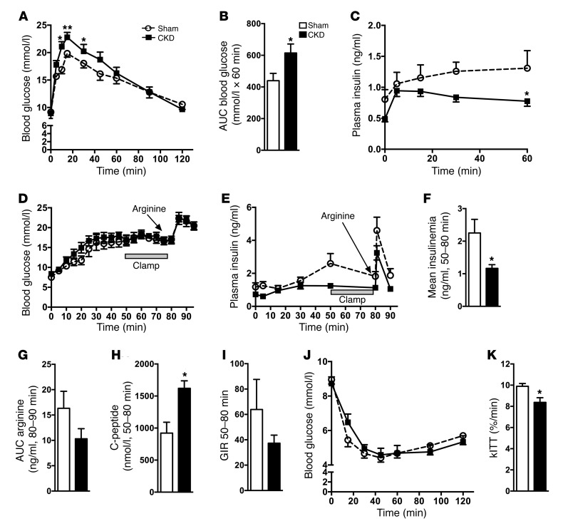
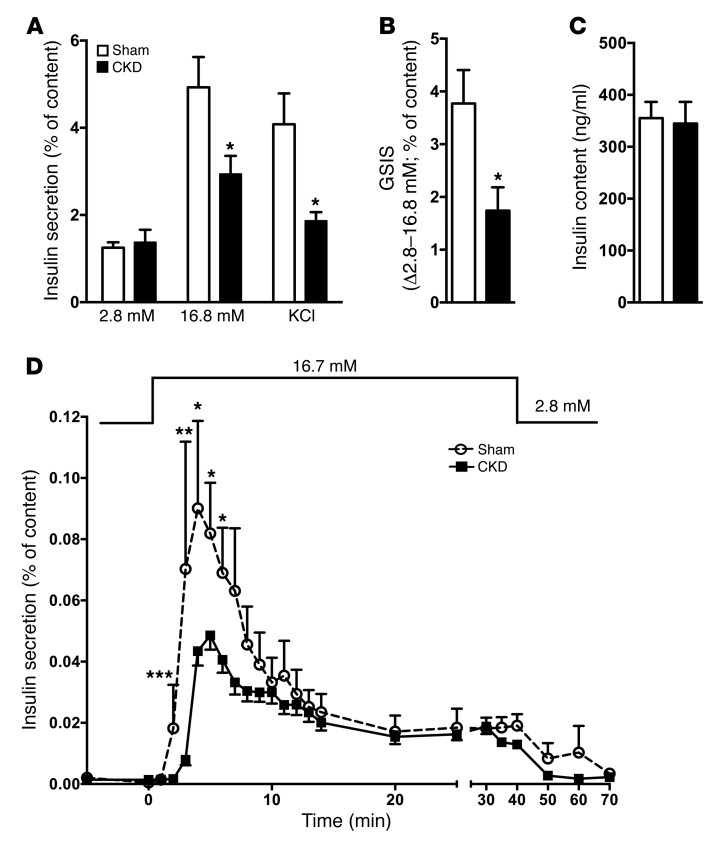
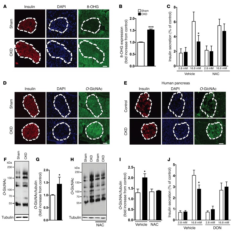
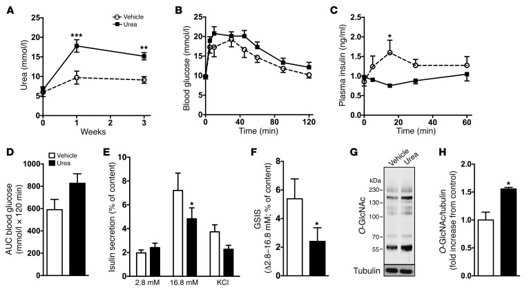
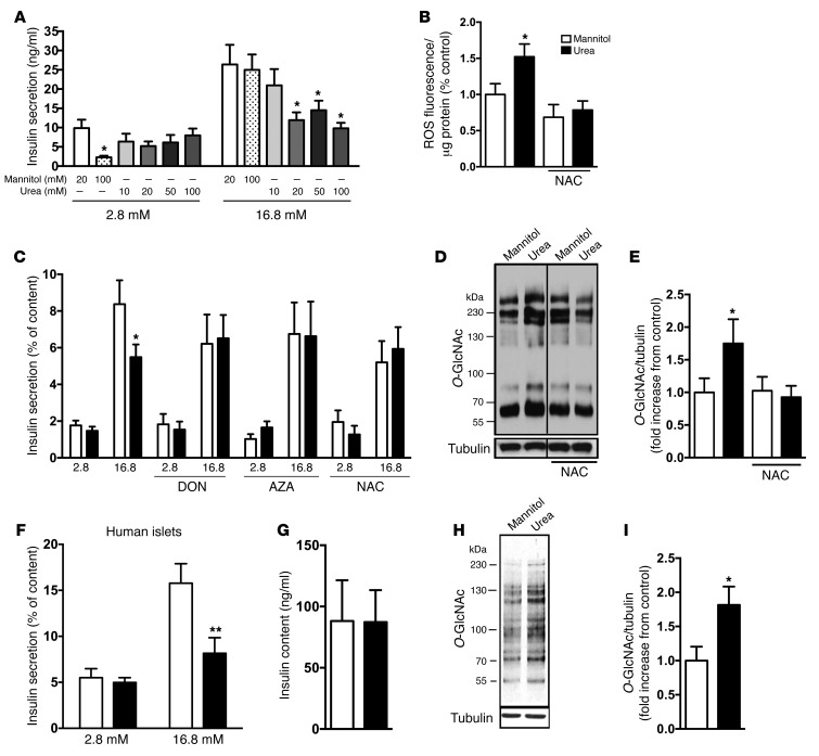
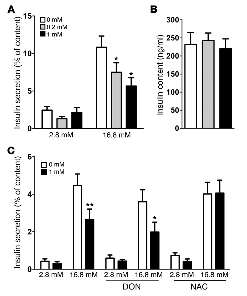
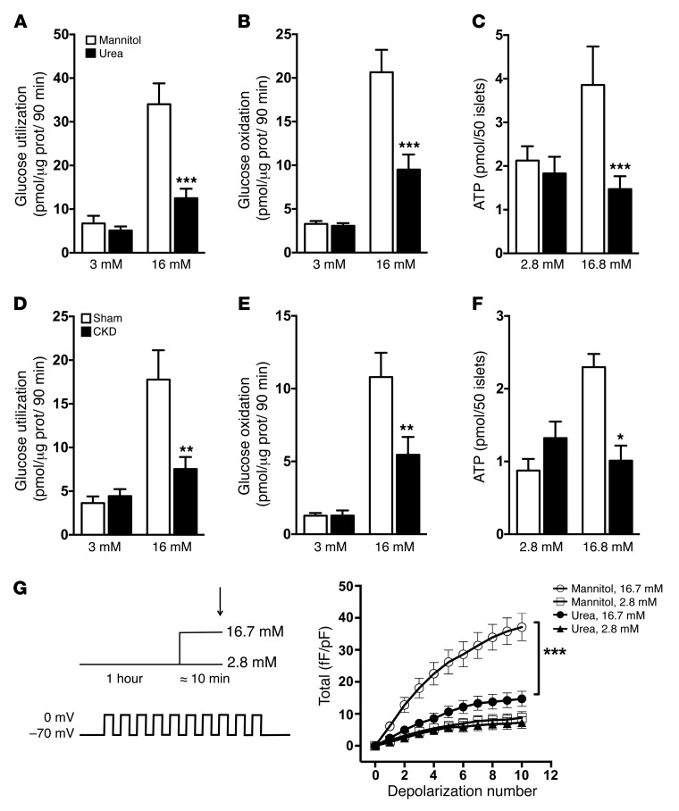
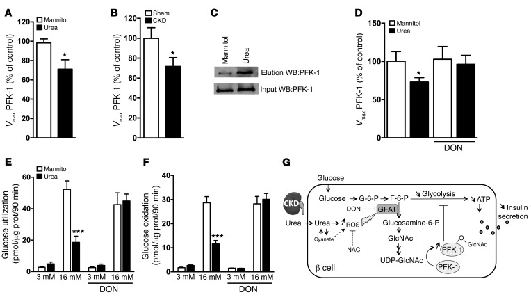
Disorders of glucose homeostasis are common in chronic kidney disease (CKD) and are associated with increased mortality, but the mechanisms of impaired insulin secretion in this disease remain unclear. Here, we tested the hypothesis that defective insulin secretion in CKD is caused by a direct effect of urea on pancreatic β cells. In a murine model in which CKD is induced by 5/6 nephrectomy (CKD mice), we observed defects in glucose-stimulated insulin secretion in vivo and in isolated islets. Similarly, insulin secretion was impaired in normal mouse and human islets that were cultured with disease-relevant concentrations of urea and in islets from normal mice treated orally with urea for 3 weeks. In CKD mouse islets as well as urea-exposed normal islets, we observed an increase in oxidative stress and protein O-GlcNAcylation. Protein O-GlcNAcylation was also observed in pancreatic sections from CKD patients. Impairment of insulin secretion in both CKD mouse and urea-exposed islets was associated with reduced glucose utilization and activity of phosphofructokinase 1 (PFK-1), which could be reversed by inhibiting O-GlcNAcylation. Inhibition of O-GlcNAcylation also restored insulin secretion in both mouse models. These results suggest that insulin secretory defects associated with CKD arise from elevated circulating levels of urea that increase islet protein O-GlcNAcylation and impair glycolysis.


Figures

Figure 1
Figure 1. CKD mice have defective glucose-stimulated insulin secretion in vivo.
(A) Blood glucose and (B) AUC during an IPGTT (1 g/kg) in CKD or sham-operated mice. (C) Insulin levels during the IPGTT (n = 9–10). (D) Glucose levels during HGCs. (E) Insulin levels during HGCs and in response to an arginine bolus (1 mM/kg). (F) Mean insulin levels during the clamp (50–80 min). (G) Arginine-induced insulin secretion (AUC) from 80 to 90 minutes (n = 7). (H) Mean C-peptide during the clamp (50–80 min). (I) GIR during the clamp (50–80 min). Blood glucose (J) and kITT (K) during the IPITT (0.5 IU/kg) (n = 6–8). Data represent the mean ± SEM. *P < 0.05 and **P < 0.01 versus CKD for the same times; 2-way ANOVA with Bonferroni’s post-hoc test for A, C, D, E, and J and Student’s t test for B, FI, and K.
Figure 2
Figure 2. Isolated islets from CKD mice have reduced insulin release in response to glucose and KCl.
(A) Insulin secretion, shown as the percentage of insulin content, was assessed in 1-hour static incubations in islets isolated from CKD and sham mice after 3 weeks in response to 2.8 or 16.8 mmol/l (mM) glucose or 2.8 mmol/l glucose plus 35 mmol/l KCl. (B) GSIS (Δ2.8–16.8 mmol/l) in sham and CKD mice after 3 weeks. (C) Total islet insulin content. (D) Insulin secretion in response to 2.8 mmol/l glucose from −5 to 0 minutes, 16.7 mmol/l glucose from 0 to 40 minutes, and 2.8 mmol/l glucose from 40 to 70 minutes in perifusion experiments. Data represent the mean ± SEM of 5 to 10 replicate experiments. *P <0.05, **P < 0.01, and ***P < 0.001 versus CKD for the same incubation condition; 1-way ANOVA with Bonferroni’s post-hoc test for A; Student’s t test for B and C; and 2-way ANOVA with Bonferroni’s post-hoc test for D.
Figure 3
Figure 3. CKD mice show an increase of O-GlcNAcylation in islets and oxidative stress markers.
(A and B) Immunostaining of 8-OHG in pancreatic sections of CKD and from sham-operated mice and quantification. (C) Insulin secretion, shown as the percentage of insulin content, was assessed in 1-hour static incubations in isolated islets from CKD or sham-operated mice in response to 2.8 or 16.8 mmol/l (mM) glucose after 3 weeks, with or without NAC treatment (200 mg/kg/day p.o., n = 6–7). (D and E) Immunostaining of O-GlcNAcylation (O-GlcNAc) in pancreatic sections from CKD and sham-operated mice (D) and from control and CKD patients (E). (F and G) Representative Western blot and quantification (n = 3) for total islet protein O-GlcNAcylation. (H and I) Representative Western blot and quantification (n = 4) for total protein O-GlcNAcylation in isolated islets from CKD and sham mice with or without NAC treatment. (J) Insulin secretion, shown as the percentage of insulin content in response to 2.8 or 16.8 mmol/l glucose, in isolated islets from CKD mice treated with or without DON (1.5 mg/kg 3 times/week, n = 5–8). Data represent the mean ± SEM. *P < 0.05 and ***P < 0.001 versus CKD for the same incubation condition; Student’s t test for B and D and 1-way ANOVA with Bonferroni’s post-hoc test for C, I, and J. Scale bars: 50 μm.
Figure 4
Figure 4. Exposure to urea in drinking water increases circulating urea levels and inhibits insulin secretion.
(A) Urea consumption in drinking water (25 g/l) elevates blood urea levels independently of renal failure (n = 4–5). (B) Blood glucose and (C) corresponding insulin levels during IPGTTs (n = 3–4). (D) AUC during IPGTTs. (E) Insulin secretion shown as the percentage of insulin content assessed in 1-hour static incubations in response to 2.8 or 16.8 mmol/l (mM) glucose or 2.8 mmol/l glucose plus 35 mmol/l KCl in islets from mice after 3 weeks with or without urea in drinking water (n = 5–7). (F) GSIS (Δ2.8–16.8 mM) (n = 5–7). (G) Representative Western blot and (H) quantification for total protein O-GlcNAcylation in islets in mice with or without addition of urea to the drinking water for 3 weeks (n = 3–4). Data represent the mean ± SEM. *P < 0.05, **P < 0.01, and ***P < 0.001 versus vehicle; 2-way ANOVA with Bonferroni’s post-hoc test for AC; 1-way ANOVA with Bonferroni’s post-hoc test for E; and Student’s t test for D, F, and H.
Figure 5
Figure 5. The insulin secretory defect resulting from exposure of normal mouse or human islets to urea is prevented by antioxidant treatment or GFAT inhibition.
(A) One-hour static insulin incubations of isolated islets from normal mice cultured for 24 hours with increasing urea levels (n = 3–5). (B) ROS accumulation in mouse islets treated with mannitol or urea, with or without NAC (500 μmol/l), for 24 hours (n = 3–8). (C) Insulin secretion shown as the percentage of insulin content as assessed in 1-hour static incubations in the presence of 20 mmol/l (mM) urea or mannitol as an osmotic control, with or without DON (20 μmol/l), AZA (5 μmol/l), or NAC (500 μmol/l) (n = 4–15). (D) Representative Western blot and (E) quantification for total protein O-GlcNAcylation in urea-treated islets with or without NAC (n = 4). (F) Insulin secretion shown as the percentage of insulin content from human islets incubated for 24 hours with 20 mmol/l urea or mannitol (n = 5). (G) Total insulin content in urea-treated human islets. (H) Representative Western blot and (I) quantification for total protein O-GlcNAcylation in urea-treated human islets (n = 5). Data represent the mean ± SEM. *P < 0.05 and **P < 0.01 versus mannitol for the same incubation condition; 1-way ANOVA with Bonferroni’s post-hoc test for AC, E, and F and Student’s t test for G and I.
Figure 6
Figure 6. Cyanate inhibits insulin secretion in islets ex vivo.
(A) Insulin secretion shown as the percentage of insulin content as assessed in 1-hour static incubations and (B) total islet insulin levels in the presence of 0, 0.2, or 1 mmol/l (mM) cyanate (n = 6). (C) Insulin secretion shown as the percentage of insulin content as assessed in 1-hour static incubations in the presence of 1 mmol/l cyanate with or without DON (20 μmol/l) or NAC (500 μmol/l) (n = 5–6). Data represent the mean ± SEM. *P < 0.05 and **P < 0.01 versus control for the same incubation condition; 1-way ANOVA with Bonferroni’s post-hoc test.
Figure 7
Figure 7. Normal mouse islets exposed to urea and islets from CKD mice have impaired glucose metabolism.
Glucose utilization (A and D) and oxidation (B and E) in islets incubated for 90 minutes in KRBH at 3 or 16 mmol/l (mM) glucose with D-[5-3H]glucose and D-[U-14C]glucose in urea-treated and CKD islets (n = 4). (C and F) Total ATP content in islets incubated for 15 minutes in KRBH at 2.8 or 16.8 mmol/l glucose (n = 3–4). (G) Averaged data of exocytotic responses of single β cells measured as increases in cell membrane capacitance by whole-cell patch clamp (arrow) performed after acute pretreatment with 2.8 or 16.7 mmol/l glucose (mean of 21 to 27 cells from 3 mice) after urea or mannitol treatment. Data represent the mean ± SEM. *P < 0.05, **P < 0.01, and ***P < 0.001 versus control; 2-way ANOVA with Bonferroni’s post-hoc test for G and 1-way ANOVA with Bonferroni’s post-hoc test for AF.
Figure 8
Figure 8. Inhibiting protein O-GlcNAcylation in islets restores glucose metabolism and PFK-1 activity.
Vmax of PFK-1 in islets (A) after urea treatment (n = 3) and (B) in CKD islets (n = 4–5). (C) Representative Western blot (WB) showing PFK-1 O-GlcNAcylation after chemoenzymatic labeling of O-GlcNAc residues with UDP-N-azidoacetylgalactosamine and the enzyme galactose-1-phosphate uridylyltransferase, followed by reaction with an alkyne-biotin derivative, streptavidin pull-down, and elution of biotinylated proteins (n = 3). (D) Vmax of PFK-1 in islets after urea treatment with or without DON (n = 4–5). Glucose utilization (E) and oxidation (F) in islets incubated for 90 minutes in KRBH at 3 or 16 mmol/l (mM) glucose with D-[5-3H]glucose and D-[U-14C]glucose, respectively, in urea-treated islets with or without DON (20 μmol/l) (n = 4). Data represent the mean ± SEM. *P < 0.05 and ***P < 0.001 versus control; 1-way ANOVA with Bonferroni’s post-hoc test for E and F and Student’s t test for A, B, and D. (G) Schematic representation of the mechanisms of impaired insulin secretion in CKD.

Comment in

Similar articles

Cited by

References

    1. Eldin WS, Ragheb A, Klassen J, Shoker A. Evidence for increased risk of prediabetes in the uremic patient. Nephron Clin Pract. 2008;108(1):c47–c55. - PubMed
    1. Lorenzo C, Nath SD, Hanley AJ, Abboud HE, Gelfond JA, Haffner SM. Risk of type 2 diabetes among individuals with high and low glomerular filtration rates. Diabetologia. 2009;52(7):1290–1297. doi: 10.1007/s00125-009-1361-4. - DOI - PMC - PubMed
    1. Sahakyan K, Lee KE, Shankar A, Klein R. Serum cystatin C and the incidence of type 2 diabetes mellitus. Diabetologia. 2011;54(6):1335–1340. doi: 10.1007/s00125-011-2096-6. - DOI - PMC - PubMed
    1. Mak RH. Insulin and its role in chronic kidney disease. Pediatr Nephrol. 2008;23(3):355–362. doi: 10.1007/s00467-007-0611-2. - DOI - PubMed
    1. Menon V, et al. Glycosylated hemoglobin and mortality in patients with nondiabetic chronic kidney disease. J Am Soc Nephrol. 2005;16(11):3411–3417. doi: 10.1681/ASN.2005050552. - DOI - PubMed

MeSH terms