. 2016 Sep 1;126(9):3389-402.
doi: 10.1172/JCI79434. Epub 2016 Aug 15.

Lymphatic vessels regulate immune microenvironments in human and murine melanoma

Lymphatic vessels regulate immune microenvironments in human and murine melanoma

Amanda W Lund et al. J Clin Invest. .

Abstract

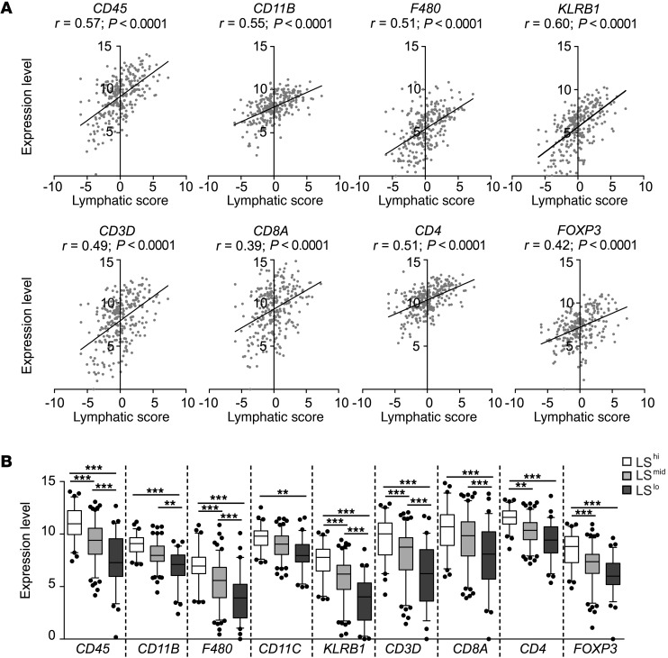
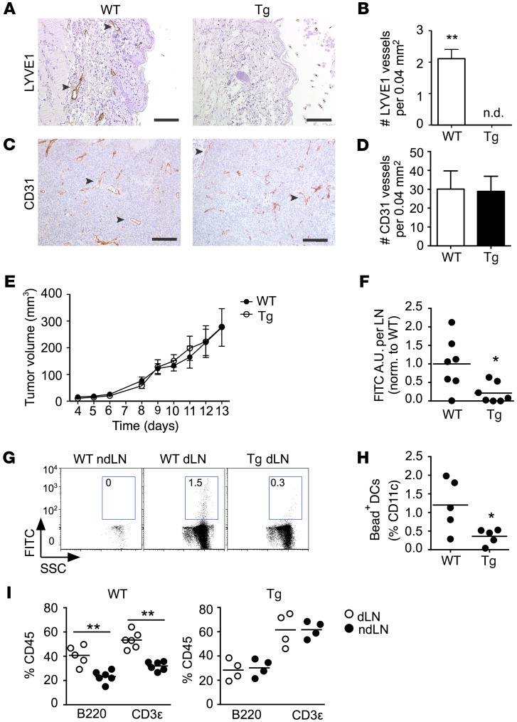
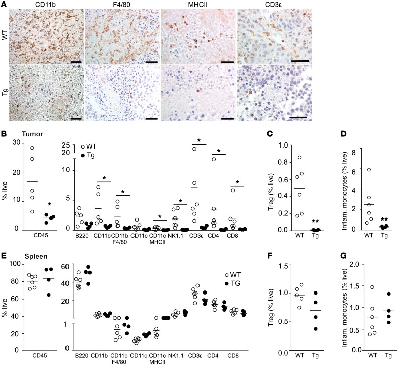
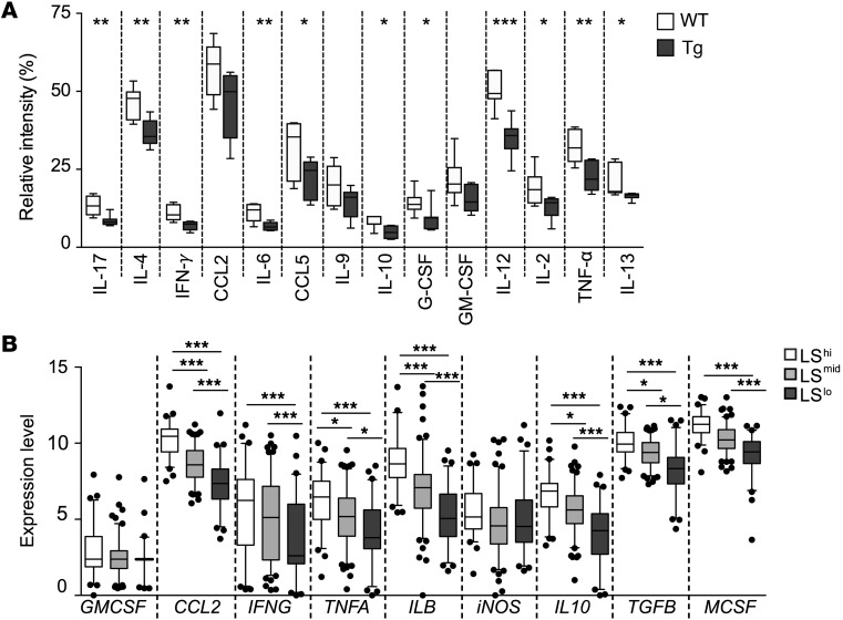
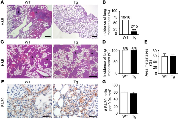
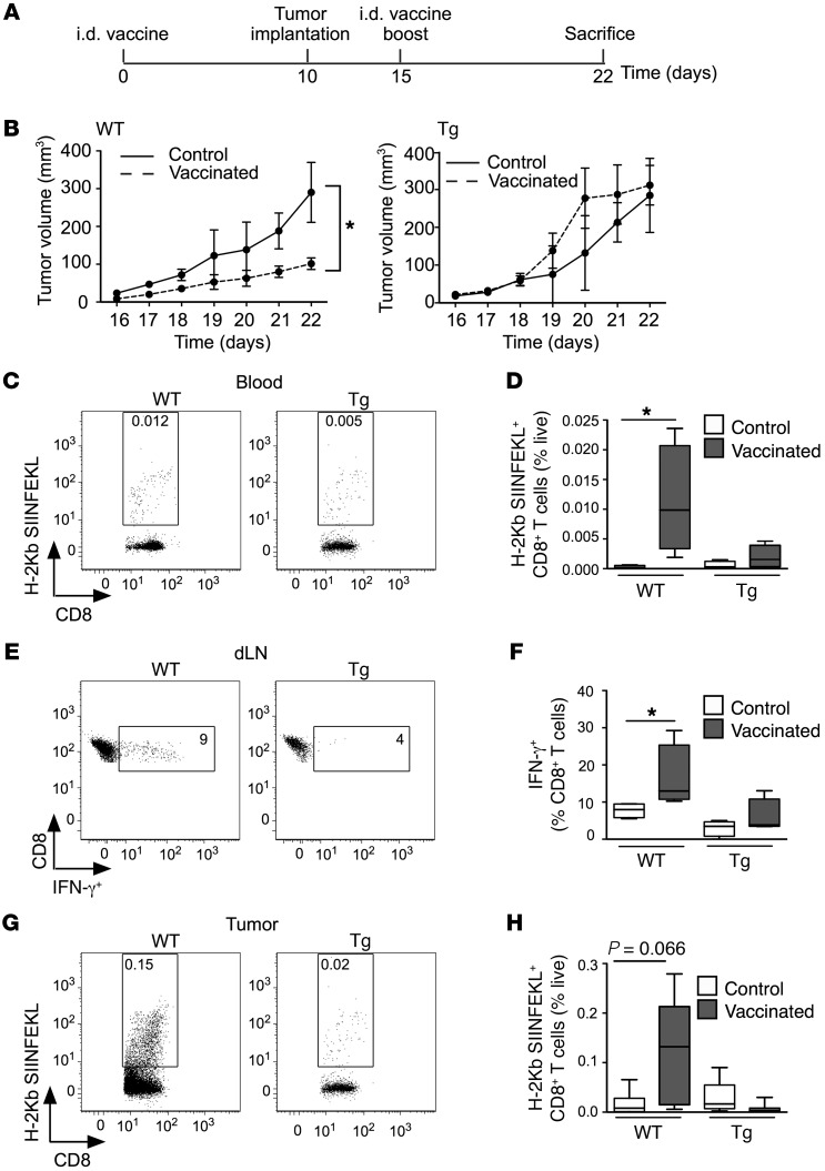
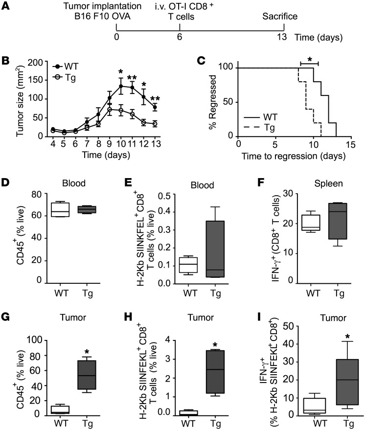
Lymphatic remodeling in tumor microenvironments correlates with progression and metastasis, and local lymphatic vessels play complex and poorly understood roles in tumor immunity. Tumor lymphangiogenesis is associated with increased immune suppression, yet lymphatic vessels are required for fluid drainage and immune cell trafficking to lymph nodes, where adaptive immune responses are mounted. Here, we examined the contribution of lymphatic drainage to tumor inflammation and immunity using a mouse model that lacks dermal lymphatic vessels (K14-VEGFR3-Ig mice). Melanomas implanted in these mice grew robustly, but exhibited drastically reduced cytokine expression and leukocyte infiltration compared with those implanted in control animals. In the absence of local immune suppression, transferred cytotoxic T cells more effectively controlled tumors in K14-VEGFR3-Ig mice than in control mice. Furthermore, gene expression analysis of human melanoma samples revealed that patient immune parameters are markedly stratified by levels of lymphatic markers. This work suggests that the establishment of tumor-associated inflammation and immunity critically depends on lymphatic vessel remodeling and drainage. Moreover, these results have implications for immunotherapies, the efficacies of which are regulated by the tumor immune microenvironment.


Figures

Figure 1
Figure 1. Lymphatic gene expression correlates with immune cell infiltrate in human metastatic cutaneous melanoma.
A lymphatic score (LS) was generated using 266 metastatic cutaneous melanoma samples from the Broad Institute’s TCGA database. LS was calculated based on relative expression levels of PDPN, LYVE1, and VEGFC in each sample. (A) Plots showing correlative expression of immune cell markers (CD45, CD11B, F480, KLRB1, CD3D, CD8A, CD4, FOXP3) with LS. Pearson’s correlation coefficient (r) is shown. (B) Samples were segregated into 3 groups; low (LSlo; < [mean – 2/3 SD]), intermediate (LSmid; [mean - 2/3 SD] < LSmid < [mean + 2/3 SD]) and high (LShi; > [mean + 2/3 SD]). Box plots are 5–95 percentile with outliers. One-way ANOVA followed by Tukey’s multiple comparisons test was used to determine significance on n = 266. **P < 0.01, ***P < 0.005.
Figure 2
Figure 2. Impaired tumor drainage and DC trafficking to local lymph nodes in K14-VEGFR3-Ig mice.
B16F10 tumors were implanted intradermally into WT or K14-VEGFR3-Ig transgenic (Tg) mice and excised at day 9. (A) Peritumoral area from WT or Tg mice stained for lymphatic endothelial cells (LYVE1; n = 3; n.d., not detected) (arrowhead). Scale bar: 50 μm. (B) Lymphatic vessel density quantified as LYVE1+ structures per 0.04 mm2. Data were compared using Student’s unpaired t test. **P < 0.01 (C) Intratumoral area stained for blood endothelial cells (CD31, n = 3) (arrowhead). Scale bar: 50 μm. (D) Blood vessel density quantification as CD31+ structures per 0.04 mm2. (E) Tumor growth profiles in WT and Tg mice over 13 days. (F) Fluid drainage from the tumor to the draining lymph node (dLN, brachial) assessed 30 minutes after intratumoral injection of 70-kDa FITC-dextran and plotted as fluorescence intensity in arbitrary units (A.U.) per LN normalized to WT (n = 7). (G) DC trafficking from the tumor to the dLN, determined by the quantity of CD11c+MHCII+FITC+ cells in the dLN 24 hours after intratumoral injection of 0.5-μm FITC-labeled latex beads and representative flow cytometry dot plots (n = 5). (H) Quantification of bead+ DCs in the LN. (I) Comparison of relative B cell (B220+) and T cell (CD3ε+) populations in the dLNs to nondraining lymph nodes (ndLN) (n ≥ 4). Data are represented as the mean ± SEM. Statistical analysis with Mann-Whitney U test. *P < 0.05, **P < 0.01.
Figure 3
Figure 3. B16F10 melanomas implanted in K14-VEGFR3-Ig mice lack a local inflammatory infiltrate.
The inflammatory infiltrate was determined on day 9 in B16F10 tumors grown in WT and K14-VEGFR3-Ig (Tg) mice. (A) Immunohistochemical analysis of tumor immune infiltrates in WT and Tg mice. Scale bars: 50 μm (CD11b, F4/80, and CD3ε) and 100 μm (MHCII). (BG) Analysis of immune cell populations in the tumor (BD) and spleen (EG) by flow cytometry. (B and E) Total leukocytes (CD45+), (C and F) Treg cells (CD3ε+CD4+CD25+FoxP3+), and (D and G) inflammatory monocytes (CD11cCD11b+F4/80Ly6chiLy6g, n ≥ 4). Statistical analysis with Man-Whitney U test. *P < 0.05, **P < 0.01.
Figure 4
Figure 4. Tumor-associated lymphatic vessels correlate with expression of local inflammatory mediators.
(A) The expression of common inflammatory mediators in tumor microenvironments was assessed by cytokine array on tumor tissue lysates from B16F10 melanomas grown in WT or K14-VEGFR3-Ig (Tg) mice (n = 7). Relative intensity as compared with internal biotin-conjugated IgG control. Results were compared using Student’s unpaired t tests. *P < 0.05, **P < 0.01, ***P < 0.001. (B) In human metastatic cutaneous melanoma, lymphatic score (LS) correlated with gene expression of inflammatory cytokines. Box plots generated by segregating samples into 3 groups: LSlo, LSmid, and LShi. Box plots show data within 5–95 percentile with outliers shown as dots. One-way ANOVA followed by Tukey’s multiple comparisons test was used to determine significance on 266 samples. *P < 0.05, ***P < 0.005.
Figure 5
Figure 5. Spontaneous lung metastasis from primary B16F10 melanomas is decreased in K14-VEGFR3-Ig mice.
(A) Histological determination of spontaneous lung metastases from orthotopically implanted B16F10 tumors. Scale bars: 1 mm. Paraformaldehyde-fixed, paraffin-embedded sections were stained with hematoxylin and eosin (H&E). (B) Quantification of incidence from 15 WT and 16 K14-VEGFR3-Ig (Tg) mice. Statistical analysis with Fisher exact test. **P < 0.01. (C) Histological determination of metastatic colonization in the lung after intravenous injection shows no differences between WT and Tg mice. Scale bars: 200 μm. (*) marks metastatic nodules. (D) Quantification of metastatic incidence and (E) area fraction of lung bearing metastatic nodules (n = 6). Data represented as the mean ± SEM. (F) Analysis of macrophage infiltrate in lung metastases 14 days following intravenous injection by immunohistochemistry on paraformaldehyde-fixed paraffin-embedded tissues (F4/80). Scale bars: 50 μm. (G) Density of F4/80+ macrophages (number per 0.04 mm2) analyzed from immunostained sections (n = 6). Data represented as the mean ± SEM.
Figure 6
Figure 6. K14-VEGFR3-Ig mice demonstrate impaired antitumor immunity in response to dermal vaccine delivery.
(A) Experimental schematic. WT or K14-VEGFR3-Ig (Tg) mice were vaccinated intradermally (i.d.) in the forelimbs with either 10 μg ovalbumin (OVA) and 10 μg LPS, or 10 μg LPS alone. After 10 days, B16F10.OVA tumor cells (0.5 × 10⁶) were injected i.d. in the back skin and the vaccine was boosted, at the same dose, on day 15. Mice were sacrificed on day 22 (12 days after tumor implantation). (B) Tumor growth was slower in vaccinated vs. control mice for melanomas in WT (left), but not Tg (right) mice. P values determined by regression analysis. *P < 0.05. (C) Representative plots and (D) quantification of circulating antigen-specific CD8+ T lymphocytes as detected by H-2Kb SIINFEKL pentamer staining on day 12. (E) Representative flow cytometry plots showing intracellular cytokine staining for IFN-γ and (F) quantification of IFN-γ+ CD8+ T cells following peptide restimulation of tumor-draining LN cells on day 12. (G) Representative flow cytometry plots and (H) quantification of tumor-infiltrating antigen-specific CD8+ T cells detected by H-2Kb SIINFEKL pentamer staining on day 12 (n = 5, in at least 2 separate experiments). Box and whisker plots show data from min to max. Statistical analysis with 1-way ANOVA. *P < 0.05.
Figure 7
Figure 7. Improved efficacy of adoptively transferred anti-OVA effector CD8+ T cells against OVA-expressing melanomas implanted in K14-VEGFR3-Ig mice.
(A) Experimental schematic. B16F10.OVA tumor cells (0.5 × 10⁶) were injected intradermally (i.d.) into either WT or K14-VEGFR3-Ig (Tg) mice, and after 6 days, activated OT-I CD8+ T cells were injected intravenously (i.v.). Mice were sacrificed 7 days following transfer. (B) Tumor growth profiles and (C) Kaplan-Meier curve showing relative times to tumor regression (n = 5). (D and E) Flow cytometric analysis of circulatory (D) CD45+ cells or (E) OT-1 cells at day 13, and (F) IFN-γ+ T cells in spleen following restimulation. (GK) Flow cytometric analysis of tumor infiltrating (G) leukocytes (CD45+), (H) antigen-specific CD8+ T cells (H-2Kb SIINFEKL+), and (I) IFN-γ+ CD8+ T cells following in vivo brefeldin A treatment. Box and whisker plots show data from min to max. Statistical analysis with Mann Whitney U test. *P < 0.05, **P < 0.01 in at least 2 separate experiments.

References

    1. Pasquali S, van der Ploeg AP, Mocellin S, Stretch JR, Thompson JF, Scolyer RA. Lymphatic biomarkers in primary melanomas as predictors of regional lymph node metastasis and patient outcomes. Pigment Cell Melanoma Res. 2013;26(3):326–337. doi: 10.1111/pcmr.12064. - DOI - PubMed
    1. Karaman S, Detmar M. Mechanisms of lymphatic metastasis. J Clin Invest. 2014;124(3):922–928. doi: 10.1172/JCI71606. - DOI - PMC - PubMed
    1. Platt AM, Randolph GJ. Dendritic cell migration through the lymphatic vasculature to lymph nodes. Adv Immunol. 2013;120:51–68. doi: 10.1016/B978-0-12-417028-5.00002-8. - DOI - PubMed
    1. Swartz MA, Lund AW. Lymphatic and interstitial flow in the tumour microenvironment: linking mechanobiology with immunity. Nat Rev Cancer. 2012;12(3):210–219. doi: 10.1038/nrc3186. - DOI - PubMed
    1. Wiig H, Swartz MA. Interstitial fluid and lymph formation and transport: physiological regulation and roles in inflammation and cancer. Physiol Rev. 2012;92(3):1005–1060. doi: 10.1152/physrev.00037.2011. - DOI - PubMed

Publication types

MeSH terms

Substances