. 2016 Aug 25;536(7617):484-7.
doi: 10.1038/nature19107. Epub 2016 Aug 15.

Diverse activation pathways in class A GPCRs converge near the G-protein-coupling region

Diverse activation pathways in class A GPCRs converge near the G-protein-coupling region

A J Venkatakrishnan et al. Nature. .

Abstract

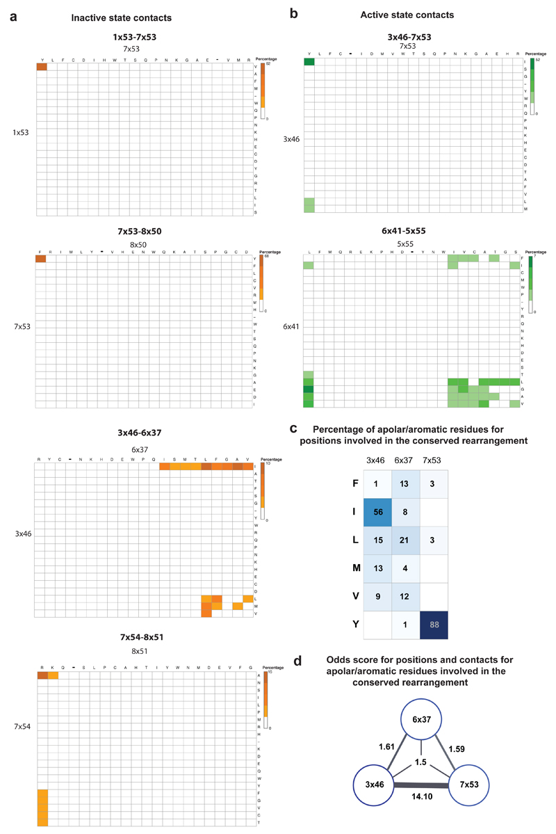
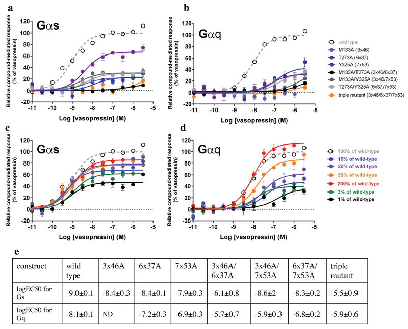
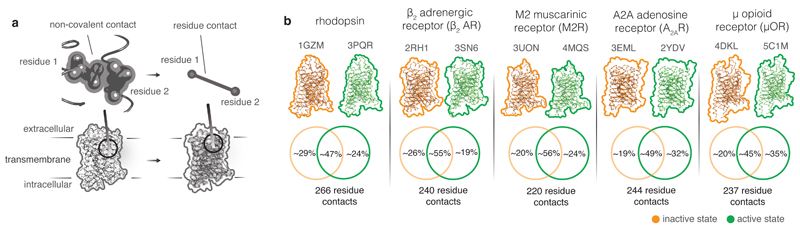
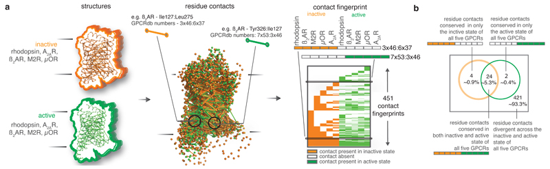
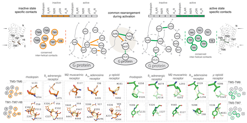
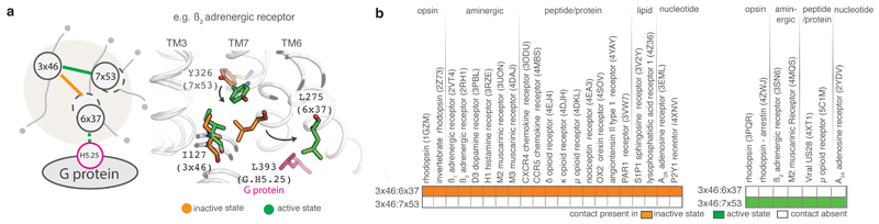
Class A G-protein-coupled receptors (GPCRs) are a large family of membrane proteins that mediate a wide variety of physiological functions, including vision, neurotransmission and immune responses. They are the targets of nearly one-third of all prescribed medicinal drugs such as beta blockers and antipsychotics. GPCR activation is facilitated by extracellular ligands and leads to the recruitment of intracellular G proteins. Structural rearrangements of residue contacts in the transmembrane domain serve as 'activation pathways' that connect the ligand-binding pocket to the G-protein-coupling region within the receptor. In order to investigate the similarities in activation pathways across class A GPCRs, we analysed 27 GPCRs from diverse subgroups for which structures of active, inactive or both states were available. Here we show that, despite the diversity in activation pathways between receptors, the pathways converge near the G-protein-coupling region. This convergence is mediated by a highly conserved structural rearrangement of residue contacts between transmembrane helices 3, 6 and 7 that releases G-protein-contacting residues. The convergence of activation pathways may explain how the activation steps initiated by diverse ligands enable GPCRs to bind a common repertoire of G proteins.


Figures

Extended Data Figure 1
Extended Data Figure 1. Sequence analysis of positions involved in the conserved rearrangement during receptor activation.
The alignment of 311 non-olfactory, Class A human GPCRs were obtained from GPCRdb. Percentage of residue pairs making a contact in the inactive state only (a; orange spectrum; five bins) and the active state only (b; green spectrum; five bins). The set of large hydrophobic/aromatic residues was defined to include the following amino acids: V, L, I, M, F, Y, W. c. The percentage of amino acids for residues in the positions (3x46, 6x37 and 7x53) involved in the conserved rearrangement is shown. d. The odds of finding a large hydrophobic/aromatic residue at a pair/triplet of positions that form a contact during the conserved rearrangement. See Methods for details.
Extended Data Figure 2
Extended Data Figure 2. Signalling profile of Vasopressin V2 receptor mutants using BRET-based biosensors.
Activation of Gs (a) and Gq (b) by Vasopressin V2 receptor wild type and mutants. The positions 3x46, 6x37 and 7x53 were mutated to alanine separately and in combination and their G protein response was measured using BRET-based biosensors. Gs is the primary and Gq is the secondary cognate G protein of the vasopressin V2 receptor. The G protein response was significantly reduced for all mutants, pointing towards a reduced receptor activity. The expression levels of the mutant receptors were quantified using a fluorescent dye bound to the SNAP tag of surface-expressed receptors. While the Y325A (Y7x53A), T273A (T6x37A) and T273A/Y325A (T6x37A/Y7x53A) proteins were expressed at wild-type levels, the expression of M133A (M3x46A) and its combinations showed reduced expression levels. As a comparison, the signalling of reduced levels of WT Vasopressin V2 receptor (% of DNA transfected) for Gs and Gq is shown in panels c and d, respectively. This indicates that the reduction in expression level alone cannot explain the reduced signalling of Y325A (Y7x53A), T273A (T6x37A) and the Y325A (Y7x53A)/T273A (T6x37A) combination. Thus, the reduced signalling activity of Y325A (Y7x53A), T273A (T6x37A) and T273A/Y325A (T6x37A/Y7x53A) is likely due to a change in receptor properties. In the case of receptor mutants (single, double and triple) involving M133A (M3x46A), both the G protein response as well as the expression level of the receptor is reduced. This maybe because the position 3x46 is involved in mediating contacts that are important for both the active and the inactive state, and mutating this position might simultaneously affect receptor biogenesis and downstream response. e. The increased EC50s are consistent with a reduced ability of the receptor to reach the G protein bound active conformation resulting in a reduced potency. This agrees well with a destabilisation of the active state of the mutants.
Figure 1
Figure 1. Comparison of residue contacts in inactive and active state structures of class A GPCR.
a. Residue contact in a GPCR. Residues are denoted as circles and the non-covalent contacts between residues (residue contacts) are denoted as lines connecting the circles. b. Similarity of residue contacts between inactive and active states of each GPCR. For five class A GPCRs, the comparison of the number of residue contacts between inactive and active states are shown using Venn diagrams.
Figure 2
Figure 2. Patterns of residue contacts across inactive and active state structures of GPCRs.
a. Contact fingerprinting. For every residue contact, the presence (or absence) of a contact between structurally equivalent residues for all structures is computed. The pattern of presence and absence (filled and empty cells respectively) across multiple structures is termed ‘contact fingerprint’. b. Distribution of the contact fingerprints representing the presence of contacts in the five inactive and five active state structures.
Figure 3
Figure 3. Conserved rearrangement of residue contacts between inactive and active state structures of GPCRs.
Considering the conserved residue contacts in the inactive (left) and the active (right) states together reveals a conserved rearrangement of residue contacts in all the five receptors. The cartoons show a schematic representation of the contacts at the secondary structure level (inter-helical) and the residue level (inter-residue). TM helices and helix 8 are shown as circles and the presence of inactive and active state specific contacts between the helices are shown as lines. Residues are shown as circles and contacts between residues are shown as lines. Inactive and active state-specific contacts are shown in orange and green respectively. Dotted circles around 6x37 and 7x53 denote the movement of helices 6 and 7 upon activation.
Figure 4
Figure 4. Conserved rearrangement of residue contacts between inactive and active state structures across diverse class A GPCRs.
a. Illustration of the conserved rearrangement of residue contacts between inactive and active state structures in β2AR. Dotted circles around 6x37 and 7x53 denote the movement of helices 6 and 7 upon activation. b. Conservation pattern of residue contacts involved in the contact rearrangement upon receptor activation in the inactive (left) and active (right) state structures of class A GPCRs.

References

    1. Bockaert J, Pin JP. Molecular tinkering of G protein-coupled receptors: an evolutionary success. EMBO J. 1999;18:1723–1729. - PMC - PubMed
    1. Lagerstrom MC, Schioth HB. Structural diversity of G protein-coupled receptors and significance for drug discovery. Nat Rev Drug Discov. 2008;7:339–357. - PubMed
    1. Rosenbaum DM, Rasmussen SG, Kobilka BK. The structure and function of G-protein-coupled receptors. Nature. 2009;459:356–363. - PMC - PubMed
    1. Venkatakrishnan AJ, et al. Molecular signatures of G-protein-coupled receptors. Nature. 2013;494:185–194. - PubMed
    1. Overington JP, Al-Lazikani B, Hopkins AL. How many drug targets are there? Nat Rev Drug Discov. 2006;5:993–996. - PubMed

Publication types

MeSH terms