. 2016 Sep 29;537(7622):634-638.
doi: 10.1038/nature19331. Epub 2016 Aug 15.

SEDS proteins are a widespread family of bacterial cell wall polymerases

Affiliations

SEDS proteins are a widespread family of bacterial cell wall polymerases

Alexander J Meeske et al. Nature. .

Abstract

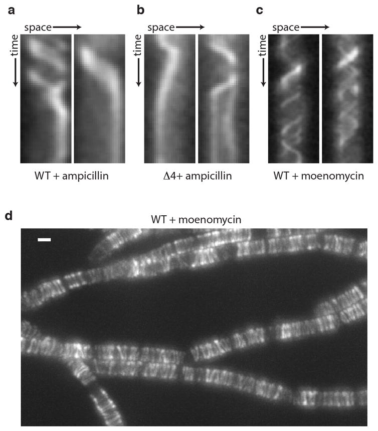
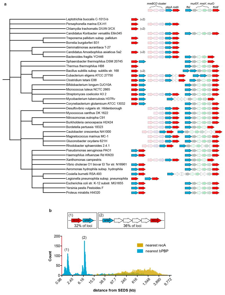
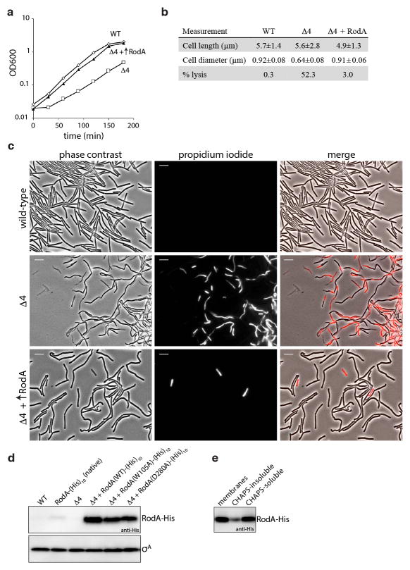
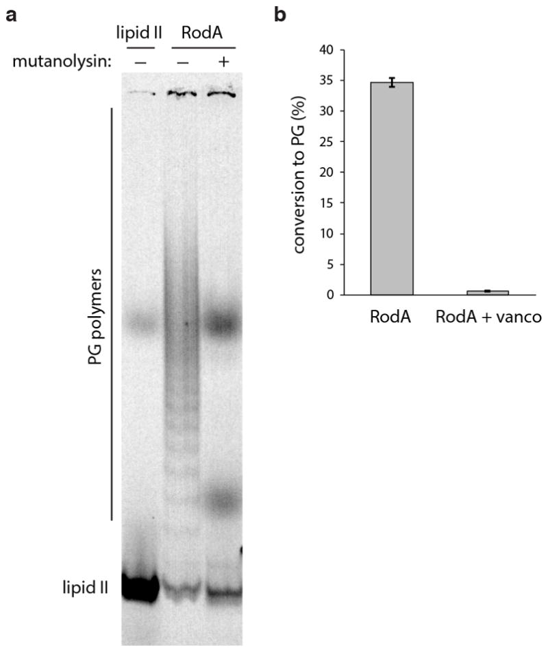
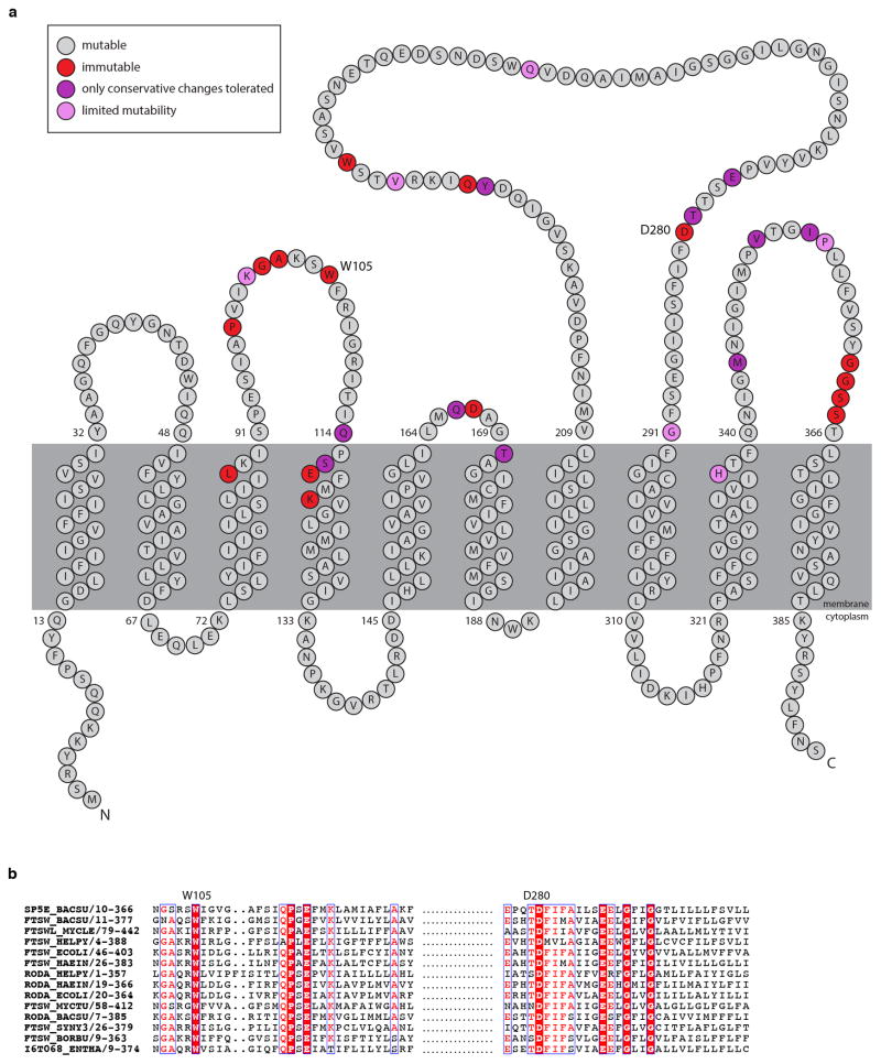
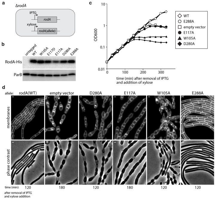
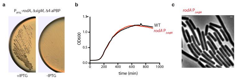
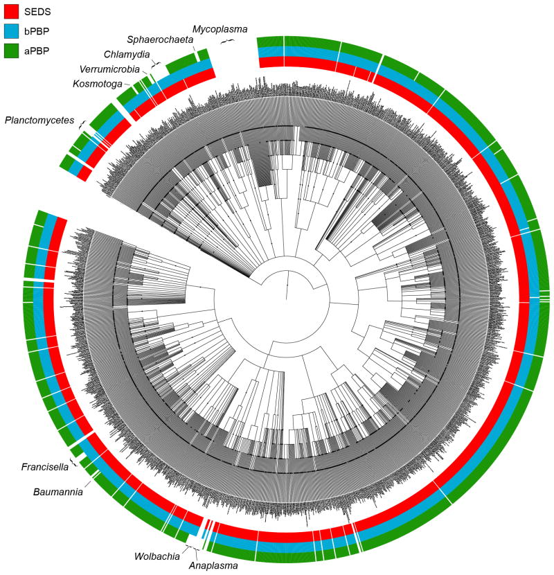
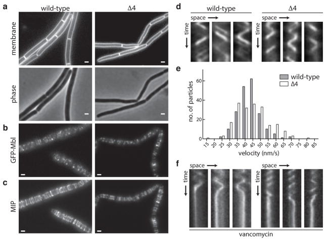
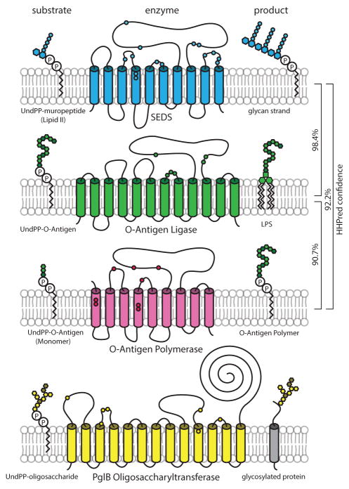
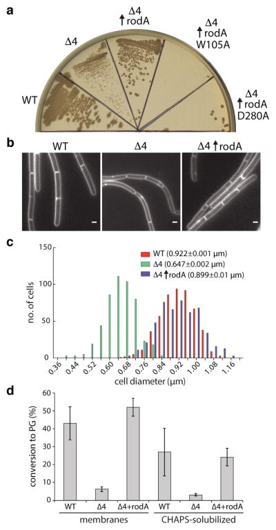
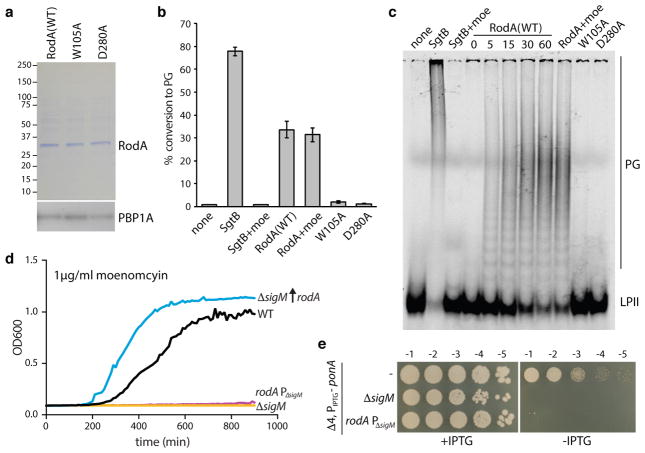
Elongation of rod-shaped bacteria is mediated by a dynamic peptidoglycan-synthetizing machinery called the Rod complex. Here we report that, in Bacillus subtilis, this complex is functional in the absence of all known peptidoglycan polymerases. Cells lacking these enzymes survive by inducing an envelope stress response that increases the expression of RodA, a widely conserved core component of the Rod complex. RodA is a member of the SEDS (shape, elongation, division and sporulation) family of proteins, which have essential but ill-defined roles in cell wall biogenesis during growth, division and sporulation. Our genetic and biochemical analyses indicate that SEDS proteins constitute a family of peptidoglycan polymerases. Thus, B. subtilis and probably most bacteria use two distinct classes of polymerase to synthesize their exoskeleton. Our findings indicate that SEDS family proteins are core cell wall synthases of the cell elongation and division machinery, and represent attractive targets for antibiotic development.


Conflict of interest statement

The authors declare no competing financial interests.

Figures

Extended Data Fig. 1
Extended Data Fig. 1. Rod complex dynamics in the presence of different antibiotics
Representative kymographs showing cessation of GFP-Mbl particle movement in a, wild-type and b, the quadruple (Δ4) aPBP mutant upon treatment with 10 μg/mL ampicillin. c, Rod complex motion is unaffected by moenomycin treatment. Cells harboring GFP-Mbl were pre-treated with 1 μg/mL moenomycin for 15 min then imaged in the presence of moenomycin. Representative kymographs of individual GFP-Mbl particles are shown. (d) Maximum intensity projection of the timelapse in Supplementary Video 5. Scale bar indicates 1 μm. Data are representative of 3 biological replicates.
Extended Data Fig. 2
Extended Data Fig. 2. Conserved neighborhood architecture for loci encoding SEDS proteins and bPBPs
a, Diagrams depicting the genomic context of genes encoding SEDS proteins (red) in a diverse set of bacterial taxa. Genes encoding bPBPs are depicted in blue and are frequently located adjacent to SEDS loci. These SEDS-bPBP pairs are often found in the context of the mreBCD operon (faded pink), suggesting that these orthologs function in cell elongation. SEDS and bPBP loci are also frequently present in the cluster of cell wall synthesis and cell division genes exemplified by the E. coli dcw cluster (faded green) and these orthologs likely function in cell division. Unrelated genes are shown as white triangles. Phylogenetic tree was constructed in PhyLoT (phylot.biobyte.de) and visualized in iToL (itol.embl.de). b, Histogram showing the genetic distance (on log10 scale) between 2,958 SEDS loci (red) and the nearest bPBP locus (blue). Two commonly observed SEDS-bPBP neighborhood architectures are depicted. Distances between SEDS and the nearest recA gene are shown in yellow as a negative control. SEDS and bPBP loci were identified using tblastn with five diverse members of each family used as the query.
Extended Data Fig. 3
Extended Data Fig. 3. RodA overexpression partially suppresses the phenotypes of the quadruple aPBP mutant
a, Growth curves of wild-type (WT), the quadruple (Δ4) aPBP mutant, and the Δ4 mutant overexpressing rodA-his10, representative of three biological replicates. b, Quantification of indicated cytological phenotypes, n=500. Error bars denote s.e.m. c, Live-dead (propidium iodide) staining of strains analyzed in a. Dead cells or cells with membrane integrity defects were visualized by fluorescence microscopy. Images representative of 3 biological replicates. Scale bar indicates 5 μm. d, Immunoblot analysis of RodA-His10 levels for the three strains in a as well as the Δ4 strain overexpressing nonfunctional mutants W105A and D280A. A fusion of his10 to rodA at its native locus was used to assess wild-type RodA levels (lane 2). Sigma A (σA) levels are shown to control for loading. e, Detergent solubilization of RodA-His10 from B. subtilis membranes using CHAPS. Anti-His immunoblot showing the relative amounts of solubilized RodA-His10 after overnight incubation with 2% CHAPS and ultracentrifugation at 100,000g.
Extended Data Fig. 4
Extended Data Fig. 4. Polymers synthesized by RodA in vitro are susceptible to muramidase digestion
a, To determine whether the products of RodA activity are glycan strands, their susceptibility to cleavage by the muramidase mutanolysin was investigated. Mutanolysin specifically cleaves the β(1,4) linkage between N-acetylmuramic acid and N-acetylglucosamine in PG glycan chains. 0.2 μM FLAG-RodA was incubated with 4 μM synthetic lipid II for 1 h, then quenched by boiling for 2 min. The products were subjected to overnight digestion with mutanolysin (0.1 mg/ml) at 37°C, and analyzed by SDS-PAGE. Lipid II, and undigested RodA products are shown for comparison. Data representative of two technical replicates. b, The reaction catalyzed by RodA can be inhibited by vancomycin (50 μg/mL), which binds and sequesters the lipid II substrate. Graph denotes the mean from 3 technical replicates, error bars show s.e.m.
Extended Data Fig. 5
Extended Data Fig. 5. Critical amino acid residues in RodA identified by MutSeq
a, Topological map of the RodA protein. The extent to which each amino acid residue tolerated mutations based on the MutSeq screen are indicated. Residues that tolerated a spectrum of amino acid changes are shown in grey. Residues that did not tolerate any mutations are shown in red. Residues that only tolerated conservative changes (conservation of charge, hydrophobicity, or functional groups) are in purple. Residues that had limited mutability but tolerated a nonconservative substitution are shown in pink. The complete dataset can be found in Supplementary Table 1. b, Multiple sequence alignment (created using ESPRIPT: http://espript.ibcp.fr/) of 14 diverse SEDS proteins with W105 and D280 residues highlighted.
Extended Data Fig. 6
Extended Data Fig. 6. Validation of critical amino acids identified in the MutSeq screen
a, Schematic of the strain used to test a subset of critical amino acid residues in rodA identified by MutSeq. A wild-type copy of rodA was placed under IPTG-inducible control at an ectopic chromosomal locus (ycgO) and the native copy of rodA was deleted. The mutant alleles to be tested were placed under xylose-inducible control at a second ectopic locus (amyE). As a positive control, a wild-type allele under xylose control was integrated at the second locus. The empty vector was used as a negative control. b, Immunoblot analysis of the RodA mutants expressed as His10-tagged fusions under xylose-inducible control, representative of two biological replicates. ParB levels are shown to control for loading. c, Growth curves of strains expressing mutant rodA alleles. Each strain was grown at 37°C in CH medium in the presence of 500 μM IPTG to maintain expression of wild-type rodA. Cultures were then washed three times in medium lacking inducer, diluted to OD600 = 0.02 in CH medium with 10mM xylose, and growth was monitored. Growth curves are representative of 2 biological replicates. d, Morphological phenotypes of the strains analyzed in (c) were examined by fluorescence microscopy at the indicated time points (below the images) after resuspension in xylose-containing medium. Fluorescent images of cell membranes stained with TMA-DPH and phase contrast images are shown. A mutation in the highly conserved residue E288 that was found to be mutable by Mutseq was included as a negative control. Consistent with the Mutseq analysis, substitution to alanine (E288A) supported wild-type growth rates and had no impact on cell morphology.
Extended Data Fig. 7
Extended Data Fig. 7. RodA overexpression suppresses the synthetic lethality of ΔsigM and the quadruple aPBP mutant
a, LB agar plates onto which a ΔsigM Δ4 aPBP strain harboring an IPTG-inducible allele of rodA was streaked in the presence and absence of 15 μM IPTG and incubated at 37°C overnight. b, The rodA allele containing mutations in its SigM-dependent promoter (rodA ΔPsigM) grows in a manner indistinguishable from wild-type in the absence of moenomycin. Wild-type (WT) and the rodA ΔP sigM mutant were grown in LB and OD600 was monitored continuously. c, The rodA ΔP sigM mutant has a normal rod-shaped morphology. Phase contrast image of cells harboring the rodA ΔPsigM promoter mutant grown to mid-exponential phase in LB medium. All data representative of two biological replicates. Scale bar indicates 1 μm.
Extended Data Fig. 8
Extended Data Fig. 8. SEDS proteins and bPBPs are more widely conserved than aPBPs
Phylogenetic tree showing distribution of SEDS proteins, bPBPs, and aPBPs in a diverse set of 1773 bacterial taxa. The amino acid sequences of five members of each family were used as queries in a BLASTp search against the NCBI nr database with an E-value cutoff of 10−4. The phylogenetic tree was constructed using PhyloT (phylot.bio-byte.de) and BLASTp results were plotted against the tree. The occurrence of a SEDS protein is indicated in red, a bPBP in blue, and an aPBP in green. The tree was visualized and annotated using iToL (itol.embl.de). Clades whose genomes contain a SEDS protein and bPBP but lack aPBPs are indicated. The PG-less Mycoplasma lack all three proteins.
Fig. 1
Fig. 1. The Rod complex is functional in the absence of all known PG polymerases
a, Morphological defects of B. subtilis cells lacking all four aPBPs. b–e, Dynamics of the Rod complex component Mbl (GFP-Mbl) in the presence and absence of the aPBPs (Δ4). b, Snapshots and c, maximum intensity projections (MIP) from timelapse microscopy. d, Kymographs showing the motion of individual GFP-Mbl particles over time. e, Histogram of particle velocities, n=200. f, Kymographs of GFP-Mbl particles upon vancomycin treatment. Data are representative of four biological replicates. Scale bars indicate 1 μm.
Fig. 2
Fig. 2. The SEDS proteins bear similarity to known glycosyltransferases
Schematic diagrams of the SEDS protein RodA and known glycosyltransferases from gram-negative bacteria with their undecaprenyl pyrophosphate-linked substrates and products. In blue, RodA from B. subtilis is shown alongside its putative substrate and product, undecaprenyl pyrophosphate-linked precursor (Lipid II) and glycan strands, respectively. O-antigen ligase WaaL (green), O-antigen polymerase Wzy (pink) and N-linked oligosaccharyltransferase PglB (yellow) are shown. Biochemical and genetic analyses indicate that functionally important residues are located in the periplasmic loops, shown schematically as small circles. Similarity between protein families detected by HHpred is indicated.
Fig. 3
Fig. 3. RodA overexpression partially suppresses the phenotypes of the aPBP mutant
a, Suppression of growth defects and b, morphological defects in the Δ4 mutant by rodA overexpression, representative of 3 biological replicates. Scale bar indicates 1 μm. c, Histogram of cell diameters for the indicated strains, n=500. Average diameter is indicated in parenthesis. d, PGT activity catalyzed by membranes and CHAPS-solubilized membrane proteins derived from the indicated strains. Data show the mean of 3 biological replicates, error bars denote standard error of the mean (s.e.m.).
Fig. 4
Fig. 4. RodA has glycosyltransferase activity in vitro
a, Purified FLAG-RodA and point mutants, and corresponding anti-PBP1A immunoblot. b, PGT activity of purified proteins and positive control SgtB in the presence and absence of moenomycin (moe). Error bars indicate s.e.m. from 2 technical and 2 biological replicates. c, SDS-PAGE of reactions from b, analyzed after 60 min or as indicated (in minutes). d and e, RodA induction is necessary and sufficient for growth in the absence of aPBPs. Representatives from 3 biological replicates. d, Moenomycin inhibition and e, depletion of ponA (encoding the major aPBP) in the indicated strains.

Comment in

References

    1. Goffin C, Ghuysen JM. Multimodular penicillin-binding proteins: an enigmatic family of orthologs and paralogs. Microbiol Mol Biol Rev. 1998;62:1079–1093. - PMC - PubMed
    1. Lombard V, Golaconda Ramulu H, Drula E, Coutinho PM, Henrissat B. The carbohydrate-active enzymes database (CAZy) in 2013. Nucleic Acids Research. 2013;42:D490–D495. - PMC - PubMed
    1. Matteï PJ, Neves D, Dessen A. Bridging cell wall biosynthesis and bacterial morphogenesis. Current Opinion in Structural Biology. 2010;20:749–755. - PubMed
    1. Packiam M, Weinrick B, Jacobs WR, Jr, Maurelli AT. Structural characterization of muropeptides from Chlamydia trachomatis peptidoglycan by mass spectrometry resolves ‘chlamydial anomaly’. Proc Natl Acad Sci USA. 2015;112:11660–11665. - PMC - PubMed
    1. McPherson DC, Popham DL. Peptidoglycan Synthesis in the Absence of Class A Penicillin-Binding Proteins in Bacillus subtilis. Journal of Bacteriology. 2003;185:1423–1431. - PMC - PubMed

MeSH terms