. 2016 Sep;22(9):987-90.
doi: 10.1038/nm.4170. Epub 2016 Aug 15.

A genome-editing strategy to treat β-hemoglobinopathies that recapitulates a mutation associated with a benign genetic condition

Affiliations

A genome-editing strategy to treat β-hemoglobinopathies that recapitulates a mutation associated with a benign genetic condition

Elizabeth A Traxler et al. Nat Med. 2016 Sep.

Abstract

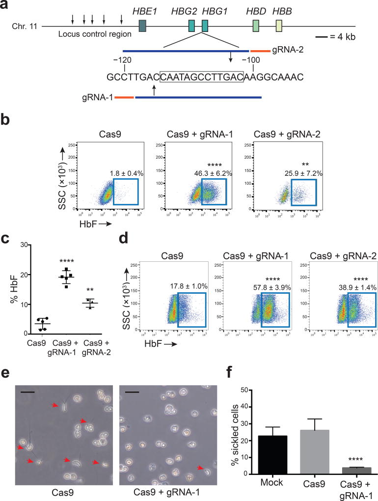
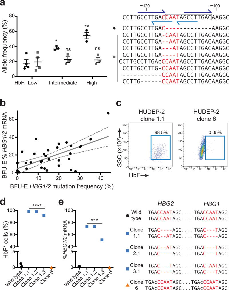
Disorders resulting from mutations in the hemoglobin subunit beta gene (HBB; which encodes β-globin), mainly sickle cell disease (SCD) and β-thalassemia, become symptomatic postnatally as fetal γ-globin expression from two paralogous genes, hemoglobin subunit gamma 1 (HBG1) and HBG2, decreases and adult β-globin expression increases, thereby shifting red blood cell (RBC) hemoglobin from the fetal (referred to as HbF or α2γ2) to adult (referred to as HbA or α2β2) form. These disorders are alleviated when postnatal expression of fetal γ-globin is maintained. For example, in hereditary persistence of fetal hemoglobin (HPFH), a benign genetic condition, mutations attenuate γ-globin-to-β-globin switching, causing high-level HbF expression throughout life. Co-inheritance of HPFH with β-thalassemia- or SCD-associated gene mutations alleviates their clinical manifestations. Here we performed CRISPR-Cas9-mediated genome editing of human blood progenitors to mutate a 13-nt sequence that is present in the promoters of the HBG1 and HBG2 genes, thereby recapitulating a naturally occurring HPFH-associated mutation. Edited progenitors produced RBCs with increased HbF levels that were sufficient to inhibit the pathological hypoxia-induced RBC morphology found in SCD. Our findings identify a potential DNA target for genome-editing-mediated therapy of β-hemoglobinopathies.


Conflict of interest statement

Competing financial interests

The authors declare no competing financial interests.

Figures

Figure 1
Figure 1. Genome editing of the HBG1 and HBG2 promoters increases erythroid fetal hemoglobin (HbF) levels
(a) Extended β-globin locus showing β-like genes as boxes. Small arrows mark DNase hypersensitive sites within the locus control region, an upstream enhancer. A region of the HBG1 promoter is shown numbered according to position upstream of the transcription start with the 13-nt HPFH deletion boxed. Guide RNA spacer sequences are blue and PAM motifs (NGG) are orange; gRNA-1 and gRNA-2 are complementary to the sense and antisense strands, respectively. Large arrows show predicted Cas9 cleavage sites. (b) Representative flow cytometry plots showing HbF+ immunostaining HUDEP-2 cells 5d after transduction with Cas9 ± gRNA-1 or gRNA-2 lentivirus. Numbers indicate mean ± standard error (SE) from four independent experiments. (c) Normal human CD34+ cells transduced with lentivirus encoding Cas9 ± gRNA-1 or gRNA-2 were cultured for 21d in erythroid cytokines, then analyzed for hemoglobin (Hb) protein by HPLC. %HbF = [HbF/(HbA + HbF) × 100]. Each dot represents a separate experiment performed with CD34+ cells from the same donor. On-target editing rates of HBG1/HBG2 in three experiments were 56%, 65% and 77%. (d) HbF+ erythroblasts, derived as described for panel (c). Numbers indicate mean ± SE from three experiments. (e) CD34+ cells from an SCD (HbSS) patient were transduced with lentivirus expressing Cas9 ± gRNA-1, differentiated into RBCs, and cultured in 2% O2.Red arrows denote cells with sickle-like morphology. Original magnification 200×. Size bars indicate 20 µm. (f) Quantification of hypoxia-induced sickled cells depicted in panel (e). Mean ± SE from three experiments using CD34+ cells from three different SCD donors (> 1,000 cells scored per experiment). The unpaired t-test was used to analyze data in panels b-d and f. **** P < 0.0001, ** P < 0.01.
Figure 2
Figure 2. Spectrum of γ-globin-inducing mutations caused by Cas9 and gRNA-1
(a) Normal adult CD34+ cells were edited with Cas9/gRNA-1 lentivirus, differentiated into RBCs, FACS-purified according to HbF immunostaining intensity (low, intermediate or high) and analyzed for on-target mutations. The wild type sequence is shown on the top left with the 13-nt HPFH deletion boxed and the CCAAT box in red. Dark blue half arrows show flanking 8-nt repeats; light blue show the DR element. The top nine mutant alleles (of more than 40 total indels identified) are shown below. Dashes indicate nucleotide deletions and lower-case letters insertions. In the graph at right, black dots denote the 13-nt HPFH deletion, which occurred at the highest frequency; gray squares show the combined frequencies of the eight next common mutations. Each symbol represents an independent experiment. * P < 0.05, ** P < 0.01 by unpaired t-test. (b) CD34+ cells were electroporated with Cas9/gRNA-1/GFP expression plasmids. GFP+ cells were FACS purified and seeded into methylcellulose. Burst forming unit-erythroid (BFU-E) colonies were analyzed for globin mRNAs and HBG1/HBG2 mutations. All colonies were mosaic for mutations; the total HBG [(HBG1 + HBG2)/2 × 100] mutation frequency for each colony is plotted against %HBG1/2 mRNA [γ / (γ+β)]. Regression analysis shows best-fit line as solid gray (y = 0.82x + 4.8, r2 = 0.41, P < 0.0001, n = 35 colonies from three experiments) and 95% confidence intervals as dashed gray. See also Supplementary Figs. 4a and 5. (c) HUDEP-2 cells were electroporated with Cas9/gRNA-1/GFP plasmid and cloned. HbF immunostaining is shown for two representative clones with different mutations (see also panel (d) and Supplementary Fig. 6). (d–e) Characterization of genome edited HUDEP-2 clones. The HBG1 and HBG2 genotypes corresponding to each clone is shown on the right, according to the convention used in panel (a); all clones are homogenous for the indicated mutations. *** P < 0.001, **** P < 0.0001, ns = not significant by unpaired t-test.

Similar articles

Cited by

References

    1. Forget BG. Molecular basis of hereditary persistence of fetal hemoglobin. Ann. N. Y. Acad. Sci. 1998;850:38–44. - PubMed
    1. Wienert B, et al. Editing the genome to introduce a beneficial naturally occurring mutation associated with increased fetal globin. Nat Commun. 2015;6:7085. - PubMed
    1. Gilman JG, et al. Distal CCAAT box deletion in the A gamma globin gene of two black adolescents with elevated fetal A gamma globin. Nucleic Acids Res. 1988;16:10635–10642. - PMC - PubMed
    1. Liberati C, et al. Cooperation and competition between the binding of COUP-TFII and NF-Y on human epsilon- and gamma-globin gene promoters. J Biol Chem. 2001;276:41700–41709. - PubMed
    1. Zhu X, et al. NF-Y recruits both transcription activator and repressor to modulate tissue- and developmental stage-specific expression of human γ-globin gene. PLoS ONE. 2012;7:e47175. - PMC - PubMed

Publication types

MeSH terms