. 2016 Aug 15;11(8):e0155201.
doi: 10.1371/journal.pone.0155201. eCollection 2016.

3D-Printing for Analytical Ultracentrifugation

Affiliations

3D-Printing for Analytical Ultracentrifugation

Abhiksha Desai et al. PLoS One. .

Abstract

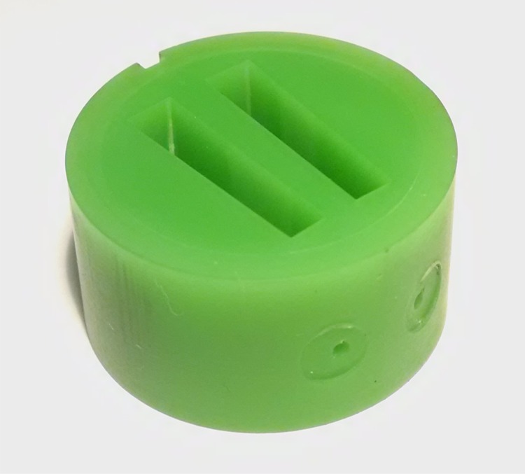
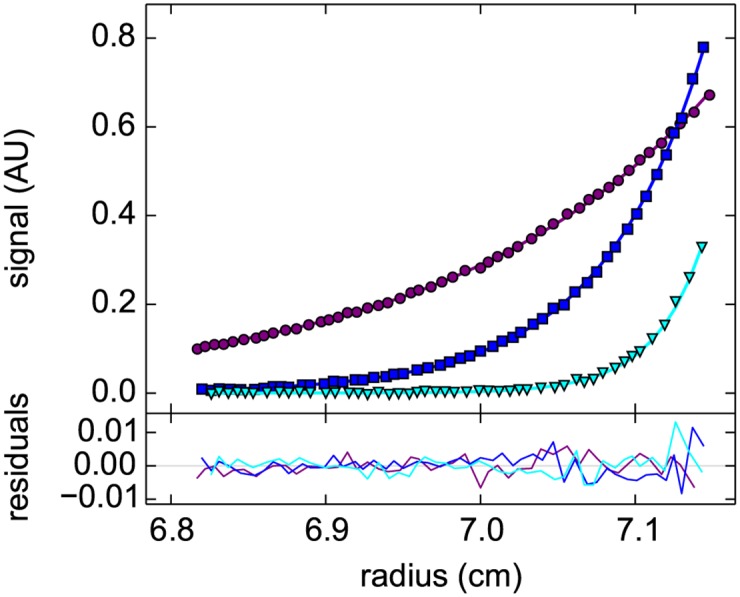
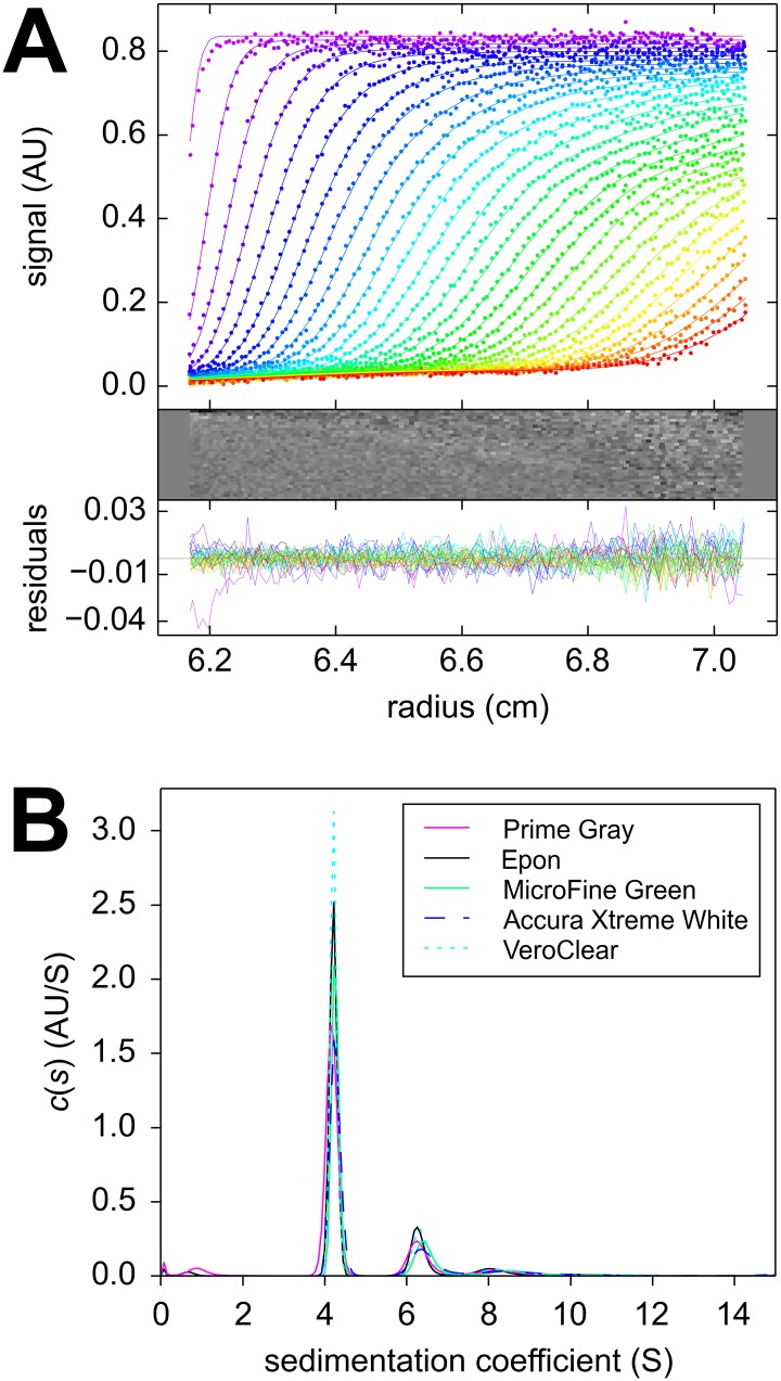
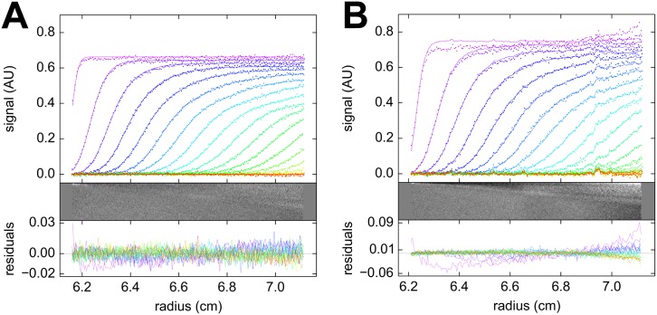
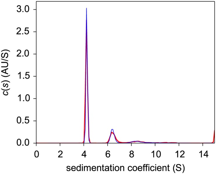
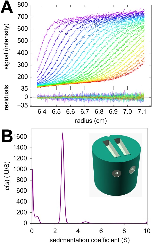
Analytical ultracentrifugation (AUC) is a classical technique of physical biochemistry providing information on size, shape, and interactions of macromolecules from the analysis of their migration in centrifugal fields while free in solution. A key mechanical element in AUC is the centerpiece, a component of the sample cell assembly that is mounted between the optical windows to allow imaging and to seal the sample solution column against high vacuum while exposed to gravitational forces in excess of 300,000 g. For sedimentation velocity it needs to be precisely sector-shaped to allow unimpeded radial macromolecular migration. During the history of AUC a great variety of centerpiece designs have been developed for different types of experiments. Here, we report that centerpieces can now be readily fabricated by 3D printing at low cost, from a variety of materials, and with customized designs. The new centerpieces can exhibit sufficient mechanical stability to withstand the gravitational forces at the highest rotor speeds and be sufficiently precise for sedimentation equilibrium and sedimentation velocity experiments. Sedimentation velocity experiments with bovine serum albumin as a reference molecule in 3D printed centerpieces with standard double-sector design result in sedimentation boundaries virtually indistinguishable from those in commercial double-sector epoxy centerpieces, with sedimentation coefficients well within the range of published values. The statistical error of the measurement is slightly above that obtained with commercial epoxy, but still below 1%. Facilitated by modern open-source design and fabrication paradigms, we believe 3D printed centerpieces and AUC accessories can spawn a variety of improvements in AUC experimental design, efficiency and resource allocation.


Conflict of interest statement

Competing Interests: The authors have declared that no competing interests exist.

Figures

Fig 1
Fig 1. Picture of a 12 mm pathlength centerpiece printed of ABS-like resin MicroFine Green.
Sedimentation velocity data of BSA at 50,000 rpm collected with this centerpiece installed into a standard cell assembly without gaskets are shown in Fig 3B.
Fig 2
Fig 2. Radial concentration distribution in a sedimentation equilibrium experiment with enhanced green fluorescent protein in a “prime gray” photopolymer centerpiece.
Data were acquired with the absorbance detection sequentially at rotor speeds of 15,000 rpm (purple), 24,000 rpm (blue), and 35,000 rpm (cyan) (symbols, only every 5th data point shown). A global model (lines) results in an apparent molar mass of 29.7 kDa with a root-mean-square deviation (rmsd) of 0.0032 OD489, and residuals as shown in the lower plot.
Fig 3
Fig 3. Temporal evolution of radial concentration profiles in a sedimentation velocity experiment with bovine serum albumin in a “prime gray” photopolymer centerpiece.
Panel A: Absorbance data acquired at a rotor speeds of 50,000 rpm at a series of time points (symbols, only every 3rd data point of every 2nd scan shown, with color temperature indicating progress of time). The c(s) fit (lines) results in an rmsd of 0.0065 OD280, with the residuals shown in the small plots as residuals bitmap and superposition. Panel B: The corresponding c(s) distribution (magenta), and for comparison the c(s) distribution from a control in the same run using a standard Epon centerpiece (black); microgreen (green); Xtreme white (blue dashed); in-house clear (cyan dotted).
Fig 4
Fig 4. Sedimentation velocity experiment in rectangular cell.
Sedimentation velocity analysis of bovine serum albumin sedimenting at 50,000 rpm in acrylic centerpieces with a sector-shaped (A) and rectangular shaped (B) solution column. The protein sample was identical in both. The upper panel shows the sedimentation boundaries (points, for clarity, only every 2nd data point of every 2nd scan is shown), along with the best-fit c(s) profiles (solid lines). Below are the residuals of the fit as bitmap and overlay plot. The c(s) distribution for both data sets are shown in Fig 5.
Fig 5
Fig 5. Sedimentation coefficient distributions from a rectangular cell.
Sedimentation coefficient distributions calculated from the data in Fig 4 for rectangular (magenta) and sectorial (blue) geometry.
Fig 6
Fig 6. Fluorescence optical data in a 3D printed carbonate centerpiece.
A centerpiece featuring a 3 mm deep sector-shaped well at the top was used, with filling and venting holes, and an embossed seal. The focal depth of the fluorescence optics was 2.0 mm. (A) Shown are sedimentation profiles acquired with 561 nm excitation for 46 nM mCherry [57] dissolved in phosphate buffered saline (dots), and best-fit c(s) sedimentation coefficient distribution with adjustments for characteristic signals of fluorescence detection [56] (solid lines). The plot appended below shows the residuals of the fit. (B) Corresponding sedimentation coefficient distribution showing a main peak at 2.68 S and diffusional boundary broadening corresponding to a species of 26.9 kDa.

References

    1. Svedberg T, Rinde H. The ultra-centrifuge, a new instrument for the determination of size and distribution of size of particle in amicroscopic colloids. J Am Chem Soc. 1924;46(1923):2677–2693. 10.1021/ja01677a011 - DOI
    1. Svedberg T, Pedersen KO. The Ultracentrifuge. London: Oxford University Press; 1940.
    1. Schachman HK. Ultracentrifugation in Biochemistry. New York: Academic Press; 1959.
    1. Scott DJ, Harding SE, Rowe AJ. Modern Analytical Ultracentrifugation: Techniques and Methods. Cambridge: The Royal Society of Chemistry; 2005.
    1. Schuck P, Zhao H, Brautigam CA, Ghirlando R. Basic Principles of Analytical Ultracentrifugation. Boca Raton, FL: CRC Press; 2015.

MeSH terms

LinkOut - more resources