. 2016 Aug 15;11(8):e0161083.
doi: 10.1371/journal.pone.0161083. eCollection 2016.

p62/Sequestosome-1 Is Indispensable for Maturation and Stabilization of Mallory-Denk Bodies

Affiliations

p62/Sequestosome-1 Is Indispensable for Maturation and Stabilization of Mallory-Denk Bodies

Pooja Lahiri et al. PLoS One. .

Abstract

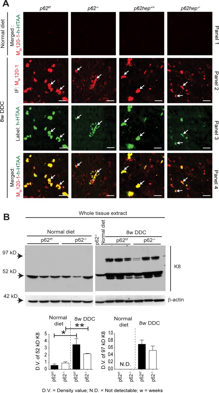
Mallory-Denk bodies (MDBs) are hepatocytic protein aggregates found in steatohepatitis and several other chronic liver diseases as well as hepatocellular carcinoma. MDBs are mainly composed of phosphorylated keratins and stress protein p62/Sequestosome-1 (p62), which is a common component of cytoplasmic aggregates in a variety of protein aggregation diseases. In contrast to the well-established role of keratins, the role of p62 in MDB pathogenesis is still elusive. We have generated total and hepatocyte-specific p62 knockout mice, fed them with 3,5-diethoxycarbonyl-1,4-dihydrocollidine (DDC) to induce MDBs and allowed the mice to recover from DDC intoxication on a standard diet to investigate the role of p62 in MDB formation and elimination. In the absence of p62, smaller, granular and less distinct MDBs appeared, which failed to mature to larger and compact inclusions. Moreover, p62 deficiency impaired the binding of other proteins such as NBR1 and Hsp25 to MDBs and altered the cellular defense mechanism by downregulation of Nrf2 target genes. Upon recovery from DDC intoxication on a standard diet, there was an enhanced reduction of p62-deficient MDBs, which was accompanied by a pronounced decrease in ubiquitinated proteins. Our data provide strong evidence that keratin aggregation is the initial step in MDB formation in steatohepatitis-related mouse models. Interaction of p62 with keratin aggregates then leads to maturation i.e., enlargement and stabilization of the MDBs as well as recruitment of other MDB-associated proteins.


Conflict of interest statement

Competing Interests: The authors have declared that no competing interests exist.

Figures

Fig 1
Fig 1. Loss of p62 impairs the formation of DDC-induced MDBs.
(A) H&E staining was performed on liver sections of p62f/f, p62-/-, p62hep+/+and p62hep-/- mice fed a normal diet (panel 1) or DDC-containing diet for 8 weeks (panel 2). Immunohistochemistry (IHC) with antibodies against p62 (panel 3) and K8/K18 (panel 4) was performed on DDC-intoxicated livers of the indicated genotypes. No morphological signs of liver injury and no MDBs were found in livers under normal diet (panel 1). Large and distinct MDBs (arrows) were only visible in DDC-fed p62f/f and p62hep+/+ mice in H&E stained sections (panel 2). These MDBs were also decorated by p62 (green; panel 3) and K8/K18 antibodies (red; panel 4). In p62-deficient mice, MDBs are small, less distinct and only decorated by K8/K18 antibodies (arrows; panel 4). MDB-containing hepatocytes display diminished or lacking keratin cytoskeleton (empty cells). Brown pigment in the livers indicates protoporphyrin accumulation. (Scale bar = 50 μm). (B) Immunoblotting revealed abundant p62 expression in livers of DDC-fed p62f/f and p62hep+/+ mice upon DDC-intoxication while no p62 signal was detected in p62-/- liver. The minimal p62 signal detected in p62hep-/- livers reflects preserved p62 expression in non-hepatocytic cells. β-actin was used as a loading control.
Fig 2
Fig 2. Loss of p62 impairs the formation of large MDBs.
Double immunofluorescence staining with K8/K18 (green) and p62 (red) antibodies was performed on liver sections of p62f/f, p62-/-, p62hep+/+and p62hep-/ mice under normal diet (panel 1). To detect MDBs, double immunofluorescence staining with MM120-1 (red) and K8/18 antibodies (green) was performed on liver sections of 8 weeks DDC-intoxicated mice (n ≥10). Note that MDBs were found in all genotypes. However, p62f/f and p62hep+/+ mice developed large aggregates while only small inclusions were observed in p62-/- and p62hep-/- mice (panels 2–5) (scale bar = 20μm). To confirm the absence of p62 in MDBs present in p62-deficient livers, various combinations of double immunofluorescence stainings with p62 (green) + MM120 (red), K8/K18 (green) + p62 (red) and ubiquitin (green) + p62 (red) antibodies were performed on DDC-treated liver of the different genotypes (panels 5–7. The arrows are used to indicate few MDBs among all the MDBs positive for keratin, MM120-1 or p62 to underline the observations (panels 2–7).
Fig 3
Fig 3
Loss of p62 influences the size but not the filament ultrastructure of MDBs (A) Morphometric scoring of the MM120-1 immunofluorescence confirmed the visual impression that p62-deficient mice contained more small MDBs but rarely developed larger inclusions. (n ≥10) Values are expressed as mean ± SEM. **p<0.01, ***p< 0.001. (B) Electron micrographs depict characteristic MDBs with densely but haphazardly arranged filaments in p62f/f mice while only small filamentous MDBs are seen in p62-/- mice. Note that the ultrastructure of the MDB filaments is identical in both genotypes. Scale bar = 500 nm.
Fig 4
Fig 4. p62 affects neither the extent of DDC-induced protein misfolding nor keratin cross-linking.
(A) A combination of immunofluorescence staining with MM120-1 antibody (red) and labeling with the luminescent conjugated oligothiophene h-HTAA (green) was performed on liver sections of p62-/-, p62f/f, p62hep-/- and p62hep+/+mice fed a normal diet (panel 1) or DDC for 8 weeks (panels 2–4). Mice fed a normal diet did not show any positive labeling with h-HTAA or MM120-1 (panel 1). DDC-intoxicated livers showed colocalization (yellow) of h-HTAA and MM120-1 indicating the presence of cross β-sheet conformation in MDBs (arrows) (panel 2; n ≥10) (scale bar = 20μm). (B) Whole liver tissue extracts of p62f/f and p62-/- mice fed a normal diet or 8weeks DDC were analyzed by western blotting with a K8-specific antibody and densitometric quantification was performed (n = 3). DDC-treatment resulted in an increased amount of K8 (52 kD; bands with lower molecular mass reflect degradation products) and formation of crosslinked K8 dimers (ca 97 kD). However, the extent of dimer formation did not differ significantly between p62-/- and p62f/f mice. β-actin was used as a loading control. *p<0.05, **p<0.01.
Fig 5
Fig 5. Loss of p62 impairs recruitment of NBR1 to MDBs.
(A). The scheme (modified from [34]) depicts the protein domain architecture of NBR1 and p62. The reported interaction partners and conserved domains are highlighted by double headed arrows and gray boxes, respectively. PB1, Phox and Bem1p domain; ZZ, zinc-binding domain; CC, coiled-coil domain; LIR, LC3-interacting region; UBA, ubiquitin-associated domain; Ub, ubiquitin. NBR1 and p62 interact via their PB1 domains [34]. (B) qPCR analyses of NBR1 and p62 mRNA expression in livers of p62f/f and p62-/- mice under normal diet or 8 weeks DDC. 18S rRNA was used as a normalization control (n = 5). (C) Whole liver tissue extracts of p62f/f and p62-/- mice fed a normal diet or 8 weeks DDC were immunoblotted for NBR1 (99 kD) and p62 (~55 kD). A sample from DDC-treated p62f/f liver was run on the same gel with liver samples from mice fed a normal diet (left panel) as common reference (right panel). Densitometric analyses of p62 and NBR1 are also shown (n = 3). *p<0.05, **p<0.01, ***p<0.001(D) Double immunofluorescence staining with MM120-1 (green) and NBR1 antibodies (red) visualized the distribution of both proteins in livers of mice treated with DDC for 8 weeks. An extensive co-localization between NBR1 and MM120-1 (C.E = colocalization efficiency) was seen in animals with intact p62 production (p62f/f C.E = 97%, and p62hep+/+ C.E = 99%) but not in the total (p62-/- C.E = 21%) and liver-specific (p62hep-/- C.E = 16%) p62-knockout animals (scale bar = 20μm). NBR1-positive and -negative MDBs are highlighted by arrows and circles, respectively.
Fig 6
Fig 6. Characterization of MDBs at 4 weeks of recovery after 8 weeks of DDC intoxication.
(A) Double immunofluorescence staining of liver sections with MM120-1 and K8/K18 antibody visualized MDBs in livers of mice fed DDC for 8 weeks (8w DDC) as well as in livers of mice that were subsequently recovered on a standard diet for four weeks (4wR). Recovery led to a substantial reduction of MDBs both in mice with intact p62 production (p62f/f and p62hep+/+) and total (p62-/-)/liver-specific (p62hep-/-) p62 knockouts. (B) Morphometrical analysis showed that after recovery there was a significant reduction of the number of MDBs in the p62-deficient groups (n = 5). (C) No significant difference in MDB size was observed after recovery in the different genotypes, which confirmed the observations of immunofluorescence staining (n = 5). (D) Immunoblotting of insoluble liver protein extracts with antibodies against keratin 8 (K8) and ubiquitin and corresponding densitometric quantification (n = 5) showed that loss of p62 did not affect the accumulation of insoluble ubiquitinated proteins or formation of cross-linked high molecular mass K8 species during DDC intoxication. However, the disappearance of ubiquitinated proteins and K8 cross-linked proteins during the recovery period was accelerated in p62-deficient mice suggesting that the inclusions were less stable. A Coomassie-stained gel for indicated genotypes and treatment shows the protein loading used for western blotting. Values are expressed as mean ± SEM. (n = 5). *p<0.05, ***p< 0.001).
Fig 7
Fig 7. Absence of p62 alters cellular defense mechanisms.
(A) Liver nuclear extracts and whole tissue extracts of p62f/f and p62-/- mice fed a normal diet and of mice DDC-intoxicated for 8 weeks were analyzed by immunoblotting with antibody against Nrf2 (65 kD). Furthermore, quantitative qPCR analyses of Nrf2 mRNA (n = 5) was performed from the same mice. Corresponding densitometric quantification is shown for nuclear extracts (n = 3). Lamin B1 (62 kD) was used as nuclear loading control. (B) Liver whole tissue extract from p62f/f and p62-/- mice fed a normal diet or 8 weeks DDC were immunoblotted for Keap1 (70 kD) and Hmox1 (32 kD) and qPCR analyses were performed for Keap1 and Hmox-1 mRNA (n = 5). Densitometric quantification for Keap1 and Hmox-1 are shown (n = 3). β-actin was used as a loading control. (C) qPCR analyses of Nqo1 and Gst mRNA levels were performed in livers of mice livers (n = 5) fed normal diet or after 8 weeks of DDC-intoxication. 18S rRNA was used as a normalization control. (D) Morphometric assessment of Ki-67-positive hepatocytes (Ki-67 score) and K19-positive bililary cells (ductular reaction score) was made by counting at least ten high power fields (400X) of five DDC-intoxicated livers of indicated genotypes stained with Ki-67 and K19 antibodies. At least five animals per genotype were used for qPCR, Ki67 and ductular reaction scoring. Values are expressed as mean ± SEM. **p< 0.01, *p<0.05.
Fig 8
Fig 8. Schematic drawing of the different role of p62 and keratin in two distinct hypothetical pathways of MDB formation.
(A1) In the first pathway alcohol abuse or metabolic alterations associated with obesity in the context of human alcoholic or non-alcoholic steatohepatitis (ASH or NASH), and DDC or griseofulvin administration in mice trigger the upregulation of keratins (K8 and K18) with, increased K8:K18 ratio and cross β-sheet conformation of K8, which leads to the formation of small ‟early” MDBs with filamentous ultrastructure and MM120-1 antigen positivity. (A2) The coalescence of small MDBs to form large, compact ‟mature” MDBs occurs via incorporation of p62. Binding of p62 to MDBs also promotes the recruitment of NBR1 and other proteins to MDBs. (B1) In the second pathway (found in hepatocellular carcinoma and idiopathic copper toxicosis) p62-containing IHBs, which are negative for keratin and do not show cross β-sheet conformation, may progress to (B2) hybrid inclusions (showing mixed features of both IHBs and MDBs) by incorporation of keratins (K8 and K18). (B3) The hybrid inclusions may transform to ‟mature” MDBs upon further incorporation of K8 and K18.

Similar articles

Cited by

References

    1. Dobson CM. Protein folding and misfolding. Nature. 2003;426(6968):884–90. - PubMed
    1. Aguzzi A, O'Connor T. Protein aggregation diseases: pathogenicity and therapeutic perspectives. Nature reviews Drug Discovery. 2010;9(3):237–48. 10.1038/nrd3050 - DOI - PubMed
    1. Naeem A, Fazili NA. Defective protein folding and aggregation as the basis of neurodegenerative diseases: the darker aspect of proteins. Cell Biochemistry and Biophysics. 2011;61(2):237–50. 10.1007/s12013-011-9200-x - DOI - PubMed
    1. Reynaud E. Protein Misfolding and Degenerative Diseases. Nature Education. 2010;3(9):28.
    1. Zatloukal K, French SW, Stumptner C, Strnad P, Harada M, Toivola DM, et al. From Mallory to Mallory-Denk bodies: what, how and why? Experimental Cell Research. 2007;313(10):2033–49. - PubMed

MeSH terms

LinkOut - more resources