. 2016 Aug 16:6:31581.
doi: 10.1038/srep31581.

Astrocytic Acid-Sensing Ion Channel 1a Contributes to the Development of Chronic Epileptogenesis

Affiliations

Astrocytic Acid-Sensing Ion Channel 1a Contributes to the Development of Chronic Epileptogenesis

Feng Yang et al. Sci Rep. .

Erratum in

Abstract

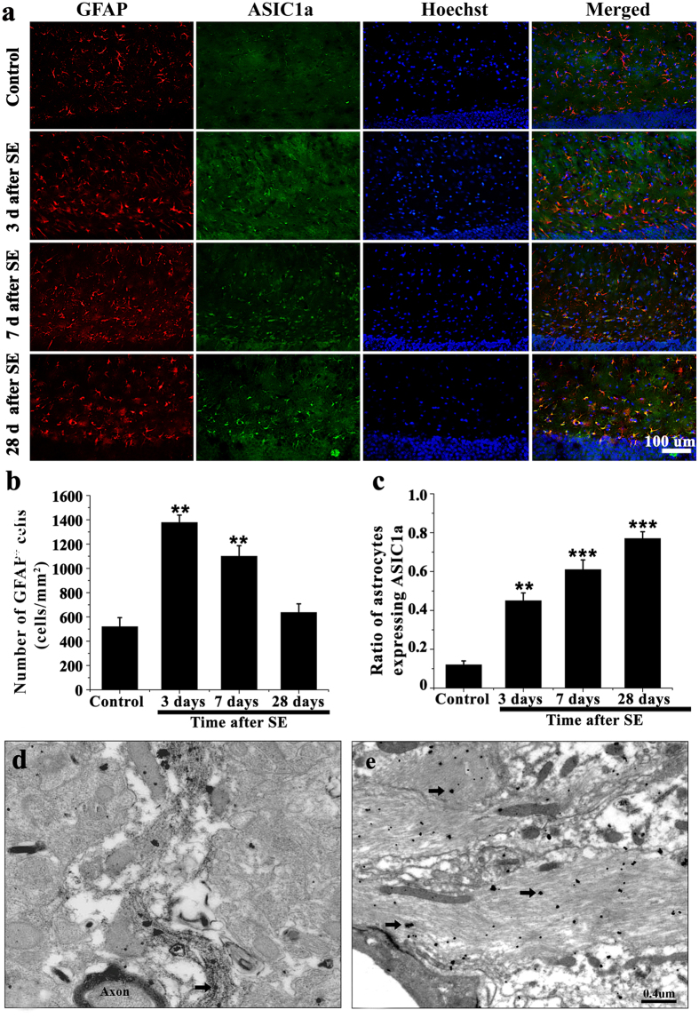
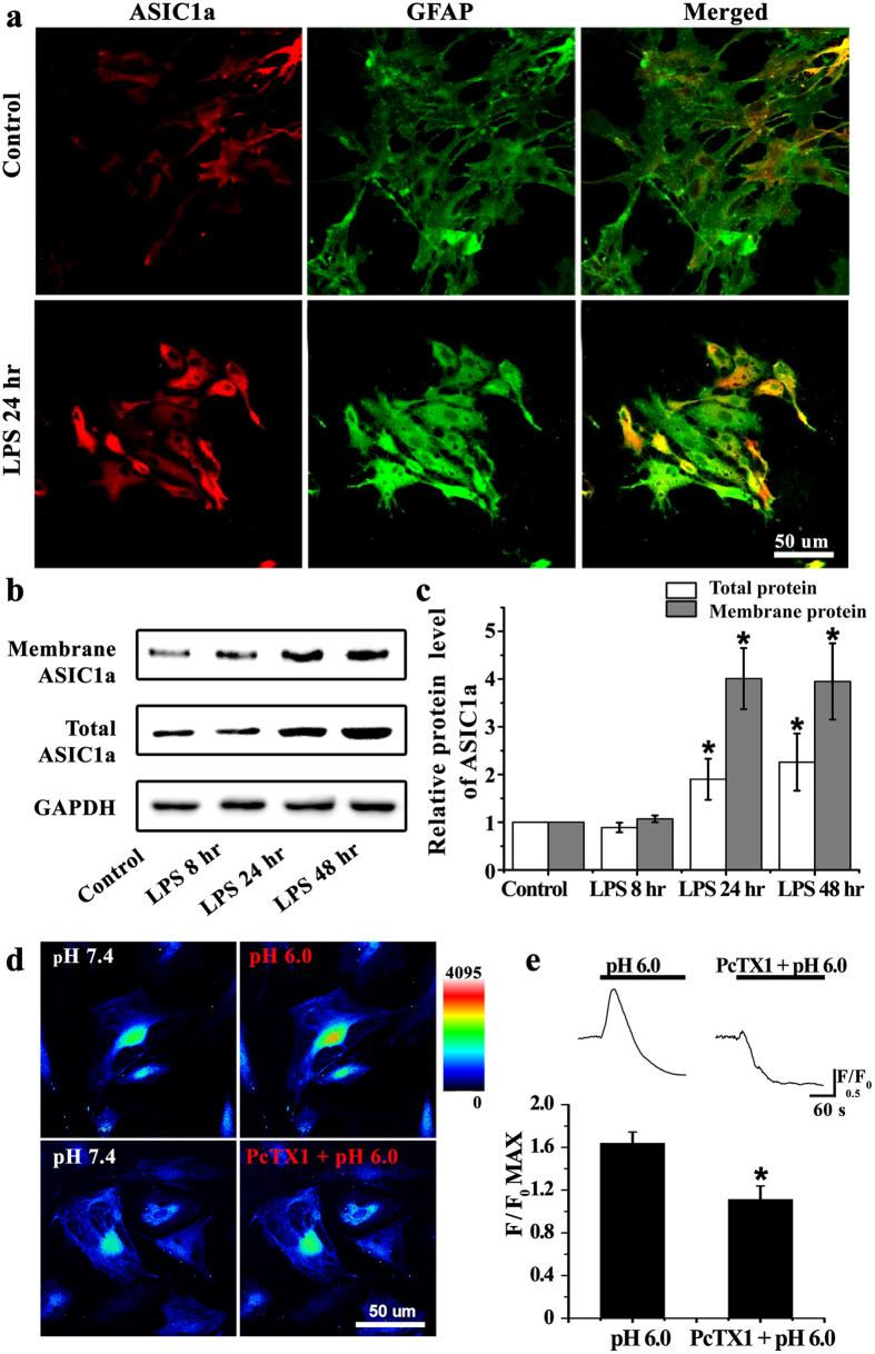
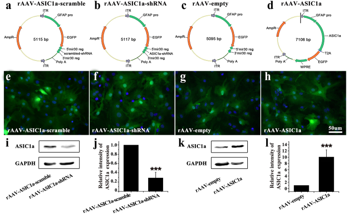
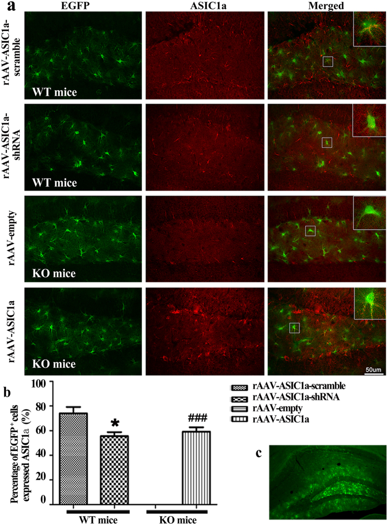
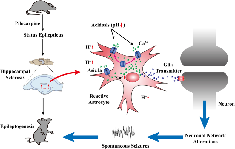
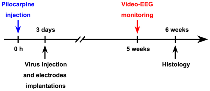
Unraveling mechanisms underlying epileptogenesis after brain injury is an unmet medical challenge. Although histopathological studies have revealed that reactive astrogliosis and tissue acidosis are prominent features in epileptogenic foci, their roles in epileptogenesis remain unclear. Here, we explored whether astrocytic acid-sensing ion channel-1a (ASIC1a) contributes to the development of chronic epilepsy. High levels of ASIC1a were measured in reactive astrocytes in the hippocampi of patients with temporal lobe epilepsy (TLE) and epileptic mice. Extracellular acidosis caused a significant Ca(2+) influx in cultured astrocytes, and this influx was sensitive to inhibition by the ASIC1a-specific blocker psalmotoxin 1 (PcTX1). In addition, recombinant adeno-associated virus (rAAV) vectors carrying a GFAP promoter in conjunction with ASIC1a shRNA or cDNA were generated to suppress or restore, respectively, ASIC1a expression in astrocytes. Injection of rAAV-ASIC1a-shRNA into the dentate gyrus of the wide type TLE mouse model resulted in the inhibition of astrocytic ASIC1a expression and a reduction in spontaneous seizures. By contrast, rAAV-ASIC1a-cDNA restored astrocytic ASIC1a expression in an ASIC1a knock-out TLE mouse model and increased the frequency of spontaneous seizures. Taken together, our results reveal that astrocytic ASIC1a may be an attractive new target for the treatment of epilepsy.


Figures

Figure 1
Figure 1. ASIC1a was abundantly expressed in hippocampal astrocytes of epileptic mice.
(a) GFAP (red), ASIC1a (green), and Hoechst (blue) immunofluorescence staining was performed on control tissue and at 3, 7, and 28 days after SE. (b) The number of GFAP positive cells peaked on day 3 after SE. **P < 0.01, ANOVA followed by Dunnett’s post-hoc test vs. control. (c) The fraction of astrocytes expressing ASIC1a increased between 3 and 28 days after SE. **P < 0.01, ***P < 0.001; ANOVA followed by Dunnett’s post-hoc test vs. control. (d,e) Electron microscopy showed astrocytic ASIC1a (black arrow) in control (d) and epileptic (28 days after SE) (e) mice.
Figure 2
Figure 2. ASIC1a was abundantly expressed in hippocampal astrocytes of TLE patients.
(a1–b4) GFAP (red), ASIC1a (green), and Hoechst (blue) immunofluorescence staining was performed on TLE patients and control (traumatic brain injury caused by car accidents) patients. (a1–a4) There was only a faint expression of ASIC1a on the hippocampal astrocyte in the 4 cases of control patients, with more seeming to be located on neuron-like cells. (b1–b4) In all the 4 cases of TLE patients, ASIC1a was abundantly expressed in activated hippocampal astrocytes, which displayed hypertrophy of cell bodies and processes.
Figure 3
Figure 3. Increased expression of astrocytic ASIC1a mediated Ca2+ elevation in LPS-treated astrocytes.
(a) ASIC1a (red) and GFAP (green) immunofluorescence staining was performed in cultured astrocytes. Compared with the control group, the number of ASIC1a-positive astrocytes markedly increased at 24 hours after LPS treatment. (b) Western blots for membrane and total ASIC1a protein in control and LPS-treated astrocytes at 8, 24, and 48 hours after treatment. (c) Densitometric quantification of the membrane and total ASIC1a protein expression, normalized to GAPDH. *P < 0.05, ANOVA followed by Dunnett’s post-hoc test vs. control. (d) Representative changes of intracellular Ca2+ concentration in response to extracellular pH reduction (from 7.4 to 6.0) in the absence and presence of 5 nM PcTX1. Warmer colors indicated higher fluorescence intensity. (e) Time course and quantification of fluorescence intensity. Peak fluorescence intensity was normalized to fluorescence intensity at baseline (F/F0 max) in the pH 6.0 (n = 24) and PcTX1 + pH 6.0 (n = 21) groups. *P < 0.05, t test vs. pH 6.0 group.
Figure 4
Figure 4. rAAV plasmids and virally induced ASIC1a expression in cultured astrocytes.
(a–d) Schematic of the four rAAV plasmids used: rAAV-ASIC1a-scramble virus carrying a GFAP-EGFP-ASIC1a-scrambled-shRNA vector, rAAV-ASIC1a virus carrying a GFAP-EGFP-ASIC1a-shRNA vector, rAAV-empty virus carrying a GFAP-EGFP vector, and rAAV-ASIC1a virus carrying GFAP-ASIC1a-EGFP vector. (e–h) EGFP and Hoechst fluorescence in astrocyte cultures at 72 hours after virus transduction. (i,k) Immunoblots for total ASIC1a protein after viral transduction. (j,l) Densitometric quantification of ASIC1a, normalized to GAPDH. ***P < 0.001, t test vs. scramble or empty vector.
Figure 5
Figure 5. The efficiency of viral vector-induced ASIC1a expression at 5 weeks after SE.
(a) EGFP and ASIC1a immunofluorescence staining in each group. (b) Percent of EGFP-positive cells expressing ASIC1a in each group. All data were shown as mean ± SEM. WT: n = 7, KO: n = 6 mice per group. *P < 0.05, t test vs. rAAV-ASIC1a-scramble; ###P < 0.001, t test vs rAAV-empty. (c) Low magnification view showing the region of virus transduction in the dentate gyrus.
Figure 6
Figure 6. Effects of rAAV vectors on spontaneous seizures in mice during chronic epileptogenesis.
(a) Ictal EEG signals in each group. (b) Spontaneous seizure frequency during the 5th week after SE. (c–e) Statistical analysis of spontaneous seizure frequency, total time in seizures, and seizure scores. *P < 0.05, t test vs. rAAV ASIC1a-scramble. #P < 0.05, t test vs. rAAV-empty. Bars represent mean ± SEM. WT: n = 7, KO: n = 6 mice/group.
Figure 7
Figure 7. Proposed role of astrocytic ASIC1a in chronic epileptogenesis.
Acute brain injury such as status epilepticus can cause hippocampal sclerosis. Reactive astrogliosis and tissue acidosis are the prominent features in sclerotic hippocampi. Reactive astrocytes express a high level of ASIC1a, which can be activated by local extracellular low pH. This would lead to excessive Ca2+ influx in astrocytes and release of gliotransmitters, thus resulting in epileptiform activities and the development of chronic epileptogenesis.
Figure 8
Figure 8. The flow chart of pilocarpine injection, virus injection and electrodes implantations, video-EEG monitoring and histology for Experiment 2 in the Materials and Methods.

References

    1. Pitkanen A. & Lukasiuk K. Mechanisms of epileptogenesis and potential treatment targets. Lancet Neurol 10, 173–186 (2011). - PubMed
    1. Franco V., French J. A. & Perucca E. Challenges in the clinical development of new antiepileptic drugs. Pharmacol Res, 103, 95–104 (2015). - PubMed
    1. Hubbard J. A., Hsu M. S., Fiacco T. A. & Binder D. K. Glial cell changes in epilepsy: overview of the clinical problem and therapeutic opportunities. Neurochem Int 63, 638–651 (2013). - PubMed
    1. Sendrowski K. & Sobaniec W. Hippocampus, hippocampal sclerosis and epilepsy. Pharmacol Rep 65, 555–565 (2013). - PubMed
    1. Gibbons M. B., Smeal R. M., Takahashi D. K., Vargas J. R. & Wilcox K. S. Contributions of astrocytes to epileptogenesis following status epilepticus: opportunities for preventive therapy? Neurochem Int 63, 660–669 (2013). - PMC - PubMed

Publication types

Substances