. 2016 Aug 3;17(8):1260.
doi: 10.3390/ijms17081260.

Bioinformatics and Microarray Analysis of miRNAs in Aged Female Mice Model Implied New Molecular Mechanisms for Impaired Fracture Healing

Affiliations

Bioinformatics and Microarray Analysis of miRNAs in Aged Female Mice Model Implied New Molecular Mechanisms for Impaired Fracture Healing

Bing He et al. Int J Mol Sci. .

Abstract

Impaired fracture healing in aged females is still a challenge in clinics. MicroRNAs (miRNAs) play important roles in fracture healing. This study aims to identify the miRNAs that potentially contribute to the impaired fracture healing in aged females. Transverse femoral shaft fractures were created in adult and aged female mice. At post-fracture 0-, 2- and 4-week, the fracture sites were scanned by micro computed tomography to confirm that the fracture healing was impaired in aged female mice and the fracture calluses were collected for miRNA microarray analysis. A total of 53 significantly differentially expressed miRNAs and 5438 miRNA-target gene interactions involved in bone fracture healing were identified. A novel scoring system was designed to analyze the miRNA contribution to impaired fracture healing (RCIFH). Using this method, 11 novel miRNAs were identified to impair fracture healing at 2- or 4-week post-fracture. Thereafter, function analysis of target genes was performed for miRNAs with high RCIFH values. The results showed that high RCIFH miRNAs in aged female mice might impair fracture healing not only by down-regulating angiogenesis-, chondrogenesis-, and osteogenesis-related pathways, but also by up-regulating osteoclastogenesis-related pathway, which implied the essential roles of these high RCIFH miRNAs in impaired fracture healing in aged females, and might promote the discovery of novel therapeutic strategies.

Keywords: bioinformatics; impaired fracture healing; miRNA.


Figures

Figure 1
Figure 1
Fracture healing in aged and adult female mice (A) representative three-dimensional (3D) images of the fracture calluses in adult and aged groups at each time point after fracture; (B) time course changes in low-density bone volume (BVL) (left), low-density bone volume fraction (BVL/TV) (middle), and bone mineral content (BMC) (right) of the callus after fracture in each group; n = 3. Note: scale bar = 1 mm. * p < 0.05 for “Aged” vs. “Adult”.
Figure 2
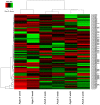
Figure 2
Heat map of differentially expressed microRNAs (miRNAs) in fracture healing. Red represents up-regulation and green represents down-regulation.
Figure 3
Figure 3
miRNA-target gene interactions for the significantly differentially expressed miRNAs in bone fracture healing.
Figure 4
Figure 4
Significantly enriched pathways of top 100 highest miRNA contribution to impaired fracture healing (RCIFH) miRNA targets at (A) 2-week after fracture and (B) 4-week after fracture.
Figure 5
Figure 5
The effect of top 100 highest RCIFH miRNAs on the key genes of nitric oxide signaling, retinoid acid receptor (RAR) activation, transforming growth factor-β (TGF-β) signaling, and interleukin 6 (IL-6) signaling pathways in impaired fracture healing. Red: gene expression and function are activated in aged female mice compared to adult ones. Green: gene expression and function are inhibited in aged female mice compared to adult ones.
Figure 6
Figure 6
The miR-494 inhibits chondrogenic differentiation in C3H10T1/2 cells. The miR-494, anti-miR-494, or inactive controls were transfected into C3H10T1/2 cells. Expressions of chondrogenic differentiation markers (Acan, Col2a1, and Col10a1) were detected by QPCR at day 1, 7, and 14 after transfection. Note: Data was represented as mean ± SD, * p < 0.05, ** p < 0.01, *** p < 0.001.

References

    1. Gruber R., Koch H., Doll B.A., Tegtmeier F., Einhorn T.A., Hollinger J.O. Fracture healing in the elderly patient. Exp. Gerontol. 2006;41:1080–1093. doi: 10.1016/j.exger.2006.09.008. - DOI - PubMed
    1. Virk M.S., Lieberman J.R. Biologic adjuvants for fracture healing. Arthritis Res. Ther. 2012;14:225. doi: 10.1186/ar4053. - DOI - PMC - PubMed
    1. Nguyen T.V., Center J.R., Eisman J.A. Osteoporosis: Underrated, underdiagnosed and undertreated. Med. J. Aust. 2004;180:S18–S22. - PubMed
    1. Kanis J.A., Johnell O., Oden A., Sembo I., Redlund-Johnell I., Dawson A., de Laet C., Jonsson B. Long-term risk of osteoporotic fracture in Malmo. Osteoporos. Int. 2000;11:669–674. doi: 10.1007/s001980070064. - DOI - PubMed
    1. Bliuc D., Nguyen N.D., Milch V.E., Nguyen T.V., Eisman J.A., Center J.R. Mortality risk associated with low-trauma osteoporotic fracture and subsequent fracture in men and women. JAMA. 2009;301:513–521. doi: 10.1001/jama.2009.50. - DOI - PubMed

LinkOut - more resources