

doi: 10.1038/srep30859.
Binding site elucidation and structure guided design of macrocyclic IL-17A antagonists
Affiliations
- PMID: 27527709
- PMCID: PMC4985813
- DOI: 10.1038/srep30859
Item in Clipboard
Binding site elucidation and structure guided design of macrocyclic IL-17A antagonists
Sci Rep.
.
Abstract
Interleukin-17A (IL-17A) is a principal driver of multiple inflammatory and immune disorders. Antibodies that neutralize IL-17A or its receptor (IL-17RA) deliver efficacy in autoimmune diseases, but no small-molecule IL-17A antagonists have yet progressed into clinical trials. Investigation of a series of linear peptide ligands to IL-17A and characterization of their binding site has enabled the design of novel macrocyclic ligands that are themselves potent IL-17A antagonists.
Figures
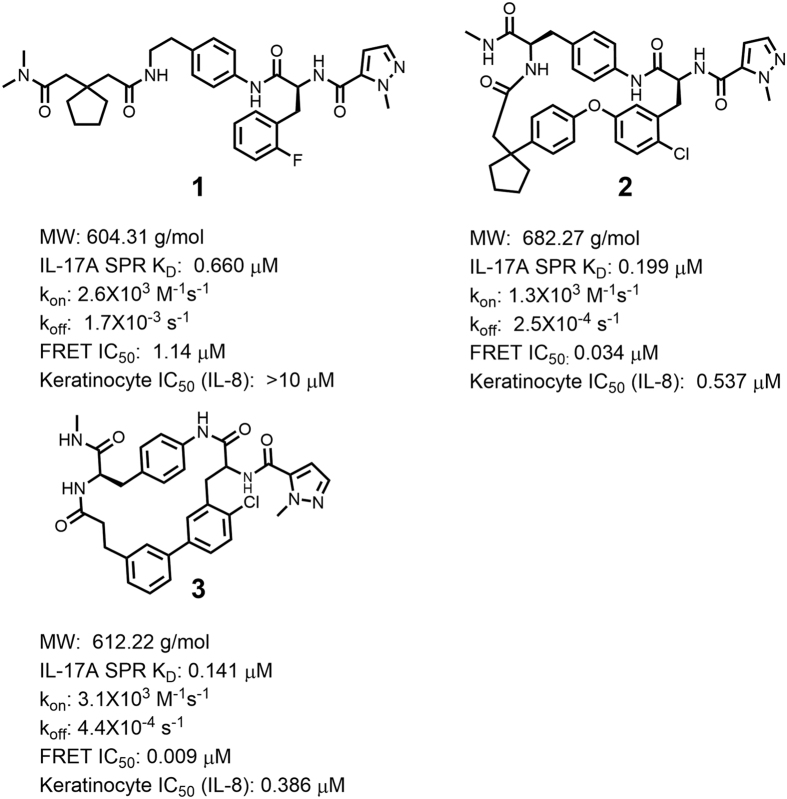
Compound 1: example of a lead IL-17A antagonist with a linear peptide motif. Compounds 2 and 3: macrocyclic IL-17A antagonists designed on basis of the structure of compound 1 complexed with IL-17A.
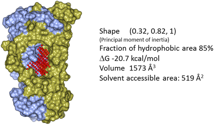
The high druggability of the pocket is manifested by the large hydrophobic cavity and the favorable druggability score (∆G) which assesses the optimal binding affinity of the binding site.
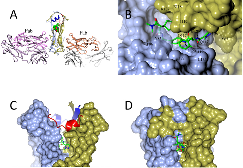
(A) Overall structure of the Fab/HAP/IL-17A/compound 1 complex. Component polypeptides appear as ribbons, with two HAP molecules in blue and red, respectively, and the two chains of IL-17A in ice blue and gold, respectively. Atoms of compound 1 appear as spheres, with carbons in green, nitrogens in blue, and oxygens in red. Hydrogen bonds are shown as dashes. Picture prepared using program CCP4MG. (B) Close view of compound 1 bound in the central pocket at the IL-17A dimer interface and interacting with both IL-17A monomers (surface representation). Dashed lines are hydrogen bonds. (C) Compound 1 binding enlarges the central pocket and makes the N-terminal half of the IL-17A dimer much less compact. (D) For comparison, the central pocket is much smaller in the apo IL-17A dimer and completely enclosed. Compound 1 would clash with the closed binding site.
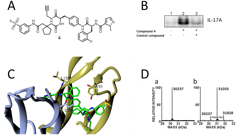
(A) Structure of compound 4, an analog of compound 1 designed as a covalent probe of the IL-17A binding site with a FRET IC50 of 0.2 μM. (B) Compound 4 specifically and covalently labeled full length IL-17A. IL-17A labeled by compound 4 was biotinylated by click chemistry, and pulled down by streptavidin. Eluates were immunoblotted. (C) In this model of compound 4 in the IL-17A binding pocket, the reactive sulfonyl fluoride is close to Tyr85. (D) Mass spectra of IL-17A (a) before and (b) after treatment with compound 4. The predicted mass shift resulting from a single modification of the protein was 795.3 Da.
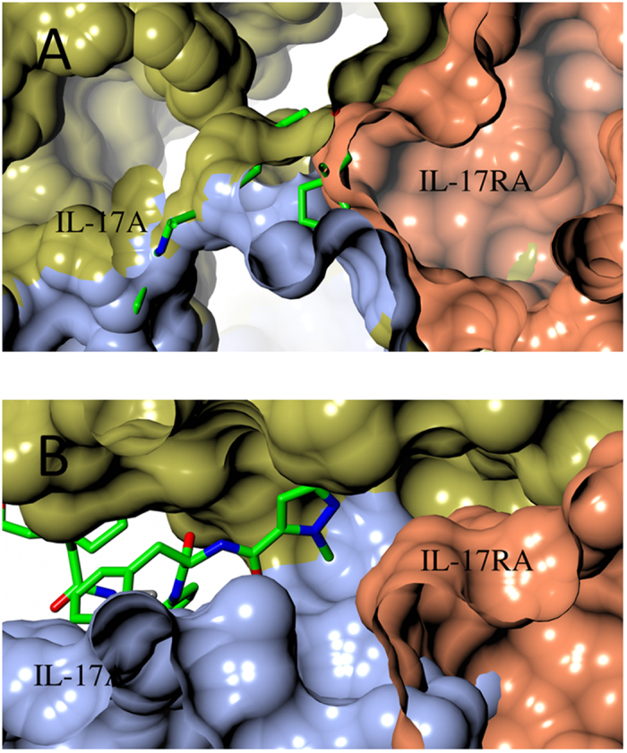
(A) IL-17A/IL-17RA binary complex (PDB accession 4HSA) is incompatible with compound 1 binding. Compound 1 (stick model) was superimposed into its IL-17A binding site in IL-17A/IL-17RA complex (surface representation, with IL-17RA colored in coral). (B) IL-17A/compound 1 binary complex is incompatible with IL-17RA binding. IL-17RA (coral surface) was superimposed onto IL-17A/compound 1 binary complex.
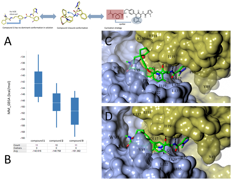
(A) In solution, lack of an NOE signal from the cyclopentyl and phenyl linker of compound 1 (left) indicated that the bound conformation was not highly populated in solution. Cyclization between the L-phenylalanine side chain and the N,N-dimethylamide (right) may reinforce the bound bioactive conformation (right). (B) Use of MD/MM-GBSA to predict binding affinities in design of macrocyclic compounds. Examples are for the discussed compounds. Box regions correspond to 50% of the distribution, lines extend to max 1.5 times of this interval, and averages are denoted by dashed lines in the boxes. (C) Compound 2 bound to IL-17A. D. Compound 3 bound to IL-17A.
References
MeSH terms
Substances
LinkOut - more resources
Full Text Sources
Other Literature Sources
