

Dynamics of the association of heat shock protein HSPA6 (Hsp70B') and HSPA1A (Hsp70-1) with stress-sensitive cytoplasmic and nuclear structures in differentiated human neuronal cells
- PMID: 27527722
- PMCID: PMC5083669
- DOI: 10.1007/s12192-016-0724-2
Dynamics of the association of heat shock protein HSPA6 (Hsp70B') and HSPA1A (Hsp70-1) with stress-sensitive cytoplasmic and nuclear structures in differentiated human neuronal cells
Abstract
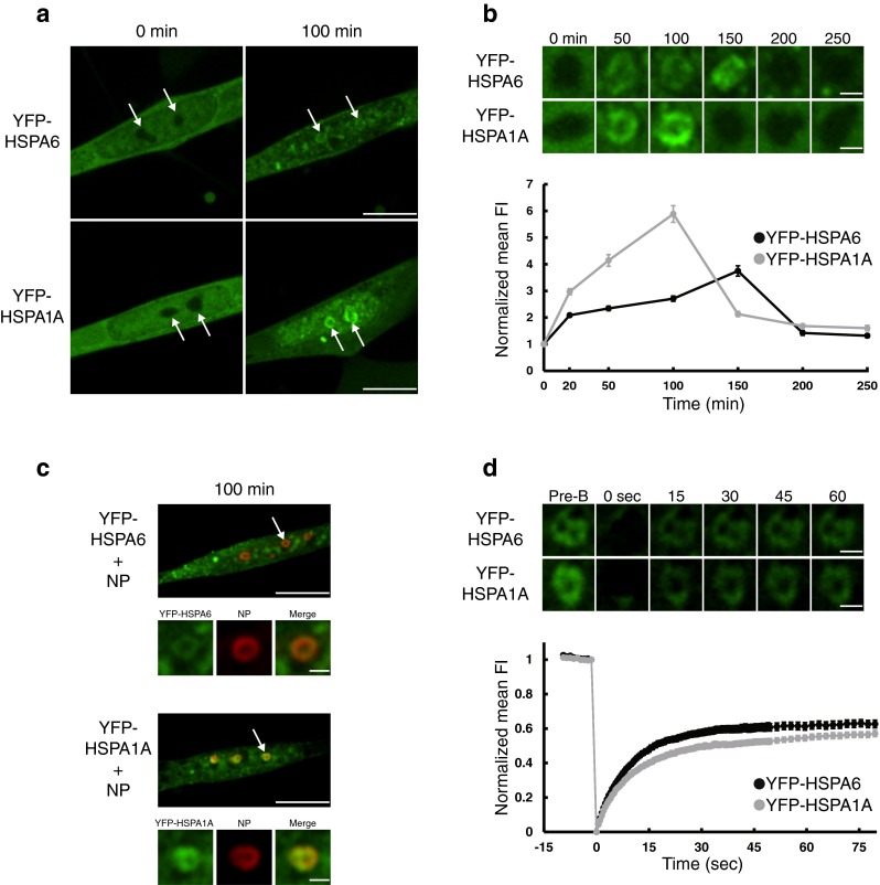
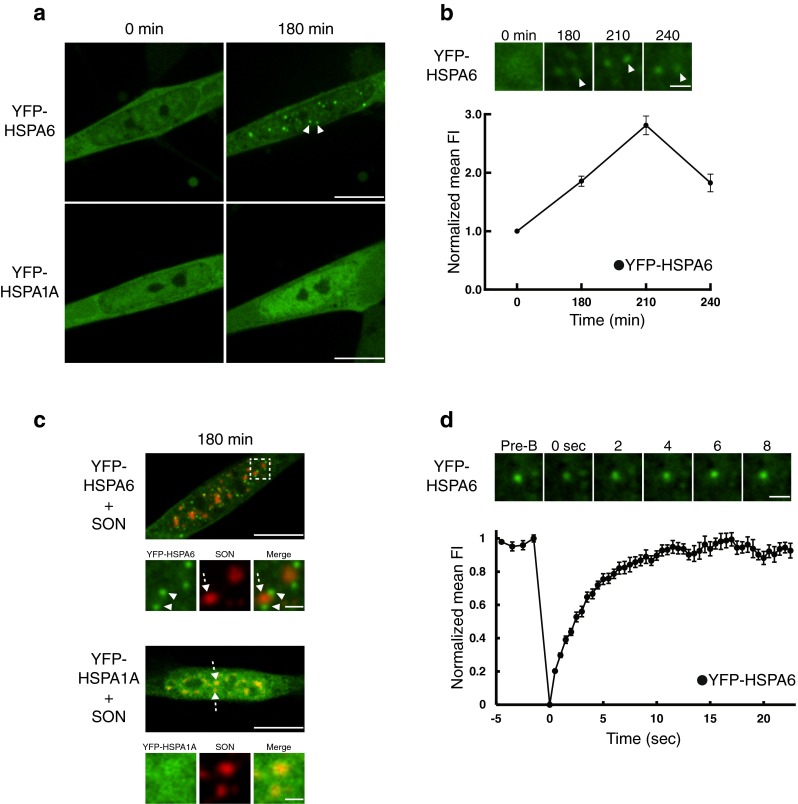
Heat shock proteins (Hsps) are cellular repair agents that counter the effects of protein misfolding that is a characteristic feature of neurodegenerative diseases. HSPA1A (Hsp70-1) is a widely studied member of the HSPA (Hsp70) family. The little-studied HSPA6 (Hsp70B') is present in the human genome and absent in mouse and rat; hence, it is missing in current animal models of neurodegenerative diseases. Differentiated human neuronal SH-SY5Y cells were employed to compare the dynamics of the association of YFP-tagged HSPA6 and HSPA1A with stress-sensitive cytoplasmic and nuclear structures. Following thermal stress, live-imaging confocal microscopy and Fluorescence Recovery After Photobleaching (FRAP) demonstrated that HSPA6 displayed a prolonged and more dynamic association, compared to HSPA1A, with centrioles that play critical roles in neuronal polarity and migration. HSPA6 and HSPA1A also targeted nuclear speckles, rich in RNA splicing factors, and the granular component of the nucleolus that is involved in rRNA processing and ribosomal subunit assembly. HSPA6 and HSPA1A displayed similar FRAP kinetics in their interaction with nuclear speckles and the nucleolus. Subsequently, during the recovery from neuronal stress, HSPA6, but not HSPA1A, localized with the periphery of nuclear speckles (perispeckles) that have been characterized as transcription sites. The stress-induced association of HSPA6 with perispeckles displayed the greatest dynamism compared to the interaction of HSPA6 or HSPA1A with other stress-sensitive cytoplasmic and nuclear structures. This suggests involvement of HSPA6 in transcriptional recovery of human neurons from cellular stress that is not apparent for HSPA1A.
Keywords: FRAP; HSPA1A (Hsp70–1); HSPA6 (Hsp70B’); Live imaging; SH-SY5Y.
Figures


Similar articles
-
Knockdown of Heat Shock Proteins HSPA6 (Hsp70B') and HSPA1A (Hsp70-1) Sensitizes Differentiated Human Neuronal Cells to Cellular Stress.Neurochem Res. 2018 Feb;43(2):340-350. doi: 10.1007/s11064-017-2429-z. Epub 2017 Oct 31. Neurochem Res. 2018. PMID: 29090408
-
Localization of heat shock protein HSPA6 (HSP70B') to sites of transcription in cultured differentiated human neuronal cells following thermal stress.J Neurochem. 2014 Dec;131(6):743-54. doi: 10.1111/jnc.12970. Epub 2014 Dec 2. J Neurochem. 2014. PMID: 25319762
-
Targeting of Heat Shock Protein HSPA6 (HSP70B') to the Periphery of Nuclear Speckles is Disrupted by a Transcription Inhibitor Following Thermal Stress in Human Neuronal Cells.Neurochem Res. 2017 Feb;42(2):406-414. doi: 10.1007/s11064-016-2084-9. Epub 2016 Oct 14. Neurochem Res. 2017. PMID: 27743288
-
HSPA6 and its role in cancers and other diseases.Mol Biol Rep. 2022 Nov;49(11):10565-10577. doi: 10.1007/s11033-022-07641-5. Epub 2022 Jun 6. Mol Biol Rep. 2022. PMID: 35666422 Review.
-
HSPA1A Protects Cells from Thermal Stress by Impeding ESCRT-0-Mediated Autophagic Flux in Epidermal Thermoresistance.J Invest Dermatol. 2021 Jan;141(1):48-58.e3. doi: 10.1016/j.jid.2020.05.105. Epub 2020 Jun 10. J Invest Dermatol. 2021. PMID: 32533962 Review.
Cited by
-
Long non-coding RNA and mRNA analysis of Ang II-induced neuronal dysfunction.Mol Biol Rep. 2019 Jun;46(3):3233-3246. doi: 10.1007/s11033-019-04783-x. Epub 2019 Apr 3. Mol Biol Rep. 2019. PMID: 30945068
-
Differential Targeting of Hsp70 Heat Shock Proteins HSPA6 and HSPA1A with Components of a Protein Disaggregation/Refolding Machine in Differentiated Human Neuronal Cells following Thermal Stress.Front Neurosci. 2017 Apr 24;11:227. doi: 10.3389/fnins.2017.00227. eCollection 2017. Front Neurosci. 2017. PMID: 28484369 Free PMC article.
-
Altered splicing factor and alternative splicing events in a mouse model of diet- and polychlorinated biphenyl-induced liver disease.Environ Toxicol Pharmacol. 2023 Oct;103:104260. doi: 10.1016/j.etap.2023.104260. Epub 2023 Sep 7. Environ Toxicol Pharmacol. 2023. PMID: 37683712 Free PMC article.
-
Components of a mammalian protein disaggregation/refolding machine are targeted to nuclear speckles following thermal stress in differentiated human neuronal cells.Cell Stress Chaperones. 2017 Mar;22(2):191-200. doi: 10.1007/s12192-016-0753-x. Epub 2016 Dec 13. Cell Stress Chaperones. 2017. PMID: 27966060 Free PMC article.
-
Knockdown of Heat Shock Proteins HSPA6 (Hsp70B') and HSPA1A (Hsp70-1) Sensitizes Differentiated Human Neuronal Cells to Cellular Stress.Neurochem Res. 2018 Feb;43(2):340-350. doi: 10.1007/s11064-017-2429-z. Epub 2017 Oct 31. Neurochem Res. 2018. PMID: 29090408
References
-
- Agholme L, Lindstrom T, Kagedal K, Marcusson J, Hallbeck M. An in vitro model for neuroscience: differentiation of SH-SY5Y cells into cells with morphological and biochemical characteristics of mature neurons. J Alzheimers Dis. 2010;20:1069–1082. - PubMed
-
- Asea AA, Brown IR (eds) (2008) Heat shock proteins and the brain: implications for neurodegenerative diseases and neuroprotection. Springer Science + Business Media B.V.
MeSH terms
Substances
LinkOut - more resources
Full Text Sources
Other Literature Sources
Miscellaneous
