. 2016 Aug 16:7:12506.
doi: 10.1038/ncomms12506.

Human TRAV1-2-negative MR1-restricted T cells detect S. pyogenes and alternatives to MAIT riboflavin-based antigens

Affiliations

Human TRAV1-2-negative MR1-restricted T cells detect S. pyogenes and alternatives to MAIT riboflavin-based antigens

Erin W Meermeier et al. Nat Commun. .

Abstract

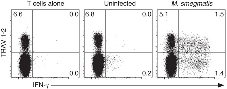
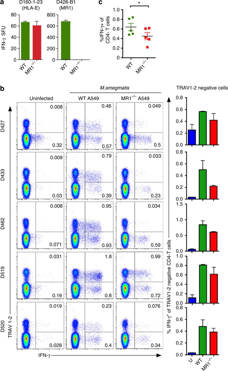
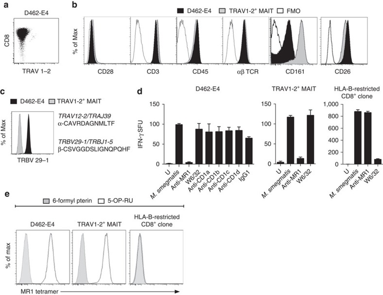
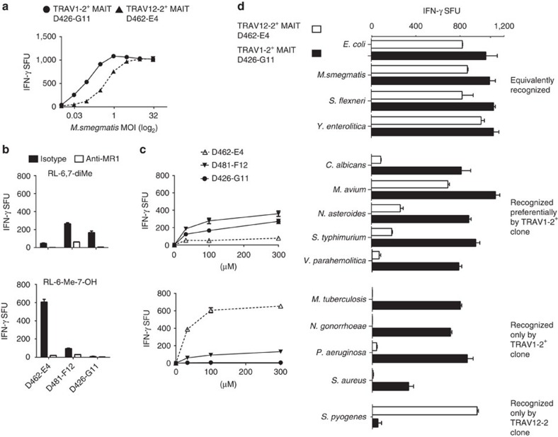
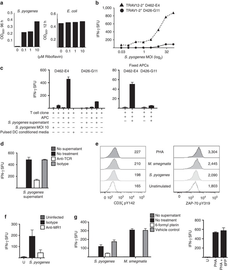
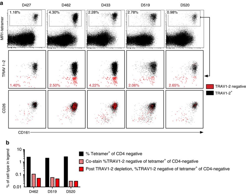
Mucosal-associated invariant T (MAIT) cells are thought to detect microbial antigens presented by the HLA-Ib molecule MR1 through the exclusive use of a TRAV1-2-containing TCRα. Here we use MR1 tetramer staining and ex vivo analysis with mycobacteria-infected MR1-deficient cells to demonstrate the presence of functional human MR1-restricted T cells that lack TRAV1-2. We characterize an MR1-restricted clone that expresses the TRAV12-2 TCRα, which lacks residues previously shown to be critical for MR1-antigen recognition. In contrast to TRAV1-2(+) MAIT cells, this TRAV12-2-expressing clone displays a distinct pattern of microbial recognition by detecting infection with the riboflavin auxotroph Streptococcus pyogenes. As known MAIT antigens are derived from riboflavin metabolites, this suggests that TRAV12-2(+) clone recognizes unique antigens. Thus, MR1-restricted T cells can discriminate between microbes in a TCR-dependent manner. We postulate that additional MR1-restricted T-cell subsets may play a unique role in defence against infection by broadening the recognition of microbial metabolites.


Figures

Figure 1
Figure 1. M. smegmatis infection elicits a response from MAIT cells and TRAV1-2 HLA-Ib-restricted CD8+ T cells.
PBMC-derived CD8+ T cells from adult donor (D462) were stimulated ex vivo by A549 cells infected with M. smegmatis to identify mycobacteria-reactive HLA-Ib T cells. Functional MAIT cells were identified by production of IFN-γ and expression of TRAV1-2 TCR. Numbers in quadrants indicate the % of live CD3+ cells. Experiments were performed three independent times with similar results. Representative results are shown.
Figure 2
Figure 2. MR1-restricted microbial-reactive CD8+ T cells from blood do not exclusively express TRAV1-2.
(a) IFN-γ production by T-cell clones D160-1-23 (restricted by HLA-E) and D426-B1 (restricted by MR1) in response to mycobacteria-infected WT and MR1−/− A549 cell line. (b) Positively selected CD8+ T cells from PBMC were tested for ex vivo IFN-γ responses to M. smegmatis-infected WT or MR1−/− A549 cell line. Events are gated on live CD3+CD4 cells. IFN-γ and TRAV1-2 expressions are shown on the x and y axes, respectively. To the right is a summary of the TRAV1-2-negative response from each donor across experiments. (c) Frequency of IFN-γ+ CD4 cells from each donor (represented by one dot) when stimulated by M. smegmatis-infected WT or MR1−\− A549s, n=5 biological replicates, with n=3 technical replicates. Statistical significance of difference between groups was determined using the nonparametric Mann–Whitney U-test. Error bars are the s.e.m. of triplicates in a,b. Experiments in this figure were performed at least twice with similar results. Representative results are shown. *P value>0.05 was considered significant.
Figure 3
Figure 3. Isolation of an MR1-restricted T-cell clone that reacts to bacteria independently of the TRAV1-2 TCR.
Generation of a CD8+ T-cell line and then T-cell clone, D462-E4, through FACS sorting on CD8+, γδ TCR, TRAV1-2-negative T cells from donor in (Fig. 1) and co-culturing for 7 days with M. smegmatis-infected autologous DCs. The line was stained with CFSE and co-cultured for 7 days with M. smegmatis-infected autologous macrophages. (a) Staining of TRAV1-2 and co-receptor CD8 on T-cell clone D462-E4. (b) Comparison of T-cell surface marker expression on TRAV1-2-negative clone D462-E4 (black) and TRAV1-2+ MAIT clone D426-G11 (grey) and (c) of TRBV 29-1 on D462-E4 and D481-A9 TRAV1-2+ MAIT clone. TCR αβ CDR3 sequences and gene names of D462-E4 are shown, right. (d) BEAS2B cell line infected with mycobacteria and tested for their ability to stimulate T-cell clones in the presence of blocking antibodies or isotype by IFN-γ ELISPOT assays, D462-E4, D481-F12 (TRAV1-2+ MAIT) and D466-A10 (HLA-B45). (e) Flow cytometry of clones with MR1 tetramer loaded with 6-formyl pterin (grey) or 5-OP-RU (clear). D462-E4, D481-A9 MAIT clone and D466-A10 HLA-B45-restricted T-cell clone. Error bars represent the s.e.m. of triplicates. Experiments were performed at least two times with similar results. Representative results are shown.
Figure 4
Figure 4. T-cell clone D462-E4 displays ligand and microbial infection selectivity.
(a) DCs were infected with M. smegmatis at a range of MOI (x axis) and then co-incubated with indicated MR1-restricted T-cell clones. (b) A549 cells were pulsed for 2 h with 100 μM RL-6,7-diMe (top) or RL-6-Me-OH (bottom) and tested for their ability to stimulate MR1-restricted T-cell clones in the presence of α-MR1 or isotype antibody by IFN-γ ELISPOT. (c) A549 cells were pulsed for 2 h with a range of concentrations of RL-6,7-diMe (top) or RL-6-Me-OH (bottom) and tested for their ability to stimulate MR1-restricted T-cell clones by IFN-γ ELISPOT. (d) DCs were infected at optimized MOIs with pathogens listed on the y axis, except for Yersinia and Shigella where the A549 cell line was used, and then co-incubated overnight with the indicated MR1-restricted T-cell clones. IFN-γ production was quantified by ELISPOT. Detection of less than 25 spot-forming unit per well was considered no response by the T-cell clone. Results are grouped by comparative response by MR1-restricted T-cell clones. Pattern of recognition was maintained over a range of MOI. Error bars are the s.e.m. of triplicates. Assays were performed twice, with similar results. Representative results are shown.
Figure 5
Figure 5. Selective recognition of S. pyogenes by a TRAV1-2-negative T-cell clone D462-E4.
(a) S. pyogenes or E. coli was cultured in minimal broth without riboflavin or with a titration of riboflavin over the course of 96 h (S. pyogenes) or 12 h (E. coli), and growth was monitored by optical density readings. (b) DCs were infected with S. pyogenes at a range of MOI (x axis) and then co-incubated with indicated MR1-restricted T-cell clones. (c) T-cell clones D462-E4 and D426-G11 were incubated with (left to right) S. pyogenes (MOI=10), S. pyogenes culture supernatant (SN) 30 μl, unloaded DCs, DCs with S. pyogenes SN or pulsed DC-conditioned media overnight. On the right, D462-E4 and D426-G11 were incubated with paraformaldehyde-fixed DCs that had been pulsed with S. pyogenes SN. IFN-γ production was quantified by ELISPOT. (d) T-cell clone D462-E4 was blocked with anti-pan TCR αβ, isotype control or not treated and then co-incubated with DCs loaded with S. pyogenes supernatant (30 μl). IFN-γ production was quantified by ELISPOT. (e) Phosphorylation of the CD3-ζ chain of the TCR/CD3 complex at tyrosine 142 or ZAP-70 at tyrosine 319 was quantified by flow cytometry after T-cell clone D462-E4 was co-incubated with DCs infected with S. pyogenes or M.smeg MOI=10, PHA (20 μg ml−1) or left untreated (unstimulated condition). Numbers on the overlay indicate the geometric mean fluorescence intensity of at least 30,000 clones. (f) DCs were infected with S. pyogenes at MOI=3, blocked with anti-MR1 or isotype control (10 μg ml−1) and then co-incubated with D462-E4 T-cell clone. IFN-γ production was quantified by ELISPOT. (g) DCs were either blocked with 6-formyl pterin (50 μg ml−1) or 0.01 M NaOH vehicle control or nothing, and then loaded with S. pyogenes or M.smeg supernatant (15 μl) or PHA at 10 μg ml−1. The DCs were then used to stimulate T-cell clone D462-E4 and IFN-γ production was quantified by ELISPOT. Error bars represent the s.e.m. of at least duplicates. Assays were performed three times, with similar results. Representative results are shown.
Figure 6
Figure 6. MR1-Ag tetramer+ CD8+ T cells from peripheral blood do not exclusively express TRAV1-2.
PBMCs were stained with antibodies to CD3, CD4, CD8, TRAV1-2, CD26, CD161, viability stain and human MR1-Ag (5-OP-RU) tetramer. All plots are gated on live, CD3+, CD4-negative cells. The x axis represents CD161 expression, and the y axis represents MR1 tetramer (top row), TRAV1-2 (middle row) and CD26 (bottom row). MR1 tetramer+ cells were gated as indicated by the gate in the top row. MR1 tetramer+ cells were subgated based on TRAV1-2 expression where TRAV1-2-negative events and their frequencies are shown in red. The second and third rows of plots are overlays of TRAV1-2+ in black and TRAV1-2-negative in red. (b) PBMCs were depleted of TRAV1-2+ cells by FACS and then stained with MR1-Ag tetramer and T-cell lineage markers (red bars). The black bars and red-patterned bars indicate frequencies presented in (a) for comparison. This experiment was performed once and (a) was performed twice with similar results. Representative results are shown.

Similar articles

Cited by

References

    1. Gold M. C. et al. Human mucosal associated invariant T cells detect bacterially infected cells. PLoS Biol. 8, e1000407 (2010). - PMC - PubMed
    1. Le Bourhis L. et al. MAIT cells detect and efficiently lyse bacterially-infected epithelial cells. PLoS Pathog. 9, e1003681 (2013). - PMC - PubMed
    1. Le Bourhis L. et al. Antimicrobial activity of mucosal-associated invariant T cells. Nat. Immunol. 11, 701–708 (2010). - PubMed
    1. Corbett A. J. et al. T-cell activation by transitory neo-antigens derived from distinct microbial pathways. Nature 509, 361–365 (2014). - PubMed
    1. Kjer-Nielsen L. et al. MR1 presents microbial vitamin B metabolites to MAIT cells. Nature 491, 717–723 (2012). - PubMed

Publication types

MeSH terms