. 2016 Sep 27;7(39):63106-63123.
doi: 10.18632/oncotarget.11056.

Phosphatidylinositide 3-kinase (PI3K) and PI3K-related kinase (PIKK) activity contributes to radioresistance in thyroid carcinomas

Affiliations

Phosphatidylinositide 3-kinase (PI3K) and PI3K-related kinase (PIKK) activity contributes to radioresistance in thyroid carcinomas

Natalie Burrows et al. Oncotarget. .

Abstract

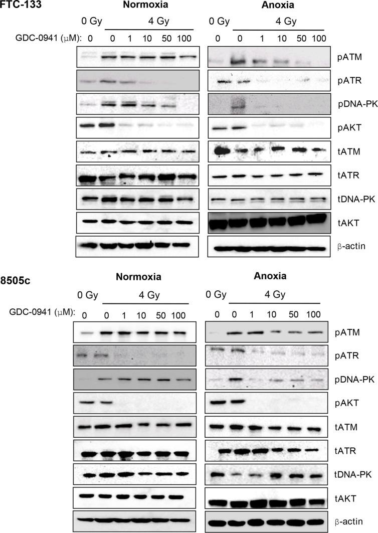
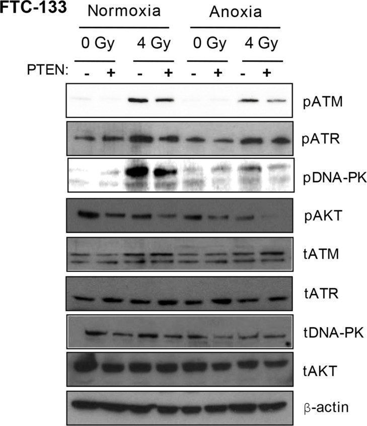
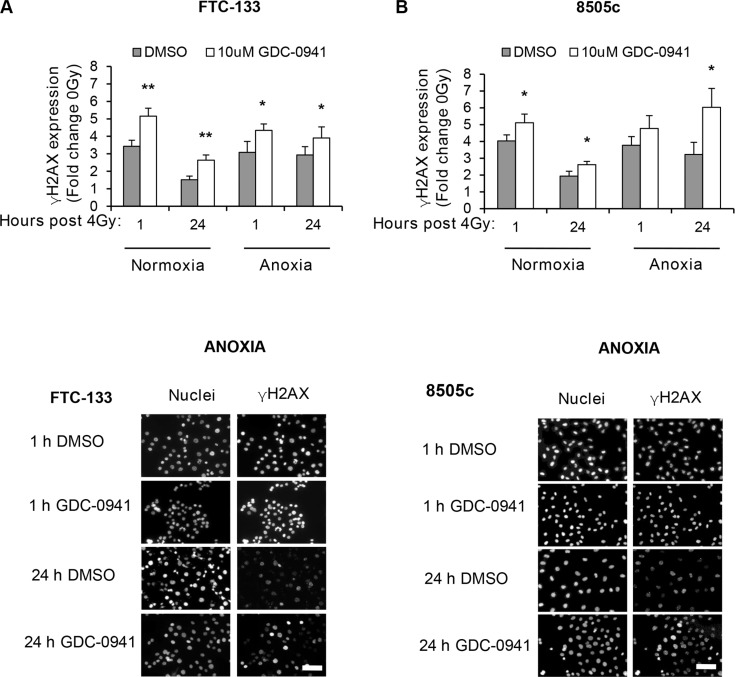
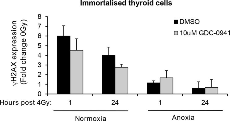
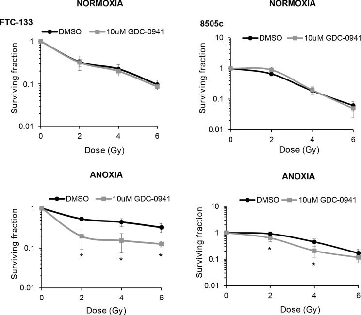
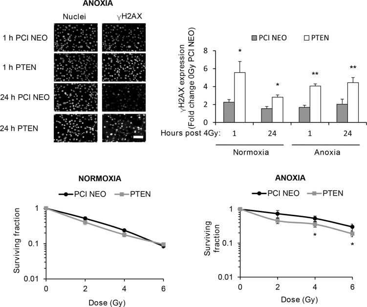
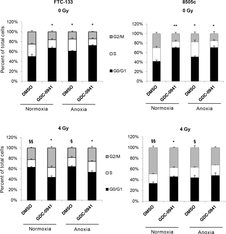
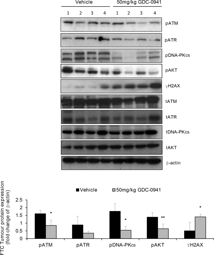
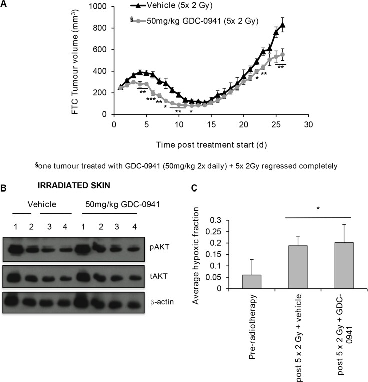
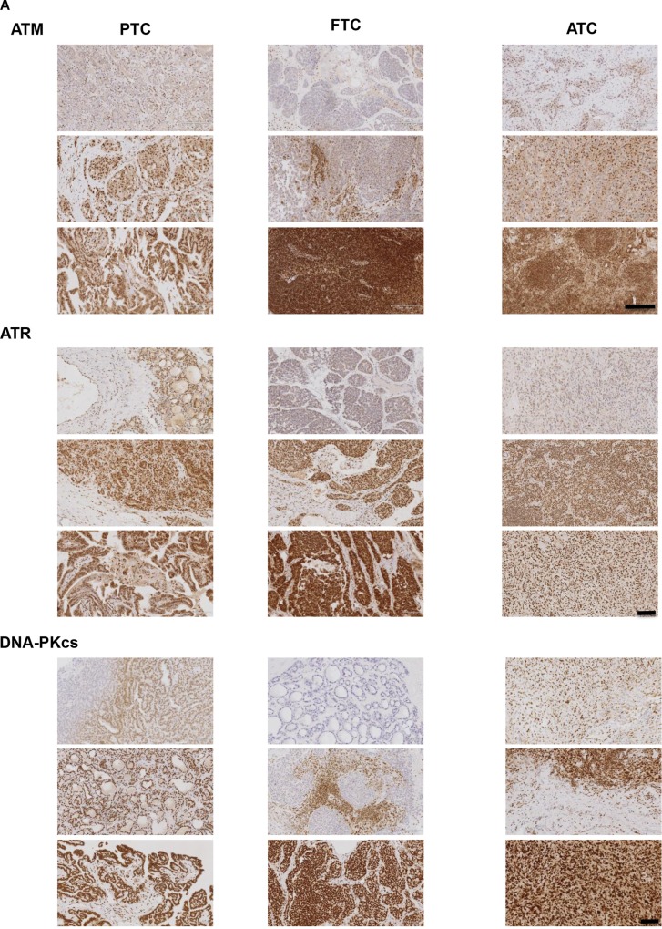
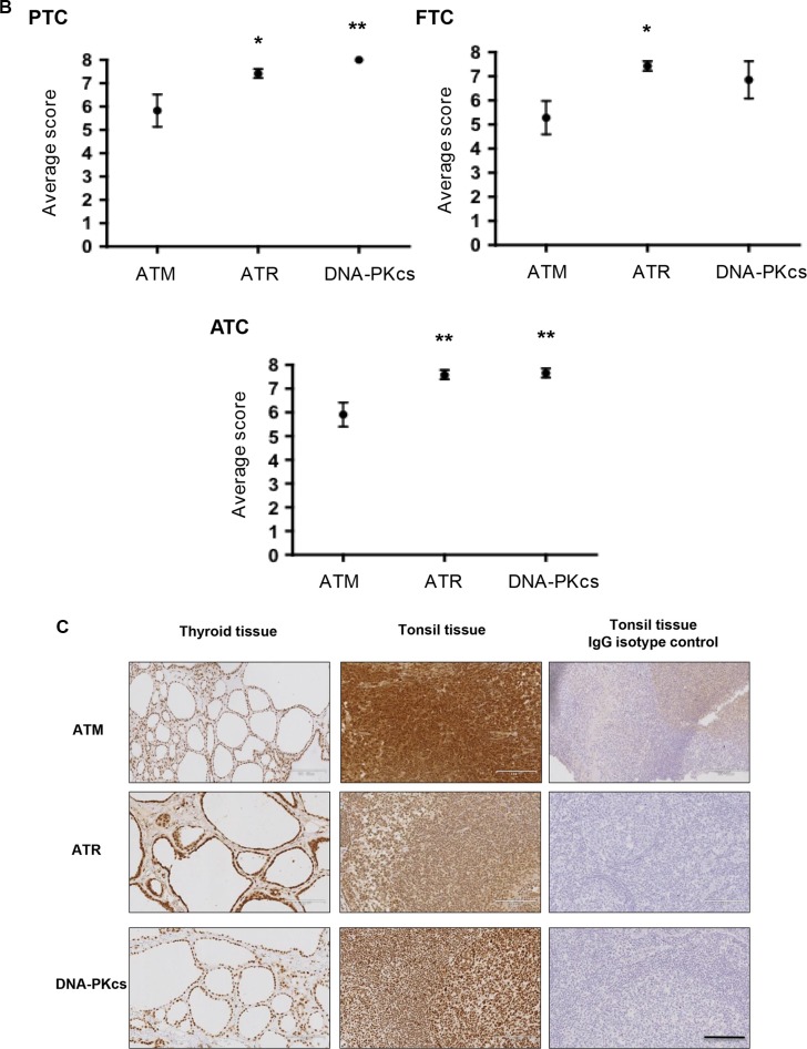
Anaplastic (ATC) and certain follicular thyroid-carcinomas (FTCs) are radioresistant. The Phosphatidylinositide 3-kinase (PI3K) pathway is commonly hyperactivated in thyroid-carcinomas. PI3K can modify the PI3K-related kinases (PIKKs) in response to radiation: How PIKKs interact with PI3K and contribute to radioresistance in thyroid-carcinomas is unknown. Further uncertainties exist in how these interactions function under the radioresistant hypoxic microenvironment. Under normoxia/anoxia, ATC (8505c) and FTC (FTC-133) cells were irradiated, with PI3K-inhibition (via GDC-0941 and PTEN-reconstitution into PTEN-null FTC-133s) and effects on PIKK-activation, DNA-damage, clonogenic-survival and cell cycle, assessed. FTC-xenografts were treated with 5 × 2 Gy, ± 50 mg/kg GDC-0941 (twice-daily; orally) for 14 days and PIKK-activation and tumour-growth assessed. PIKK-expression was additionally assessed in 12 human papillary thyroid-carcinomas, 13 FTCs and 12 ATCs. GDC-0941 inhibited radiation-induced activation of Ataxia-telangiectasia mutated (ATM), ATM-and Rad3-related (ATR) and DNA-dependent protein kinase catalytic subunit (DNA-PKcs). Inhibition of ATM and DNA-PKcs was PI3K-dependent, since activation was reduced in PTEN-reconstituted FTC-133s. Inhibition of PIKK-activation was greater under anoxia: Consequently, whilst DNA-damage was increased and prolonged under both normoxia and anoxia, PI3K-inhibition only reduced clonogenic-survival under anoxia. GDC-0941 abrogated radiation-induced cell cycle arrest, an effect most likely linked to the marked inhibition of ATR-activation. Importantly, GDC-0941 inhibited radiation-induced PIKK-activation in FTC-xenografts leading to a significant increase in time taken for tumours to triple in size: 26.5 ± 5 days (radiation-alone) versus 31.5 ± 5 days (dual-treatment). PIKKs were highly expressed across human thyroid-carcinoma classifications, with ATM scoring consistently lower. Interestingly, some loss of ATM and DNA-PKcs was observed. These data provide new insight into the mechanisms of hypoxia-associated radioresistance in thyroid-carcinoma.

Keywords: ATM; ATR; DNA-PKcs; PI3K; radioresistance.


Conflict of interest statement

CONFLICTS OF INTEREST

The authors have nothing to disclose.

Figures

Figure 1
Figure 1. Under anoxia, radiation-induced PIKK activation is markedly inhibited by GDC-0941
PIKK activation was assessed via analysis of phospho(p)PIKK protein expression. In FTC-133 and 8505c cells, GDC-0941 inhibited expression of pATR at concentrations ≤ 1 μM under both normoxia and anoxia. Under normoxia, GDC-0941 inhibited pDNA-PKcs expression at higher concentrations; ≤ 10 μM in FTC-133′s and 100 μM 8505c's. Under anoxia, pDNA-PKcs was much more sensitive to GDC-0941, with almost complete inhibition observed at 1 μM GDC-0941 in both cell lines. Under normoxia, GDC-0941 had little effect on pATM, whilst a clear inhibition was observed in both cell lines under anoxia. Cells were exposed to GDC-0941 in normoxia/anoxia for 18 h, irradiated (4 Gy) whilst remaining in the specified conditions and lysates collected 1 h later for analysis. Cropped blots are representative of 4 independent experiments.
Figure 2
Figure 2. Under anoxia, genetic inhibition of PI3K inhibits radiation-induced ATM and DNA-PKcs activation
PIKK activation was assessed via analysis of phospho(p)PIKK protein expression. Genetic inhibition of PI3K (via PTEN reconstitution into PTEN-null FTC-133 cells) had little effect on pATR expression under normoxia and anoxia. Radiation-induced pATM and pDNA-PKcs were inhibited in anoxic PTEN-expressing cells. PCI NEO and PTEN reconstituted FTC-133 cells were incubated in normoxia/anoxia for 18 h irradiated (4 Gy) whilst remaining in the specified conditions and lysates collected 1 h later for analysis. Cropped blots are representative of 4 independent experiments.
Figure 3
Figure 3. Pharmacological inhibition of PI3K increases and prolongs radiation-induced DNA damage across oxygen environments
(AB) GDC-0941 increases γH2AX expression 1 and 24 h post irradiation under normoxia and anoxia (*p < 0.05, **p < 0.01 compared to vehicle (DMSO) in same condition). Examples of immuno-fluorescence images of γH2AX under anoxia are shown. FTC-133 and 8505c cells were incubated under normoxia/anoxia for 18 h with DMSO or 10 μM GDC-0941 and irradiated (4 Gy) in the specified conditions. Samples remained in the specified conditions until fixation 1 and 24 h post 4 Gy. DNA damage was assessed by expression of γH2AX that was displayed as fold change of 0 Gy DMSO for each condition. Nuclei were counterstained with DAPI. Scale-bar 10 μm. Data represents the mean ± S.D. of 3 independent experiments.
Figure 4
Figure 4. GDC-0941 does not affect radiation-induced DNA damage in immortalised thyroid cells
Radiotherapy increased γH2AX expression to similar levels to that observed in thyroid carcinoma cell lines but had little effect under anoxia. GDC-0941 had little effect on radiation-induced DNA damage under normoxia or anoxia. Immortalised thyroid cells were treated and analysed as described in figure legend 3. Data represents the mean ± S.D. of 3 experimental repeats.
Figure 5
Figure 5. GDC-0941 reduces clonogenic survival of anoxic cells
GDC-0941 had little effect on clonogenic survival under normoxia but significantly inhibited survival under anoxia (*p < 0.05 versus DMSO). For clonogenic assays, cells were incubated under normoxia/anoxia for 18 h with DMSO or 10 μM GDC-0941and irradiated (4 Gy) in the specified conditions. Samples remained in the specified conditions for 24 h post irradiation, then plated in serial dilution and cultured until visible colonies formed. Data represents the mean ± S.D. of 3 independent experiments.
Figure 6
Figure 6. Genetic inhibition of PI3K increases and prolongs radiation-induced DNA damage across oxygen environments but selectively inhibits clonogenic survival of anoxic cells
PTEN reconstitution in FTC-133 cells significantly increased γH2AX expression 1 and 24 h post irradiation under normoxia and anoxia (*p < 0.05, **p < 0.001) and reduced clonogenic survival under anoxia (*p < 0.05 versus DMSO), whilst having little effect under normoxia. PCI NEO and PTEN reconstituted FTC-133 cells were incubated under normoxia/anoxia for 18 h and irradiated (4 Gy) under condition. Cells then remained in normoxia/anoxia and were either fixed 1 and 24 h post 4 Gy for analysis of γH2AX expression or seeded in serial dilution 24 h post 4 Gy, and cultured until visible colonies formed for clonogenic survival assays. DNA damage was assessed by expression of γH2AX and displayed as fold change of 0 Gy PCI NEO for each condition. Nuclei were counterstained with DAPI. Scale-bar 10 μm. Data represents the mean ± S.D. of 3 independent experiments.
Figure 7
Figure 7. GDC-0941 abrogates radiation-mediated effects on cell cycle
In un-irradiated cell lines, GDC-0941 and anoxia independently increased percentage cells in G0/G1 (*p < 0.05, **p < 0.01). Radiation had differential effects across cell lines; significantly increasing percentage cells in G0/G1 (FTC-133) and G2/M (8505c) under normoxia and anoxia (§p < 0.05, §§p < 0.01). In irradiated FTC-133 cells, GDC-0941 significantly reduced percentage cells in G0/G1 under normoxia and anoxia (*p < 0.05). In irradiated 8505c cells, GDC-0941 significantly reduced percentage cells in G2/M under normoxia (*p < 0.05), with similar affects observed in anoxia. As for the clonogenic assays, FTC-133 and 8505c cells were incubated under normoxia/anoxia for 18 h with DMSO or 10 μM GDC-0941 and irradiated (4 Gy) in the specified conditions. Samples remained in the specified conditions for 24 h post irradiation, but instead of seeding for clonogenicity, samples were fixed and cell cycle distribution was assessed by flow cytometry analysis of propidium iodide stained cells. Data represents the mean ± S.E.M. of 3 independent experiments.
Figure 8
Figure 8. GDC-0941 inhibits PIKK activation and increases DNA damage in irradiated human FTC xenografts
In irradiated FTC133 xenografts, GDC-0941 reduces pATR expression and significantly reduces pATM and pDNA-PKcs expression (*p < 0.05), whilst γH2AX expression is significantly increased (*p < 0.05). Inhibition of PI3K was confirmed by analysis of pAKT expression, which was reduced in GDC-0941 versus vehicle treated tumours (**p < 0.01). Mice received vehicle or 50 mg/kg GDC-0941 (p.o.) 4 h prior and immediately after radiotherapy (10 Gy fractionated (2 Gy) over 5 consecutive days). 1 h after the final dose, tumours were rapidly excised and immediately snap frozen for analysis of PIKK activity by western-blot of pPIKK expression. Cropped blot represents irradiated tumours from 4 independent mice that received either vehicle or GDC-0941. Graph represents densitometry data for the calculated mean ± S.D of the depicted western-blot.
Figure 9
Figure 9. GDC-0941 increases tumour regression post-radiotherapy
Mice were dosed twice daily (p.o.) with vehicle or 50 mg/kg GDC-0941 for 14 d. Tumours received 10 Gy radiation fractionated into 2 Gy over 5 days (treatment days 1-5). On these days vehicle/GDC-0941 was administered 4 h prior and immediately after radiation. (A) Mice dosed with GDC-0941 had significantly smaller tumour volumes on days 4–12, 22,23,25 and 26 (*p < 0.05, **p < 0.01, ***p < 0.001 versus vehicle treated mice). Data displayed shows up until the time when vehicle treated mice reached the designated end-point of three times the relative tumour volume (RTV3: 26.5 ± 5 days for radiation alone versus 31.5 ± 5 days for GDC-0941/radiation; p < 0.05). Data shown is for 11 vehicle and 9 GDC-0941 treated mice, Data represents the mean ± S.D. §One co-treated tumour regressed completely and was excluded from analysis. (B) Mice received vehicle or 50 mg/kg GDC-0941 (p.o.) 4 h prior and immediately after radiotherapy (10 Gy fractionated (2 Gy) over 5 consecutive days). 1 h after the final dose, skin exposed to radiation (that covering the tumour) was excised, snap frozen and homogenised in lysis buffer for western-blot analysis. GDC-0941 did not affect pAKT expression in skin tissue exposed to radiotherapy. Cropped blot shows pAKT, tAKT and the loading control β-actin for mice that received radiation with either vehicle (n = 4) or with GDC-0941 (n = 4). (C) Tumour hypoxia was measured by IHC analysis of pimonidazole, dosed at 60 mg/kg (i.p.) 2 h prior to tumour excision. A significant increase in hypoxic fraction was observed in irradiated vehicle and GDC-0941 treated tumours (*p < 0.05 versus un-irradiated). GDC-0941 had no significant effect on hypoxic-fraction compared to vehicle in irradiated xenografts. Tumour volume did not significantly differ between groups. Data represents the mean ± S.D. of 4-5 mice per group.
Figure 10
Figure 10. The PIKKS are a viable target in human thyroid cancer
(A) Representative images of PIKK staining (brown). Intensity and distribution of PIKK proteins varied within each tumour classification. Representative images from three independent tumours are shown. Scale bar 1700 μm (for ATR and DNA-PKcs images) and 2000 μm (for ATM images). (B) PIKK expression was scored based on intensity and proportion of staining (described in methods). ATR and DNA-PKcs expression was significantly higher versus ATM in PTCs/ATCs and ATR expression significantly higher versus ATM in FTCs (*p < 0.05, **p < 0.01). Data represents mean ± S.E.M. of 12 PTC/ATC and 13 FTC. (C) Thyroid tissue stained positive for ATM, ATR and DNA-PKcs. Human tonsil was used as a known positive control for PIKK expression. Additionally tonsil tissue was exposed to the appropriate concentration matched IgG isotype antibodies as a negative control. See methods for full details. Scale bar 119 μm.

References

    1. Xing M, Haugen BR, Schlumberger M. Progress in molecular-based management of differentiated thyroid cancer. Lancet. 2013;381:1058–1069. - PMC - PubMed
    1. Patel KN, Shaha AR. Poorly differentiated and anaplastic thyroid cancer. Cancer control. 2006;13:119–128. - PubMed
    1. Busaidy NL, Cabanillas ME. Differentiated thyroid cancer: management of patients with radioiodine nonresponsive disease. J Thyroid Res. 2012;2012:618985. - PMC - PubMed
    1. Rouse J, Jackson SP. Interfaces between the detection signaling and repair of DNA damage. Science. 2002;297:547–551. - PubMed
    1. Lovejoy CA, Cortez D. Common mechanisms of PIKK regulation. DNA repair. 2009;8:1004–1008. - PMC - PubMed

MeSH terms