Celecoxib and sulindac inhibit TGF-β1-induced epithelial-mesenchymal transition and suppress lung cancer migration and invasion via downregulation of sirtuin 1
- PMID: 27528025
- PMCID: PMC5302984
- DOI: 10.18632/oncotarget.11127
Celecoxib and sulindac inhibit TGF-β1-induced epithelial-mesenchymal transition and suppress lung cancer migration and invasion via downregulation of sirtuin 1
Abstract
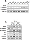
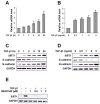
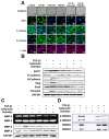
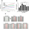
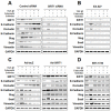
The non-steroidal anti-inflammatory drugs (NSAIDs) celecoxib and sulindac have been reported to suppress lung cancer migration and invasion. The class III deacetylase sirtuin 1 (SIRT1) possesses both pro- and anticarcinogenic properties. However, its role in inhibition of lung cancer cell epithelial-mesenchymal transition (EMT) by NSAIDs is not clearly known. We attempted to investigate the potential use of NSAIDs as inhibitors of TGF-β1-induced EMT in A549 cells, and the underlying mechanisms of suppression of lung cancer migration and invasion by celecoxib and sulindac. We demonstrated that celecoxib and sulindac were effective in preventing TGF-β1-induced EMT, as indicated by upregulation of the epithelial marker, E-cadherin, and downregulation of mesenchymal markers and transcription factors. Moreover, celecoxib and sulindac could inhibit TGF-β1-enhanced migration and invasion of A549 cells. SIRT1 downregulation enhanced the reversal of TGF-β1-induced EMT by celecoxib or sulindac. In contrast, SIRT1 upregulation promoted TGF-β1-induced EMT. Taken together, these results indicate that celecoxib and sulindac can inhibit TGF-β1-induced EMT and suppress lung cancer cell migration and invasion via downregulation of SIRT1. Our findings implicate overexpressed SIRT1 as a potential therapeutic target to reverse TGF-β1-induced EMT and to prevent lung cancer cell migration and invasion.
Keywords: EMT; SIRT1; celecoxib; lung cancer; sulindac.
Conflict of interest statement
No potential conflicts of interest were disclosed.
Figures


Similar articles
-
Resveratrol inhibits TGF-β1-induced epithelial-to-mesenchymal transition and suppresses lung cancer invasion and metastasis.Toxicology. 2013 Jan 7;303:139-46. doi: 10.1016/j.tox.2012.09.017. Epub 2012 Nov 9. Toxicology. 2013. PMID: 23146760
-
Salinomycin suppresses TGF-β1-induced EMT by down-regulating MMP-2 and MMP-9 via the AMPK/SIRT1 pathway in non-small cell lung cancer.Int J Med Sci. 2021 Jan 1;18(3):715-726. doi: 10.7150/ijms.50080. eCollection 2021. Int J Med Sci. 2021. PMID: 33437206 Free PMC article.
-
Stereospecific effects of ginsenoside 20-Rg3 inhibits TGF-β1-induced epithelial-mesenchymal transition and suppresses lung cancer migration, invasion and anoikis resistance.Toxicology. 2014 Aug 1;322:23-33. doi: 10.1016/j.tox.2014.04.002. Epub 2014 May 2. Toxicology. 2014. PMID: 24793912
-
Narrower insight to SIRT1 role in cancer: A potential therapeutic target to control epithelial-mesenchymal transition in cancer cells.J Cell Physiol. 2018 Jun;233(6):4443-4457. doi: 10.1002/jcp.26302. Epub 2018 Jan 15. J Cell Physiol. 2018. PMID: 29194618 Review.
-
Roles of Dietary Phytoestrogens on the Regulation of Epithelial-Mesenchymal Transition in Diverse Cancer Metastasis.Toxins (Basel). 2016 May 24;8(6):162. doi: 10.3390/toxins8060162. Toxins (Basel). 2016. PMID: 27231938 Free PMC article. Review.
Cited by
-
Oridonin inhibits migration, invasion, adhesion and TGF-β1-induced epithelial-mesenchymal transition of melanoma cells by inhibiting the activity of PI3K/Akt/GSK-3β signaling pathway.Oncol Lett. 2018 Jan;15(1):1362-1372. doi: 10.3892/ol.2017.7421. Epub 2017 Nov 15. Oncol Lett. 2018. PMID: 29399187 Free PMC article.
-
Reciprocal Regulation of Cancer-Associated Fibroblasts and Tumor Microenvironment in Gastrointestinal Cancer: Implications for Cancer Dormancy.Cancers (Basel). 2023 Apr 27;15(9):2513. doi: 10.3390/cancers15092513. Cancers (Basel). 2023. PMID: 37173977 Free PMC article. Review.
-
Retraction Note: The suppressing role of miR-622 in renal cell carcinoma progression by down-regulation of CCL18/MAPK signal pathway.Cell Biosci. 2023 Feb 9;13(1):27. doi: 10.1186/s13578-023-00976-x. Cell Biosci. 2023. PMID: 36759851 Free PMC article. No abstract available.
-
Celecoxib as a Valuable Adjuvant in Cutaneous Melanoma Treated with Trametinib.Int J Mol Sci. 2021 Apr 22;22(9):4387. doi: 10.3390/ijms22094387. Int J Mol Sci. 2021. PMID: 33922284 Free PMC article.
-
Artemisinin induced reversal of EMT affects the molecular biological activity of ovarian cancer SKOV3 cell lines.Oncol Lett. 2019 Sep;18(3):3407-3414. doi: 10.3892/ol.2019.10608. Epub 2019 Jul 11. Oncol Lett. 2019. PMID: 31452821 Free PMC article.
References
-
- Cavellaro U, Christofori G. Cell adhesion and signalling by cadherins and Ig-CAMs in cancer. Nature reviews. Cancer. 2004;4:118–132. - PubMed
-
- Deckers M, van Dinther M, Buijs J, Que I, Löwik C, van der Pluijm G, ten Dijke P. The tumor suppressor Smad4 is required for transforming growth factor beta-induced epithelial to mesenchymal transition and bone metastasis of breast cancer cells. Cancer Research. 2006;66:2202–2209. - PubMed
MeSH terms
Substances
LinkOut - more resources
Full Text Sources
Other Literature Sources
Medical
Miscellaneous

