. 2016 Sep 20;7(38):61469-61484.
doi: 10.18632/oncotarget.11129.

Stromal remodeling by the BET bromodomain inhibitor JQ1 suppresses the progression of human pancreatic cancer

Affiliations

Stromal remodeling by the BET bromodomain inhibitor JQ1 suppresses the progression of human pancreatic cancer

Keisuke Yamamoto et al. Oncotarget. .

Abstract

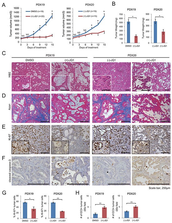
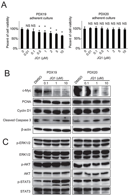
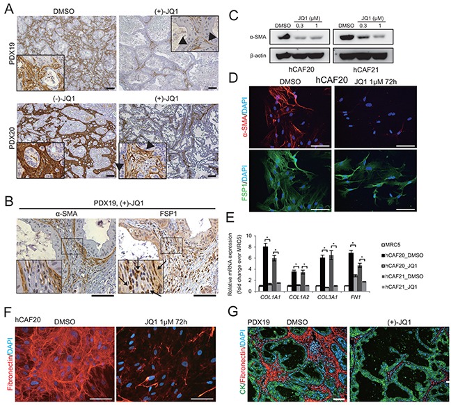
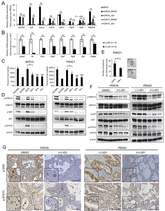
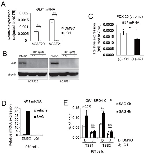
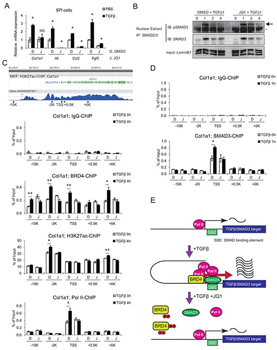
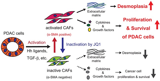
Inhibitors of bromodomain and extraterminal domain (BET) proteins, a family of chromatin reader proteins, have therapeutic efficacy against various malignancies. However, the detailed mechanisms underlying the anti-tumor effects in distinct tumor types remain elusive. Here, we show a novel antitumor mechanism of BET inhibition in pancreatic ductal adenocarcinoma (PDAC). We found that JQ1, a BET inhibitor, decreased desmoplastic stroma, a hallmark of PDAC, and suppressed the growth of patient-derived tumor xenografts (PDX) of PDACs. In vivo antitumor effects of JQ1 were not always associated with the JQ1 sensitivity of respective PDAC cells, and were rather dependent on the suppression of tumor-promoting activity in cancer-associated fibroblasts (CAFs). JQ1 inhibited Hedgehog and TGF-β pathways as potent regulators of CAF activation and suppressed the expression of α-SMA, extracellular matrix, cytokines, and growth factors in human primary CAFs. Consistently, conditioned media (CM) from CAFs promoted the proliferation of PDAC cells along with the activation of ERK, AKT, and STAT3 pathways, though these effects were suppressed when CM from JQ1-treated CAFs was used. Mechanistically, chromatin immunoprecipitation experiments revealed that JQ1 reduced TGF-β-dependent gene expression by disrupting the recruitment of the transcriptional machinery containing BET proteins. Finally, combination therapy with gemcitabine plus JQ1 showed greater efficacy than gemcitabine monotherapy against PDAC in vivo. Thus, our results reveal BET proteins as the critical regulators of CAF-activation and also provide evidence that stromal remodeling by epigenetic modulators can be a novel therapeutic option for PDAC.

Keywords: JQ1; bromodomain and extraterminal domain (BET) proteins; cancer-associated fibroblast (CAF); epigenetics; pancreatic ductal adenocarcinoma (PDAC).


Conflict of interest statement

No potential conflicts of interest were disclosed.

Figures

Figure 1
Figure 1. JQ1 attenuates tumor growth and desmoplasia in PDX of human PDAC
Mice bearing PDX tumors were treated daily with (+)-JQ1 or control reagents (DMSO or (−)-JQ1) at 50 mg/kg for 2 wk. A. Average volumes of subcutaneous PDX tumors. *, P < .05; NS, not significant. B. Tumor weight at the end of the treatment period. Bars represent means ± SEM; *, P < .05. (C and D) H & E staining C. and Azan staining D. of PDX tumors at the end of the treatment. Scale bars represent 250 μm. Insets show higher magnification pictures. E and F. Representative IHC images stained for Ki-67 (E) and cleaved caspase-3 (CC3) (F). Scale bars represent 250 μm. Insets show higher magnification pictures. G and H. Percentage of Ki-67 (E) and CC3 (F) positive tumor cells per 20x field (average of five random fields per tumor) are shown. Four tumors per group were analyzed. Bars represent mean ± SEM (n = 4); *, P < .05 and **, P < .01.
Figure 2
Figure 2. JQ1 exhibits minimal effects on the growth of primary human PDAC cells in vitro
Primary human PDAC cells were isolated from PDX tumors and cultured under adherent conditions in DMEM/F12 containing 10% FBS. A. PDAC cells were incubated at indicated doses of JQ1 for 72 h, and then cell growth was quantified. *, P <.05 compared to vehicle by Student's t-test. B and C. Western blot of whole cell lysates from primary PDAC cells treated with JQ1 for 48 h in vitro.
Figure 3
Figure 3. JQ1 suppresses α-SMA expression and ECM synthesis in CAFs
A. α-SMA immunohistochemistry was performed on PDX tumors from mice treated with control reagents or JQ1 for 2 wks. In JQ1-treated tumors, positive α-SMA staining was confined to the stromal cells adjacent to the tumor epithelia (arrow heads). Scale bars represent 100 μm. B. Serial sections of JQ1-treated PDX tumors were stained for α-SMA and FSP1. Most stromal cells that were negative for α-SMA expression were stained positively for FSP1 (arrows), suggesting that fibroblasts not in direct contact with cancer cells lost α-SMA expression by JQ1 treatment. Scale bars represent 100 μm. C. Western blot of lysates from primary human CAFs (hCAF20 and hCAF21) treated with JQ1 for 48 h. JQ1 treatment reduced α-SMA expression in hCAFs in a dose-dependent manner. D. Immunofluorescence images of hCAF20 cells stained for α-SMA (red) and FSP1 (green) following JQ1 treatment. Nuclei were stained with DAPI (blue). Scale bars represent 100 μm. E. The expression levels of ECM related genes were analyzed by qRT-PCR. hCAFs were treated with 1 μM JQ1 for 48 h. MRC-5, a human lung fibroblast cell line, was used as normal fibroblasts. Values were normalized to ACTB and expressed as fold change against MRC-5. Bars represent means ± SEM (n = 3); *, P < .01, compared to respective DMSO controls. F. Representative immunofluorescence images of hCAF20 cells. After treatment with DMSO or 1 μM JQ1 for 72 h, hCAF20 cells were stained with antibodies against fibronectin (red). Nuclei were stained with DAPI (blue). Scale bars represent 100 μm. G. Representative immunofluorescence images of PDX19 tumors that were co-stained with antibodies against pan-cytokeratin (CK, green) and fibronectin (red). Nuclei were stained with DAPI (blue). Scale bars represent 100 μm.
Figure 4
Figure 4. JQ1 alters the secretome of CAFs, reducing PDAC proliferation
A-B. The expression levels of activated-CAF related genes, including inflammatory cytokines and growth factors, were analyzed by qRT-PCR in hCAFs in vitro (A) and in the stromal cells of PDX tumors in vivo (B). (A) hCAFs were treated with 1μM JQ1 for 48 h. Values were normalized to ACTB and expressed as fold change over MRC-5. For FGF7, the value of MRC5 was set to 10. Bars represent means ± SEM (n = 3); *, P < .05 and **, P < .01, compared to the respective DMSO controls. (B) Bulk RNA from PDX20 tumors treated with (−)-JQ1 or (+)-JQ1 for 2 weeks were used for qRT-PCR. Bars represent means ± SEM (n = 6); *, P < .05 and **, P < .01, compared to (−)-JQ1-treated tumors. C-D. CM from hCAF20 cells enhances pancreatic cancer cell proliferation, accompanied by the activation of ERK, Akt, and STAT3 pathways. CM-D and CM-J represent CM collected from hCAF20 cells treated with DMSO or JQ1, respectively. Negative control medium without CAF culture was prepared in the same manner either with DMSO or JQ1 (referred to as M-D and M-J). C. The effects of CM on PDAC cell proliferation. BxPC3 and PANC1 cells were cultured under the indicated conditions for 72 h, and viable cells were quantified. D. Western blot was performed using lysates from cells after 15 min incubation with the indicated CM. E. A tumor sphere formation assay was performed. Bars represent means ± SEM (n = 5); *, P = .007. Representative images of tumor spheres are shown. Bars represent 100 μm. F. Western blot using whole lysates from PDX tumors treated with either JQ1 or control reagents. Analyses of three tumors from each group are shown. G. Representative immunohistochemistry images stained for p-ERK and p-STAT3. Positive p-ERK staining was observed exclusively in cancer cells, while positive p-STAT3 staining was observed both in cancer cells and stromal cells. Scale bar represents 250 μm.
Figure 5
Figure 5. JQ1 inhibits Hh target gene GLI1 expression in both human and murine CAFs
A. qRT-PCR analysis showed that JQ1 reduced GLI1 mRNA expression in human CAFs. hCAFs were treated with DMSO or 1μM JQ1 for 24 h. Bars represent means ± SEM (n = 3); **, P < .01, compared to DMSO treated controls. B. Western blots showed that JQ1 reduced GLI1 expression in human CAFs. hCAFs were treated with DMSO or JQ1 for 24 h. C. qRT-PCR showed that Gli1 mRNA expression was reduced in murine stromal cells of PDX20 tumors that were treated with (+)-JQ1 for 14 days. Bars represent means ± SEM (n = 6); **, P < .01, compared with (−)-JQ1-treated tumors. D. qRT-PCR analysis showing that JQ1 reduced Gli1 mRNA expression in murine CAFs (97f cells). 97f cells were treated with DMSO (D) or 1μM JQ1 (J) for 2 hours, followed by addition of DMSO or 0.3 μM SAG for 24 h. E. ChIP-qPCR revealed that SAG-induced BRD4 recruitment to Gli1 promoters were suppressed by JQ1 treatment. After pretreatment with DMSO (D) or JQ1 (J) for 2 h, 97f cells were stimulated with SAG for 0 or 4 h and subjected to ChIP-qPCR. Bars represent means ± SD (n = 3); *, P < .05; **, P < .01.
Figure 6
Figure 6. JQ1 inhibits the TGF-β pathway by disrupting BRD4 binding to promoter regions of target genes
A. qRT-PCR shows TGF-β-induced upregulation of activated CAF related genes, which was suppressed by JQ1 pre-treatment. Mouse CAF (97f) cells were pretreated with DMSO (D) or 1 μM JQ1 (J) for 2 h, followed by TGF-β1 (10ng/mL) stimulation for 24 h. Values were normalized to Actb and expressed as fold change against control. Bars represent means ± SEM (n = 3); *, P < .05, compared to respective DMSO controls. B. JQ1 did not affect phosphorylation or nuclear translocation of Smad3. Nuclear extracts were isolated from 97f cells after pretreatment with DMSO or 1 μM JQ1 followed by TGF-β1 (10ng/mL) stimulation for the indicated times. Nuclear extracts were immunoprecipitated with an anti-Smad2/3 antibody followed by immunoblot. The arrow indicates bands for pSmad3 and the asterisk indicates nonspecific bands. C. Tracks are H3K27ac ChIP-seq data from MEF (ENCODE). Schematic representations of mouse Col1a1 genes and ChIP primers are shown. Localization of primers is depicted as distances from the TSS. After pretreatment with DMSO (D) or JQ1 (J) for 2 h, 97f cells were stimulated with TGF-β1 (10ng/mL) for 0 or 4 h and subjected to ChIP-qPCR. D. SMAD3-ChIP was also performed on 97f cells after TGF-β stimulation for 0 or 1 h. Bars represent means ± SEM (n = 3); *, P < .05. E. Model schematic for JQ1-mediated inhibition of TGF-β/Smad3 gene transcription.
Figure 7
Figure 7. Graphical summary

Similar articles

Cited by

References

    1. Tang D, Wang D, Yuan Z, Xue X, Zhang Y, An Y, Chen J, Tu M, Lu Z, Wei J, Jiang K, Miao Y. Persistent activation of pancreatic stellate cells creates a microenvironment favorable for the malignant behavior of pancreatic ductal adenocarcinoma. Int J Cancer. 2013;132:993–1003. doi: 10.1002/ijc.27715. - DOI - PubMed
    1. Qi J. Bromodomain and extraterminal domain inhibitors (BETi) for cancer therapy: chemical modulation of chromatin structure. Cold Spring Harb Perspect Biol. 2014;6:a018663. doi: 10.1101/cshperspect.a018663. - DOI - PMC - PubMed
    1. Filippakopoulos P, Qi J, Picaud S, Shen Y, Smith WB, Fedorov O, Morse EM, Keates T, Hickman TT, Felletar I, Philpott M, Munro S, McKeown MR, et al. Selective inhibition of BET bromodomains. Nature. 2010;468:1067–73. doi: 10.1038/nature09504. - DOI - PMC - PubMed
    1. Nicodeme E, Jeffrey KL, Schaefer U, Beinke S, Dewell S, Chung CW, Chandwani R, Marazzi I, Wilson P, Coste H, White J, Kirilovsky J, Rice CM, et al. Suppression of inflammation by a synthetic histone mimic. Nature. 2010;468:1119–23. doi: 10.1038/nature09589. - DOI - PMC - PubMed
    1. Dawson MA, Prinjha RK, Dittmann A, Giotopoulos G, Bantscheff M, Chan WI, Robson SC, Chung CW, Hopf C, Savitski MM, Huthmacher C, Gudgin E, Lugo D, et al. Inhibition of BET recruitment to chromatin as an effective treatment for MLL-fusion leukaemia. Nature. 2011;478:529–33. doi: 10.1038/nature10509. - DOI - PMC - PubMed

MeSH terms