

Microsporidial stromal keratitis and endophthalmitis in an immunocompetent patient
- PMID: 27528053
- PMCID: PMC5007235
- DOI: 10.1186/s12348-016-0099-7
Microsporidial stromal keratitis and endophthalmitis in an immunocompetent patient
Abstract
Purpose: The purpose of this study is to report a case of microsporidial endophthalmitis after penetrating keratoplasty in a healthy patient and discuss the management.
Methods: This is a case report.
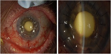
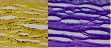
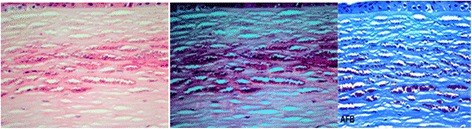
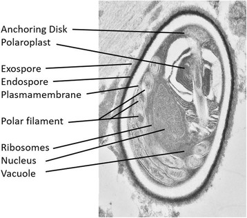
Results: A 69-year-old healthy male underwent penetrating keratoplasty for corneal scar secondary to herpes stromal keratitis. He presented with features of acute graft rejection 3 years later. After failure of medical management, a repeat full thickness keratoplasty was performed. Pathologic examination of the corneal specimen showed microsporidia. The patient then developed a chronic endophthalmitis, and a vitreous tap and injection followed by pars plana vitrectomy were performed. Pathologic examination of tissue showed microsporidia.
Conclusions: Microsporidia are being increasingly identified as the cause of stromal keratitis. This is the first report of microsporidial endophthalmitis in a patient without underlying systemic illness.
Figures
Similar articles
-
Case Report: Microsporidial Endophthalmitis after Penetrating Eye Trauma.Optom Vis Sci. 2022 Nov 1;99(11):830-832. doi: 10.1097/OPX.0000000000001951. Epub 2022 Oct 25. Optom Vis Sci. 2022. PMID: 36413632
-
Stromal Keratitis with Endophthalmitis Caused by Vittaforma Corneae in an Immunocompetent Patient: A Case Report.Ocul Immunol Inflamm. 2019;27(5):826-828. doi: 10.1080/09273948.2018.1455875. Epub 2018 Apr 19. Ocul Immunol Inflamm. 2019. PMID: 29672246
-
Is microsporidial keratitis an emerging cause of stromal keratitis? A case series study.BMC Ophthalmol. 2005 Aug 17;5:19. doi: 10.1186/1471-2415-5-19. BMC Ophthalmol. 2005. PMID: 16105181 Free PMC article.
-
Granulicatella Adiacens as an Unusual Cause of Microbial Keratitis and Endophthalmitis: A Case Series and Literature Review.Ocul Immunol Inflamm. 2022 Jul;30(5):1181-1185. doi: 10.1080/09273948.2020.1860233. Epub 2021 Jan 10. Ocul Immunol Inflamm. 2022. PMID: 33426987 Review.
-
Burkholderia gladioli keratitis associated with consecutive recurrent endophthalmitis.Cornea. 2002 Aug;21(6):602-3. doi: 10.1097/00003226-200208000-00014. Cornea. 2002. PMID: 12131039 Review.
Cited by
-
Practical Guidance for Clinical Microbiology Laboratories: Laboratory Diagnosis of Parasites from the Gastrointestinal Tract.Clin Microbiol Rev. 2017 Nov 15;31(1):e00025-17. doi: 10.1128/CMR.00025-17. Print 2018 Jan. Clin Microbiol Rev. 2017. PMID: 29142079 Free PMC article. Review.
-
Case Report: The Use of In Vivo Confocal Microscopy for Diagnosis and Monitoring in a Rare Case of Ancaliia algerae Microsporidial Keratitis in New South Wales, Australia.Am J Trop Med Hyg. 2023 Nov 20;110(1):87-89. doi: 10.4269/ajtmh.23-0474. Print 2024 Jan 3. Am J Trop Med Hyg. 2023. PMID: 37983904 Free PMC article.
-
Microsporidial stromal keratitis: treatment outcomes, clinical manifestations, confocal microscopy and histopathology findings: a retrospective observational study.BMJ Open Ophthalmol. 2024 Sep 16;9(1):e001581. doi: 10.1136/bmjophth-2023-001581. BMJ Open Ophthalmol. 2024. PMID: 39284677 Free PMC article.
-
Case Report: Ocular Microsporidiosis: Case in a Patient Returning from India and Review of the Literature.Am J Trop Med Hyg. 2018 Jul;99(1):90-93. doi: 10.4269/ajtmh.18-0015. Epub 2018 Apr 19. Am J Trop Med Hyg. 2018. PMID: 29692301 Free PMC article. Review.
-
A Narrative Review of Microsporidial Infections of the Cornea.Ophthalmol Ther. 2020 Jun;9(2):265-278. doi: 10.1007/s40123-020-00243-z. Epub 2020 Mar 10. Ophthalmol Ther. 2020. PMID: 32157613 Free PMC article. Review.
References
-
- Mietz H, Franzen C, Hoppe T, et al. Microsporidia-induced sclerouveitis with retinal detachment. Arch Ophthalmol. 2002;120(6):864–5. - PubMed
LinkOut - more resources
Full Text Sources
Other Literature Sources
Miscellaneous
