. 2016 Aug 16:6:31524.
doi: 10.1038/srep31524.

The ontogeny of Butyrophilin-like (Btnl) 1 and Btnl6 in murine small intestine

Affiliations

The ontogeny of Butyrophilin-like (Btnl) 1 and Btnl6 in murine small intestine

Cristina Lebrero-Fernández et al. Sci Rep. .

Abstract

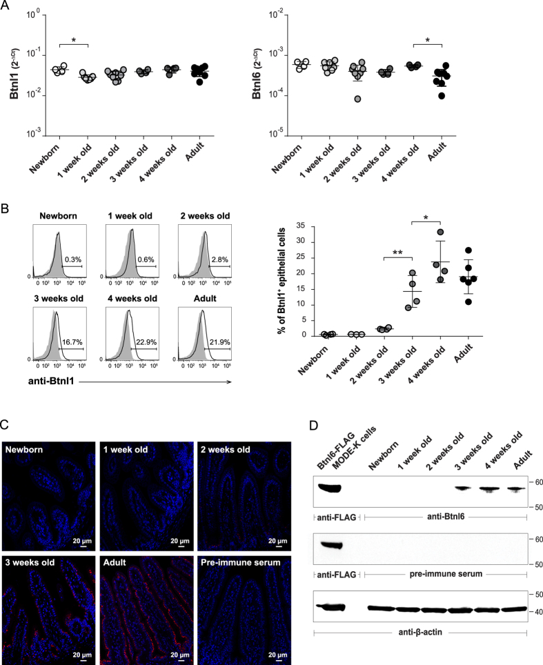
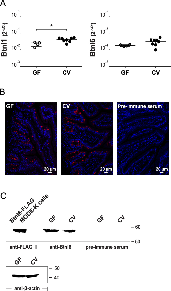
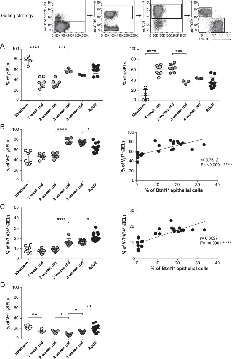
Murine Butyrophilin-like (Btnl) 1 and Btnl6 are primarily restricted to intestinal epithelium where they regulate the function of intraepithelial T lymphocytes. We recently demonstrated that Btnl1 and Btnl6 can form an intra-family heterocomplex and that the Btnl1-Btnl6 complex selectively expands Vγ7Vδ4 TCR IELs. To define the regulation of Btnl expression in the small intestine during ontogeny we examined the presence of Btnl1 and Btnl6 in the small bowel of newborn to 4-week-old mice. Although RNA expression of Btnl1 and Btnl6 was detected in the small intestine at day 0, Btnl1 and Btnl6 protein expression was substantially delayed and was not detectable in the intestinal epithelium until the mice reached 2-3 weeks of age. The markedly elevated Btnl protein level at week 3 coincided with a significant increase of γδ TCR IELs, particularly those bearing the Vγ7Vδ4 receptor. This was not dependent on gut microbial colonization as mice housed in germ-free conditions had normal Btnl protein levels. Taken together, our data show that the expression of Btnl1 and Btnl6 is delayed in the murine neonatal gut and that the appearance of the Btnl1 and Btnl6 proteins in the intestinal mucosa associates with the expansion of Vγ7Vδ4 TCR IELs.


Figures

Figure 1
Figure 1. Btnl1 and Btnl6 expression in murine intestine during ontogeny.
Expression of Btnl1 and Btnl6 genes (A), and Btnl1 (B,C) and Btnl6 proteins (D) was examined in small intestinal tissue of newborn (day 0), 1, 2, 3, and 4-week-old and adult C57BL/6 mice. (A) Btnl1 and Btnl6 gene expression was assessed by qPCR, run in duplicates, and normalized against β-actin. (B) Murine intestinal epithelial cells, gated on CD45 epithelial cells and 7AAD negative cells to exclude non-viable cells, were stained with anti-Btnl1 rabbit polyclonal antiserum (solid black line) or pre-immune serum (shaded histogram) which served as a negative control. Representative histogram for each time-point is shown. 4–8 mice/time-point were analyzed and One-Way ANOVA followed by Holm-Sidak´s multiple comparisons test was used for statistical analysis (*P ≤ 0.05, **P ≤ 0.01, ***P ≤ 0.001 and ****P ≤ 0.0001) (A,B). (C) Small intestinal sections were immunostained with anti-Btnl1 rabbit polyclonal antiserum (red) and counterstained with DAPI (blue) to visualize nuclei. No staining was detected using pre-immune serum. Original magnification 20x. Four mice were stained for each time-point and representative stainings are shown. (D) Isolated iECs from small intestinal tissue of C57BL/6 mice (20 μg) were analyzed for Btnl6 protein expression. Lysates from MODE-K cells transfected with FLAG-tagged Btnl6 cDNA pMX-IRES-GFP served as a positive control. The predicted protein migrating under reducing conditions at the theoretical molecular weight of ~59 kDa for FLAG-tagged Btnl6 and ~58 kDa for non-tagged Btnl6 was detected with anti-FLAG antibody or Btnl6-specific polyclonal antibody. No bands were detected on gels immunoblotted with pre-immune serum. The β-actin immunoblot acts as a loading control. Data are representative of four experiments.
Figure 2
Figure 2. Btnl1 and Btnl6 expression in germ-free mice.
Expression of Btnl1 and Btnl6 transcripts (A), and Btnl1 (B) and Btnl6 proteins (C) was analyzed in small intestinal tissue of GF and CV adult C57BL/6 mice. (A) Btnl1 and Btnl6 gene expression was assessed by qPCR, run in duplicates, and normalized against β-actin. 4–8 mice/group were included and unpaired two-tailed t test was used for statistical analysis (*P ≤ 0.05, **P ≤ 0.01, ***P ≤ 0.001 and ****P ≤ 0.0001). (B) Small intestinal sections were immunostained with anti-Btnl1 rabbit polyclonal antiserum (red) and counterstained with DAPI (blue) to visualize nuclei. No staining was detected using pre-immune serum. Original magnification 20x. Four mice were stained for each group and representative stainings are shown. (C) Lysates from small intestinal tissue of GF and CV adult C57BL/6 mice (20 μg) were analyzed for Btnl6 protein expression. Lysates from MODE-K cells transfected with FLAG-tagged Btnl6 cDNA pMX-IRES-GFP served as a positive control. The predicted protein migrating under reducing conditions at the theoretical molecular weight of ~59 kDa for FLAG-tagged Btnl6 and ~58 kDa for non-tagged Btnl6 was detected with anti-FLAG antibody or Btnl6-specific polyclonal antibody. No bands were detected on gels immunoblotted with pre-immune serum. The β-actin immunoblot acts as a loading control. Data are representative of two experiments. GF: germ-free; CV: conventional.
Figure 3
Figure 3. Btnl1 expression correlates with the presence of IELs bearing the Vγ7Vδ4 TCR.
Small intestinal IELs from newborn (day 0), 1, 2, 3, and 4-week-old and adult C57BL/6 mice were analyzed for the expression of αβ and γδ TCR (A), and the γδ TCR IELs for the expression of Vγ7, Vγ1, and Vδ4 chains (B–D). 3–11 mice/group (A) and 7–16 mice/group (B–D) were analyzed and One-Way ANOVA followed by Holm-Sidak´s multiple comparisons test was used for statistical analysis. Correlation between Btnl1 expression and the percentage of Vγ7 TCR IELs (B) or Vγ7Vδ4 TCR IELs (C) during the mouse ontogeny was determined using the Pearson correlation test (*P ≤ 0.05, **P ≤ 0.01, ***P ≤ 0.001 and ****P ≤ 0.0001). Each dot in the correlation analysis in Fig. 3B,C represents % of Btnl1+ epithelial cells vs % of Vγ7 (Vδ4) TCR IELs of one mouse at a particular time-point. Newborn −8-week-old mice were used in this correlation analysis.

References

    1. Nguyen T., Liu X. K., Zhang Y. & Dong C. BTNL2, a butyrophilin-like molecule that functions to inhibit T cell activation. J Immunol. 176, 7354–7360, 176/12/7354 (2006). - PMC - PubMed
    1. Yamazaki T. et al.. A butyrophilin family member critically inhibits T cell activation. J Immunol. 185, 5907–5914, 10.4049/jimmunol.1000835 (2010). - DOI - PubMed
    1. Arnett H. A. et al.. BTNL2, a butyrophilin/B7-like molecule, is a negative costimulatory molecule modulated in intestinal inflammation. J Immunol. 178, 1523–1533, 178/3/1523 (2007). - PubMed
    1. Chapoval A. I. et al.. BTNL8, a butyrophilin-like molecule that costimulates the primary immune response. Mol Immunol 56, 819–828, 10.1016/j.molimm.2013.08.003 (2013). - DOI - PubMed
    1. Bas A. et al.. Butyrophilin-like 1 encodes an enterocyte protein that selectively regulates functional interactions with T lymphocytes. Proc Natl Acad Sci USA 108, 4376–4381, 10.1073/pnas.1010647108 (2011). - DOI - PMC - PubMed

Publication types

LinkOut - more resources