. 2016 Aug 15;17(1):634.
doi: 10.1186/s12864-016-2841-9.

Genomic and expression analyses of Tursiops truncatus T cell receptor gamma (TRG) and alpha/delta (TRA/TRD) loci reveal a similar basic public γδ repertoire in dolphin and human

Affiliations

Genomic and expression analyses of Tursiops truncatus T cell receptor gamma (TRG) and alpha/delta (TRA/TRD) loci reveal a similar basic public γδ repertoire in dolphin and human

Giovanna Linguiti et al. BMC Genomics. .

Erratum in

Abstract

Background: The bottlenose dolphin (Tursiops truncatus) is a mammal that belongs to the Cetartiodactyla and have lived in marine ecosystems for nearly 60 millions years. Despite its popularity, our knowledge about its adaptive immunity and evolution is very limited. Furthermore, nothing is known about the genomics and evolution of dolphin antigen receptor immunity.

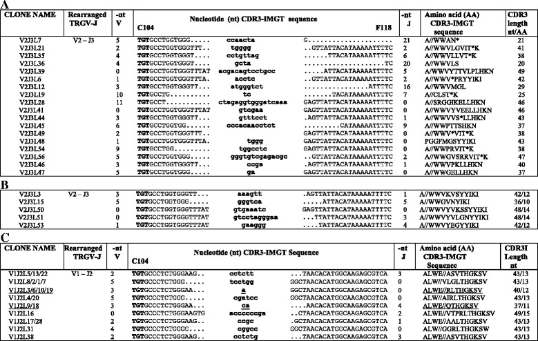
Results: Here we report a evolutionary and expression study of Tursiops truncatus T cell receptor gamma (TRG) and alpha/delta (TRA/TRD) genes. We have identified in silico the TRG and TRA/TRD genes and analyzed the relevant mature transcripts in blood and in skin from four subjects. The dolphin TRG locus is the smallest and simplest of all mammalian loci as yet studied. It shows a genomic organization comprising two variable (V1 and V2), three joining (J1, J2 and J3) and a single constant (C), genes. Despite the fragmented nature of the genome assemblies, we deduced the TRA/TRD locus organization, with the recent TRDV1 subgroup genes duplications, as it is expected in artiodactyls. Expression analysis from blood of a subject allowed us to assign unambiguously eight TRAV genes to those annotated in the genomic sequence and to twelve new genes, belonging to five different subgroups. All transcripts were productive and no relevant biases towards TRAV-J rearrangements are observed. Blood and skin from four unrelated subjects expression data provide evidence for an unusual ratio of productive/unproductive transcripts which arise from the TRG V-J gene rearrangement and for a "public" gamma delta TR repertoire. The productive cDNA sequences, shared both in the same and in different individuals, include biases of the TRGV1 and TRGJ2 genes. The high frequency of TRGV1-J2/TRDV1- D1-J4 productive rearrangements in dolphins may represent an interesting oligo-clonal population comparable to that found in human with the TRGV9- JP/TRDV2-D-J T cells and in primates.

Conclusions: Although the features of the TRG and TRA/TRD loci organization reflect those of the so far examined artiodactyls, genomic results highlight in dolphin an unusually simple TRG locus. The cDNA analysis reveal productive TRA/TRD transcripts and unusual ratios of productive/unproductive TRG transcripts. Comparing multiple different individuals, evidence is found for a "public" gamma delta TCR repertoire thus suggesting that in dolphins as in human the gamma delta TCR repertoire is accompanied by selection for public gamma chain.

Keywords: Dolphin genome; Expression analysis; IMGT; T cell receptor; TRA/TRD locus; TRAV and TRDV genes; TRG locus; TRGJ and TRGC genes; TRGV.


Figures

Fig. 1
Fig. 1
Schematic representation of the genomic organization of the dolphin TRG locus as deduced from the genome assembly Ttru_1.4. a The diagram shows the position of all V, J and C genes according to the IMGT® nomenclature. The AMPH (located 15.5 kb upstream of the first TRGV gene) and the STARD3NL (located 11.6 kb downstream of the unique TRGC gene, in the inverted transcriptional orientation) genes at the 5′ end and at the 3′ end, respectively of the TRG locus are shown. Boxes representing genes are not to scale. Exons are not shown. b Description of the TRGV, TRGJ, and TRGC genes in the dolphin genome. The position of all genes in the JH473572.1 scaffold and their classification are reported. The bottlenose dolphin (Tursiops truncatus) TRG genes and alleles have been approved by the WHO/IUIS/IMGT nomenclature subcommittee for IG, TR and MH [66, 67]. a From L-PART1 to 3′ end of V-REGION. c IMGT Protein display of the dolphin, human and sheep TRGC genes. The description of the strands and loops is according to the IMGT unique numbering for C-DOMAIN [68]. The extracellular region is shown with black letters, the connecting region is in orange, the transmembrane-region is in purple, and the cytoplasmic region is in pink. 1st-CYS C23, CONSERVED-TRP W41 and hydrophobic AA L89 and 2nd-CYS C104 are colored (IMGT color menu) and in bold
Fig. 2
Fig. 2
Schematic representation of the genomic organization of the bottlenose dolphin (Tursiops truncatus) TRA/TRD locus as deduced from the genome assembly Ttru_1.4. a The diagram shows the position of the TRDV, TRDD, TRDJ and TRDC genes and of the TRAV, TRAJ and TRAC genes according to the IMGT® nomenclature. The retrieval of the relevant contigs from the GenBank (JH484271.1 and JH481615.1) and Ensembl databases (S_742; S_97; S_89; S_123 and S_112178), has allowed the identification, starting from the 5′ end of the locus, of 16 TRAV (including 5 pseudogenes), 5 TRDV (including 1 pseudogene), 2 TRDD, 4 TRDJ (including one ORF and 1 pseudogene), 1 TRDC, 59 TRAJ and 1 TRAC genes, located in a genomic region spanning approximatively 450 Kb (Additional file 5). Boxes representing genes are not to scale. Exons are not shown. Arrow head indicates the transcriptional orientation of the TRDV4 gene. The arrows above the line of the TRAJ genes indicate the 70 kb region that has been magnified in the lower part of the figure. b IMGT Protein display for the dolphin TRAV and TRDV functional genes. The description of the strands and loops is according to the IMGT unique numbering for V-REGION [25]. 1st-CYS C23, CONSERVED-TRP W41, hydrophobic L89 and 2nd-CYS C104 are colored (IMGT color menu) and in bold
Fig. 3
Fig. 3
IMGT Protein display of the TRA (a) and TRD (b) cDNA clones. The TRAV and TRAJ genes are listed respectively at the left and the right of the figure. Leader region (L-Region), complementary determining regions (CDR-IMGT) and framework regions (FR-IMGT) are also indicated, according to the IMGT unique numbering for V-REGION [25]. The TRAV allele amino acid changes, if any are green boxed. The name of the clones are also reported. The TRDV and TRDJ genes are listed respectively at the left and the right of the figure. In (b), for TRDV2, TRDV1-1 N and TRDV1-1, the CDR-IMGT lengths are of [8.3.13], [7.3.20] and [7.3.21], respectively. 5R1D15 clone (TRDV2) lacks the TRDD gene. 5R1D3 clone (TRDV1-1) has a new TRDD gene (TRDD7S1) with respect to the available genomic sequence. The name of the clones are also reported
Fig. 4
Fig. 4
CDR3-IMGT nucleotide sequences retrieved from the cDNA clones with productive (a) and unproductive (b) rearrangements. Nucleotide sequences are shown from codon 104 (2nd-CYS) to codon 118 (J-PHE) (a and b). N-nucleotides added by the deoxynucleotidyltransferase terminal (DNTT, TdT) are indicated in lower cases. Numbers in the left and right columns indicate the number of nucleotides that are trimmed from the 3′V-REGION and 5′J-REGION, respectively; the germline region of the TRGV and TRGJ genes coincides with 0 in the nt V and nt J columns, respectively. In A, clonotypes with the same CDR3-IMGT nucleotide sequence deriving from two or more animals (letters M, L, K and C) are underlined. A shared clonotype (AA) between individuals has per definition a given V and J gene and allele and a given AA sequence for the junction. Three individuals (M, K and C) share the same CDR3 (AA) sequence with the V1-J2 rearrangement (RTV1M1/C1/K2/K3) however the junction in K3 differs from the junction in the other shared clonotypes by a nucleotide difference in the 5′J-REGION which may represent an allele of the TRGJ2 gene. Similarly, for the K and M shared clonotypes with a V1-J3 rearrangement (RTV1K7/5RV1M1), the junction in M1 differs from the junction in K7 by a nucleotide in the 3′V-REGION, which may represent an allele of the TRGV1 gene. This has been described as “convergent recombination” in which a given “public” TR amino acid sequence may be encoded by different nucleotide sequences both within the same and in different individuals [28]. In B, unproductive rearrangements (non-redundant out-of-frame clonotypes column of Table 2) for the presence of a stop codon (*) and for frameshifts in the CDR3, are indicated
Fig. 5
Fig. 5
CDR3 nucleotide sequences retrieved from genomic rearranged clones. Nucleotide sequences are shown from codon 104 (2nd-CYS) to codon 118 (J-PHE) (a and b) and from codon 104 (2nd-CYS) to codon 115 (c). They are grouped on the basis of their rearrangement (a) and (b) TRGV2-TRGJ3 or (c) TRGV1-TRGJ2. N-nucleotides added by TdT are indicated in lower cases. Numbers in the left and right columns indicate the number of nucleotides that are trimmed from the 3'V-REGION and 5'J-REGION, respectively. The germline region of the TRGV and TRGJ genes coincides with 0 in the nt V and nt J columns, respectively. Clones with the same CDR3-IMGT nucleotide sequence deriving from two or more animals (L, K and C) are underlined (see also Fig. 4). In (a), unproductive rearrangements for the presence of a stop codon (*) and for frameshifts in the CDR3, are indicated
Fig. 6
Fig. 6
Computationally inferred interaction between RTV1M1 V-gamma domain (TRGV1 – J2) and 5R1D8 V-delta domain (TRDV1-1 N – J4) (a) and between 5RV1M1 V-gamma domain (TRGV1 – J3) and 5R1D15 V-delta domain (TRDV2 – J4) (b) cDNA clonotypes. In RTV1M1 and 5RV1M1 V-gamma domain CDR-IMGT are blue-green-green; in 5R1D8 and 5R1D15 V-delta domain CDR-IMGT are red-orange-purple. IMGT Collier de Perles of RTV1M1/5R1D8 and 5RV1M1/5R1D15 clones are shown [25, 65]. The protein complex interface were computed by the online tool PDBePISA at the EBI server. (http://www.ebi.ac.uk/msd-srv/prot_int/) and visualized by UCSF Chimera tool (http://www.cgl.ucsf.edu/chimera/) (Additional file 14)

Similar articles

Cited by

References

    1. Gingerich PD, Haq M, Zalmout IS, Khan IH, Malkani MS. Origin of whales from early artiodactyls: hands and feet of Eocene Protocetidae from Pakistan. Science. 2001;293(5538):2239–2242. doi: 10.1126/science.1063902. - DOI - PubMed
    1. Price SA, Bininda-Emonds OR, Gittleman JL. A complete phylogeny of the whales, dolphins and even-toed hoofed mammals (Cetartiodactyla) Biol Rev Camb Philos Soc. 2005;80(3):445–473. doi: 10.1017/S1464793105006743. - DOI - PubMed
    1. Boisserie JR, Lihoreau F, Brunet M. The position of Hippopotamidae within Cetartiodactyla. Proc Natl Acad Sci U S A. 2005;102(5):1537–1541. doi: 10.1073/pnas.0409518102. - DOI - PMC - PubMed
    1. Bininda-Emonds OR, Cardillo M, Jones KE, MacPhee RD, Beck RM, Grenyer R, Price SA, Vos RA, Gittleman JL, Purvis A. The delayed rise of present-day mammals. Nature. 2007;446(7135):507–512. doi: 10.1038/nature05634. - DOI - PubMed
    1. Geisler JH, Theodor JM. Hippopotamus and whale phylogeny. Nature. 2009;458(7236):E1–E4. doi: 10.1038/nature07776. - DOI - PubMed

Publication types

MeSH terms

Substances