. 2016 Sep 23;291(39):20295-20302.
doi: 10.1074/jbc.M116.729731. Epub 2016 Aug 15.

Activated G Protein Gαs Samples Multiple Endomembrane Compartments

Affiliations

Activated G Protein Gαs Samples Multiple Endomembrane Compartments

Brent R Martin et al. J Biol Chem. .

Abstract

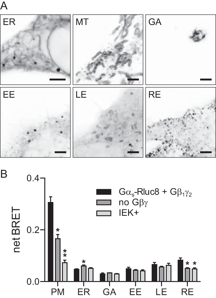
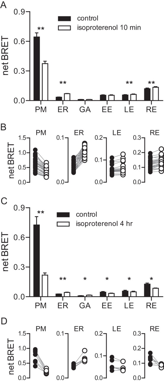
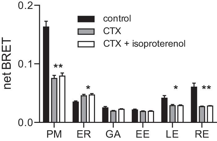
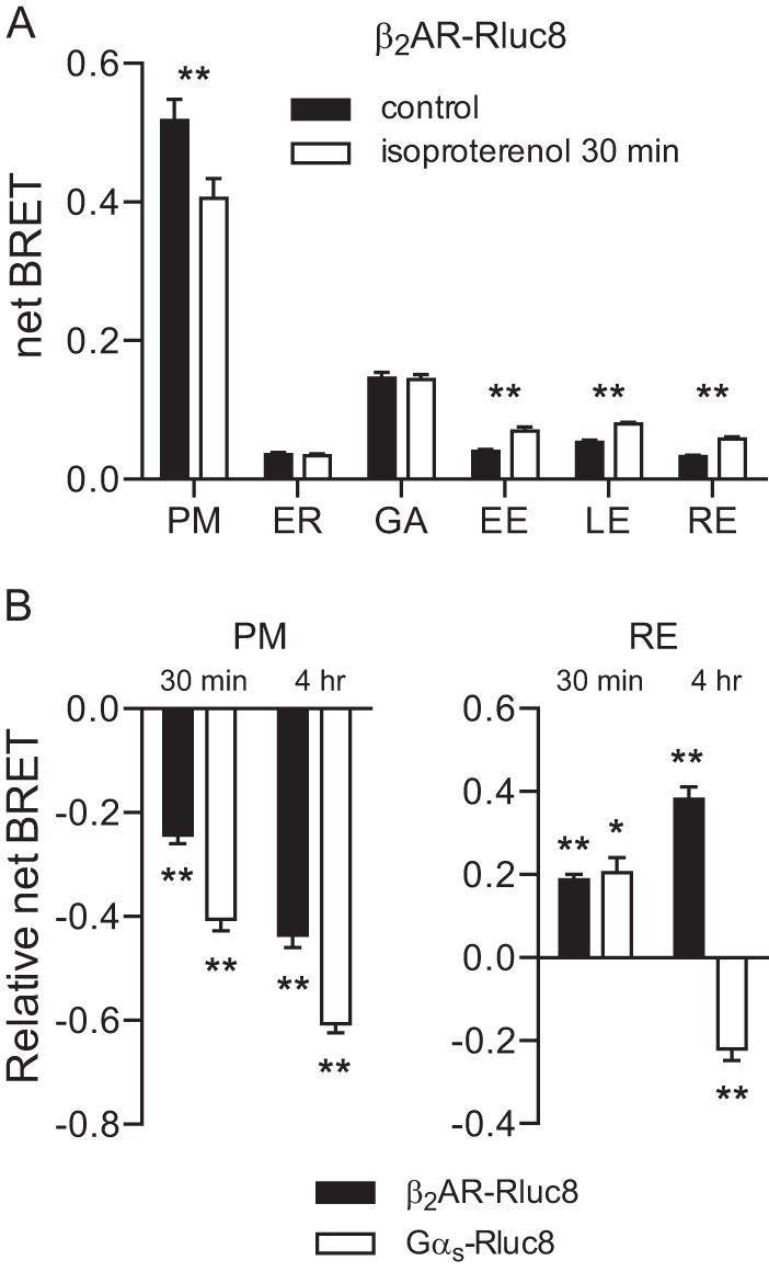
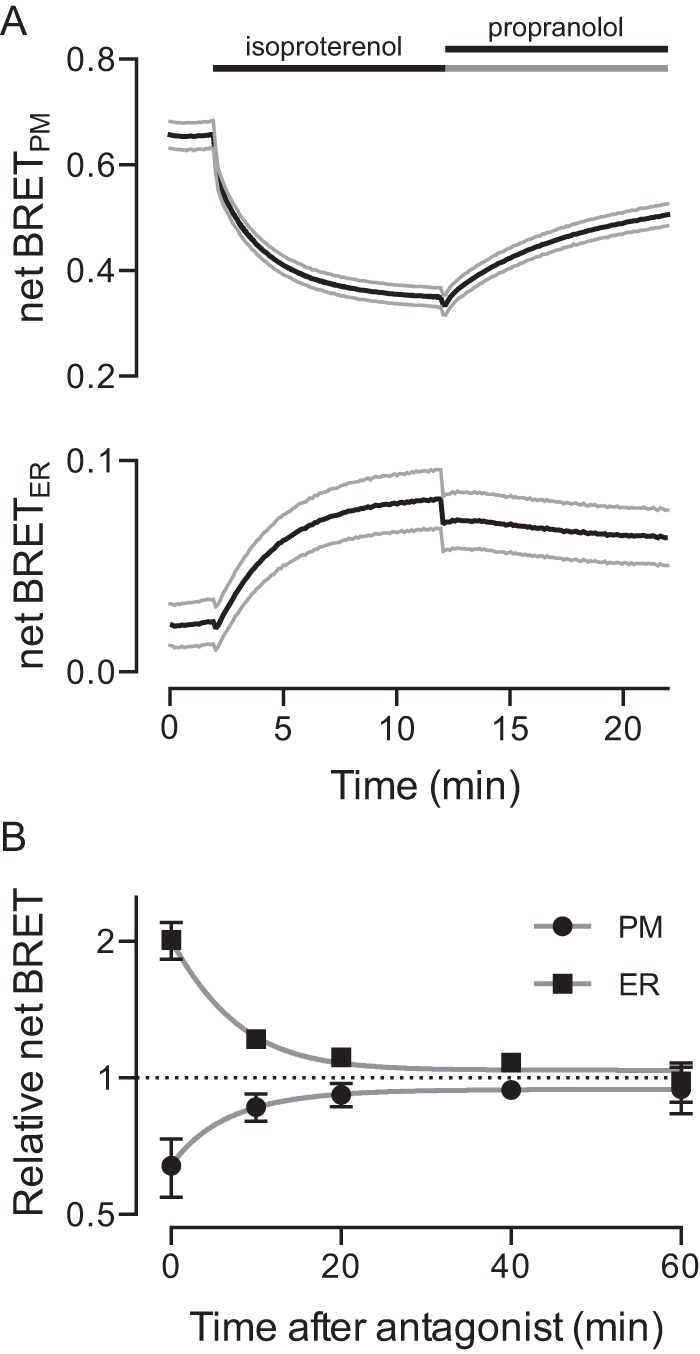
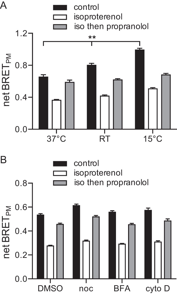
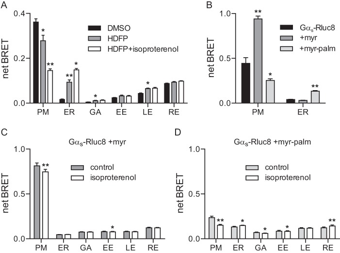
Heterotrimeric G proteins are localized to the plasma membrane where they transduce extracellular signals to intracellular effectors. G proteins also act at intracellular locations, and can translocate between cellular compartments. For example, Gαs can leave the plasma membrane and move to the cell interior after activation. However, the mechanism of Gαs translocation and its intracellular destination are not known. Here we use bioluminescence resonance energy transfer (BRET) to show that after activation, Gαs rapidly associates with the endoplasmic reticulum, mitochondria, and endosomes, consistent with indiscriminate sampling of intracellular membranes from the cytosol rather than transport via a specific vesicular pathway. The primary source of Gαs for endosomal compartments is constitutive endocytosis rather than activity-dependent internalization. Recycling of Gαs to the plasma membrane is complete 25 min after stimulation is discontinued. We also show that an acylation-deacylation cycle is important for the steady-state localization of Gαs at the plasma membrane, but our results do not support a role for deacylation in activity-dependent Gαs internalization.

Keywords: G protein-coupled receptor (GPCR); endosome; endosomes; heterotrimeric G protein; intracellular trafficking; lipase; protein acylation; protein palmitoylation.


Figures

FIGURE 1.
FIGURE 1.
BRET acceptors targeted to membrane compartments indicate the subcellular localization of Gαs-Rluc8. A, confocal images of HEK 293 cells expressing Venus-tagged markers of the endoplasmic reticulum (ER), mitochondria (MT), Golgi apparatus (GA), early endosomes (EE), late endosomes (LE), and recycling endosomes (RE). Approximately half of each panel is occupied by the cell interior. Scale bars, 3 μm. B, net BRET between Gαs-Rluc8 (with and without exogenous Gβ1 and Gγ2) and Gαs-Rluc8 IEK+ and acceptors localized to the plasma membrane (PM) or various intracellular compartments. Bars, mean ± S.E. of n = 3–5 independent experiments; **, p < 0.001; *, p < 0.05 (one-way ANOVA versus control, Dunnett's multiple comparisons).
FIGURE 2.
FIGURE 2.
Activity-dependent redistribution of Gαs-Rluc8. A, net BRET between Gαs-Rluc8 expressed with unlabeled Gβ1, Gγ2, β2AR, and acceptors localized to membrane compartments in control cells and in cells stimulated for 10 min with the β2AR agonist isoproterenol (10 μm). PM, plasma membrane; ER, endoplasmic reticulum; GA, Golgi apparatus; EE, early endosomes; LE, late endosomes; RE, recycling endosomes. Bars, mean ± S.E. of n = 18–25 independent experiments; **, p < 0.001 (paired t test). B, data points from individual experiments shown in panel A. C, a similar experiment to that shown in panel A with cells stimulated for 4 h with isoproterenol. Bars, mean ± S.E. of n = 7 independent experiments; **, p < 0.001; *, p < 0.05 (paired t test). D, data points from individual experiments shown in panel C.
FIGURE 3.
FIGURE 3.
Cholera toxin redistributes Gαs-Rluc8. Shown is net BRET between Gαs-Rluc8 and membrane markers in control cells, in cells treated for 2–3 h with cholera toxin (CTX; 10 μg ml−1), and in cells treated with cholera toxin and then isoproterenol for 10 min. PM, plasma membrane; ER, endoplasmic reticulum; GA, Golgi apparatus; EE, early endosomes; LE, late endosomes; RE, recycling endosomes. Bars, mean ± S.E. of n = 4 independent experiments; **, p < 0.001; *, p < 0.05 (one-way ANOVA comparing CTX or CTX + isoproterenol to control, Tukey's multiple comparisons). No significant difference was observed between CTX and CTX + isoproterenol.
FIGURE 4.
FIGURE 4.
Activity-dependent internalization of β2AR-Rluc8 and Gαs-Rluc8. A, net BRET between β2AR-Rluc8 and acceptors localized to membrane compartments in control cells and in cells stimulated for 30 min with isoproterenol. PM, plasma membrane; ER, endoplasmic reticulum; GA, Golgi apparatus; EE, early endosomes; LE, late endosomes; RE, recycling endosomes. Bars, mean ± S.E. of n = 6 independent experiments; **, p < 0.001 (paired t test). B, relative net BRET compared with vehicle-treated controls between β2AR-Rluc8 or Gαs-Rluc8 and acceptors localized to the plasma membrane or recycling endosomes in cells stimulated for 30 min or 4 h with isoproterenol. Bars, mean ± S.E. of n = 9 independent experiments; **, p < 0.001; *, p < 0.05 (one-way ANOVA versus vehicle, Dunnett's multiple comparisons).
FIGURE 5.
FIGURE 5.
The time course of Gαs-Rluc8 internalization and recycling. A, net BRET at the plasma membrane (PM, top) and the endoplasmic reticulum (ER, bottom) during sequential addition of isoproterenol (10 μm) and the β2AR antagonist propranolol (20 μm). Lines, mean ± S.E. of n = 13 experiments. B, normalized net BRET (plotted on a log 2 scale) at the plasma membrane and the endoplasmic reticulum in cells that were sequentially exposed to isoproterenol (10 μm) and the β2AR antagonist alprenolol (20 μm). Symbols, mean ± S.E. of n = 4 independent experiments. Lines, least squares fits to a single exponential function.
FIGURE 6.
FIGURE 6.
The sensitivity of Gαs-Rluc8 internalization and recycling to temperature and disruption of microtubules, actin, and the Golgi apparatus. A, net BRET at the plasma membrane in cells treated with vehicle (control), isoproterenol for 10 min (isoproterenol), or isoproterenol followed by propranolol for 15 min (iso then propranolol) in cells maintained throughout at 37 °C, room temperature (RT, ∼25 °C), and 15 °C. Bars, mean ± S.E. of n = 9 independent experiments; **, p < 0.001; (one-way ANOVA, Tukey's multiple comparisons). The percentages of isoproterenol-induced decreases were not significantly different between any of the groups. The percentages of propranolol-induced recoveries were significantly different between all three groups (p < 0.001; one-way ANOVA, Tukey's multiple comparisons). B, similar experiments to those shown in panel A, all performed at room temperature, in cells treated with vehicle (DMSO; n = 28), nocodazole (noc; 10 μm for 1 h; n = 12), brefeldin A (BFA; 10 μg ml−1 for 4 h; n = 13), and cytochalasin D (cyto D; 1 μm for 4 h; n = 13). Bars, mean ± S.E. The percentages of isoproterenol-induced decreases were not significantly different between any of the groups. The percentages of propranolol-induced recoveries were significantly different from DMSO only for brefeldin A (p < 0.001; one-way ANOVA, Dunnett's multiple comparisons).
FIGURE 7.
FIGURE 7.
The role of palmitate cycling in constitutive localization and activity-dependent redistribution of Gαs-Rluc8. A, net BRET at the plasma membrane and the endoplasmic reticulum generated by Gαs-Rluc8, Gαs-Rluc8 +myr, or Gαs-Rluc8 +myr-palm. PM, plasma membrane; ER, endoplasmic reticulum; GA, Golgi apparatus; EE, early endosomes; LE, late endosomes; RE, recycling endosomes. Bars, mean ± S.E. of n = 5 independent experiments; **, p < 0.001; *, p < 0.05 (one-way ANOVA versuss-Rluc8, Dunnett's multiple comparisons). B, net BRET between Gαs-Rluc8 expressed with unlabeled Gβ1, Gγ2, β2AR, and acceptors localized to membrane compartments in control cells, in cells treated with HDFP (20 μm) for 4 h, and in HDFP-treated cells stimulated for 10 min with isoproterenol (10 μm). Bars, mean ± S.E. of n = 4 independent experiments; **, p < 0.001; *, p < 0.05 (one-way ANOVA versus DMSO (for HDFP) or versus HDFP (for HDFP + isoproterenol), Tukey's multiple comparisons). C and D, net BRET between Gαs-Rluc8 +myr (C) or Gαs-Rluc8 +myr-palm (D) and acceptors localized to membrane compartments in control cells and in cells stimulated for 10 min with isoproterenol. Bars, mean ± S.E. of n = 8–10 independent experiments; **, p < 0.001; *, p < 0.05 (paired t test).

References

    1. Gilman A. G. (1987) G proteins: transducers of receptor-generated signals. Annu. Rev. Biochem. 56, 615–649 - PubMed
    1. Hewavitharana T., and Wedegaertner P. B. (2012) Non-canonical signaling and localizations of heterotrimeric G proteins. Cell. Signal. 24, 25–34 - PMC - PubMed
    1. Slepak V. Z., and Hurley J. B. (2008) Mechanism of light-induced translocation of arrestin and transducin in photoreceptors: interaction-restricted diffusion. IUBMB Life 60, 2–9 - PMC - PubMed
    1. Wedegaertner P. B. (2012) G protein trafficking. Subcell Biochem. 63, 193–223 - PMC - PubMed
    1. Saini D. K., Chisari M., and Gautam N. (2009) Shuttling and translocation of heterotrimeric G proteins and Ras. Trends Pharmacol. Sci. 30, 278–286 - PMC - PubMed

Publication types

MeSH terms

Substances

LinkOut - more resources