. 2016 Oct 13;36(21):2668-2680.
doi: 10.1128/MCB.00160-16. Print 2016 Nov 1.

BAR Domain-Containing FAM92 Proteins Interact with Chibby1 To Facilitate Ciliogenesis

Affiliations

BAR Domain-Containing FAM92 Proteins Interact with Chibby1 To Facilitate Ciliogenesis

Feng-Qian Li et al. Mol Cell Biol. .

Abstract

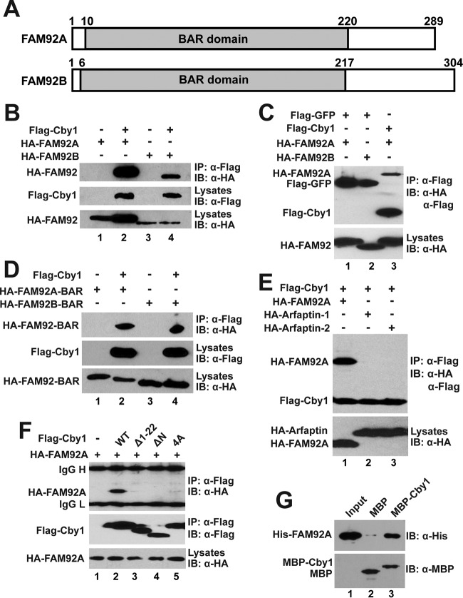
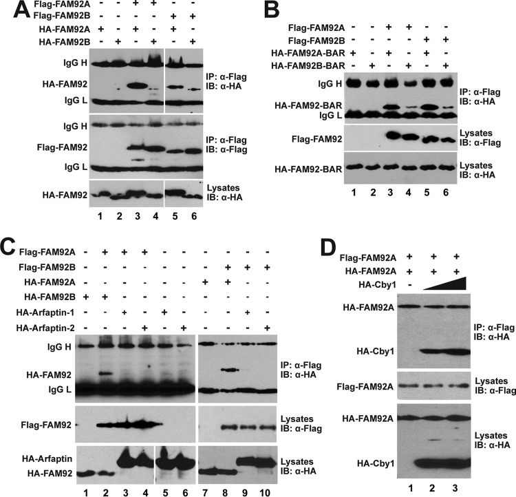
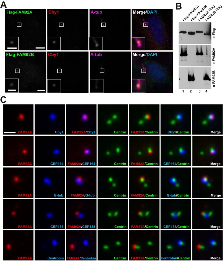
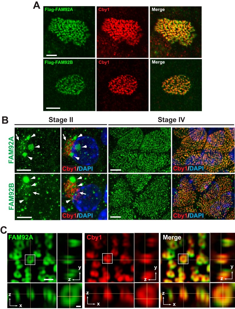
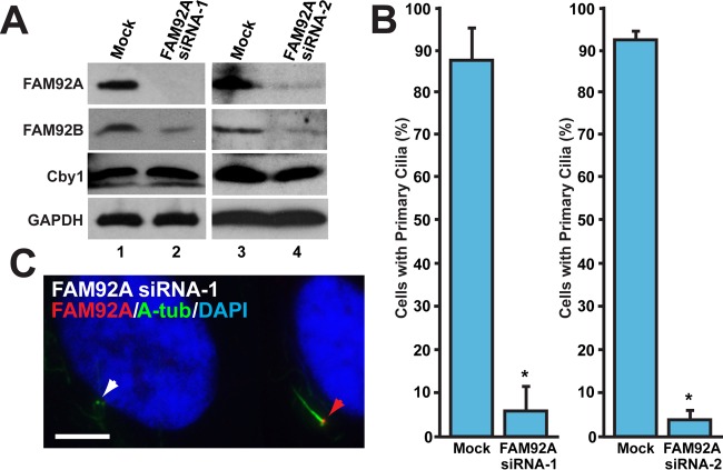
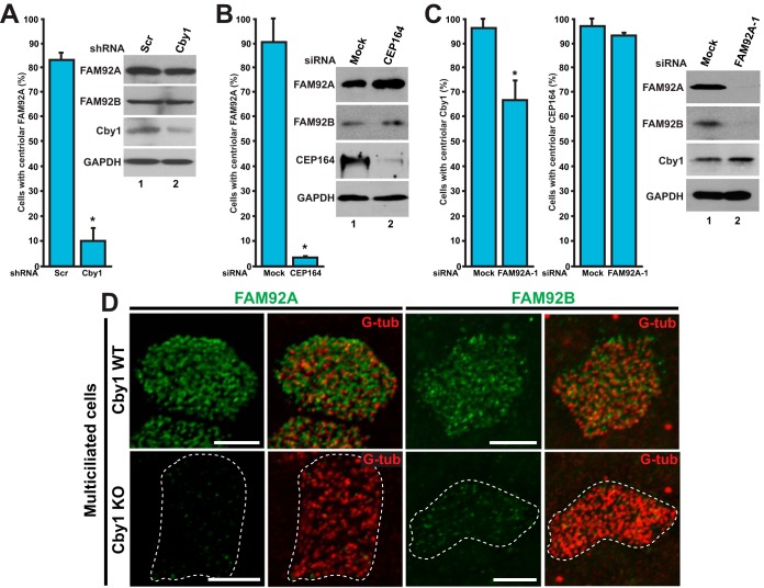
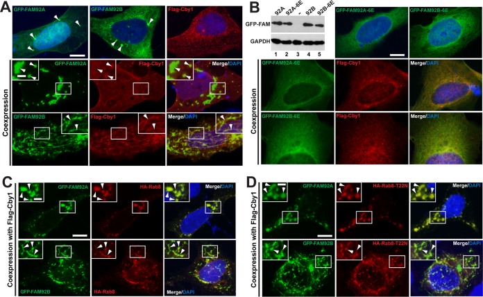
Chibby1 (Cby1) is a small, conserved coiled-coil protein that localizes to centrioles/basal bodies and plays a crucial role in the formation and function of cilia. During early stages of ciliogenesis, Cby1 is required for the efficient recruitment of small vesicles at the distal end of centrioles to facilitate basal body docking to the plasma membrane. Here, we identified family with sequence similarity 92, member A (FAM92A) and FAM92B, which harbor predicted lipid-binding BAR domains, as novel Cby1-interacting partners using tandem affinity purification and mass spectrometry. We found that in cultured cell lines, FAM92A colocalizes with Cby1 at the centrioles/basal bodies of primary cilia, while FAM92B is undetectable. In airway multiciliated cells, both FAM92A and -92B colocalize with Cby1 at the base of cilia. Notably, the centriolar localization of FAM92A and -92B depends largely on Cby1. Knockdown of FAM92A in RPE1 cells impairs ciliogenesis. Consistent with the membrane-remodeling properties of BAR domains, FAM92A and -92B in cooperation with Cby1 induce deformed membrane-like structures containing the small GTPase Rab8 in cultured cells. Our results therefore suggest that FAM92 proteins interact with Cby1 to promote ciliogenesis via regulation of membrane-remodeling processes.


Figures

FIG 1
FIG 1
Interactions between Cby1 and FAM92 proteins. (A) Schematic representation of the domain structures of human FAM92A and -92B. The conserved BAR domains are shaded with the amino acid positions indicated above. (B to F) HEK293T cells were transfected with the indicated plasmids, and cell lysates were immunoprecipitated with Flag antibody and detected with HA antibody. Cell lysates and immunoprecipitates were also probed with the indicated HA and Flag antibodies to confirm stable protein expression. (B) FAM92A and -92B interact with Cby1. (C) FAM92 proteins specifically bind to Cby1 but not to GFP. (D) The BAR domains of FAM92A and -92B are sufficient for Cby1 binding. (E) Cby1 does not interact with the BAR domain-containing proteins arfaptin-1 and arfaptin-2. (F) Both the N- and C-terminal regions of Cby1 are involved in FAM92 binding. (G) FAM92A directly binds to Cby1. Bacterially produced MBP or an MBP-Cby1 fusion was incubated with His-tagged FAM92A. The protein complex was then pulled down with amylose resin and subjected to Western blotting with FAM92A antibody. The input lane was loaded with 1/10 of the amount of His-FAM92A used in the binding reaction mixture. Western blotting with MBP antibody indicated that similar amounts of MBP and MBP-Cby1 were pulled down by amylose resin. IB, immunoblotting.
FIG 2
FIG 2
FAM92A and -92B homo- and heterodimerize through their BAR domains. (A to C) HEK293T cells were cotransfected with the indicated Flag-FAM92 and HA-FAM92 (A), HA-FAM92 BAR domain (B), or HA-arfaptin (C) expression constructs for co-IP assays. IgG H and IgG L denote IgG heavy and light chains, respectively. (D) Overexpression of Cby1 does not interfere with FAM92A homodimer formation. HEK293T cells were cotransfected with constant amounts of Flag-FAM92A and HA-FAM92A and increasing amounts of HA-Cby1, and cell lysates were immunoprecipitated with Flag antibody and detected with HA antibody.
FIG 3
FIG 3
FAM92 proteins colocalize with Cby1 at mother centrioles/basal bodies. (A) RPE1 cells were infected with lentiviruses expressing Flag-tagged FAM92A or -92B and fixed after 48 h of serum starvation to induce ciliogenesis. These cells were triple stained with antibodies against the Flag tag, Cby1, and the centriolar/ciliary axoneme marker A-tub. Merged images are shown with DAPI nuclear stain. The insets show enlarged views of the boxed areas. Bars, 10 μm; bars in the insets, 3 μm. (B) Specificity of the FAM92A and -92B antibodies. Cell lysates were prepared from HEK293T cells expressing Flag-tagged FAM92A or -92B and subjected to Western blotting with FAM92A or -92B antibody as shown. Both N- and C-terminally tagged proteins were used to ensure that tagging did not interfere with antibody binding. The blot was also probed with Flag antibody to confirm equal loading of Flag-tagged proteins. (C) Cycling RPE1 cells expressing GFP-tagged centrin1 were costained for FAM92A and Cby1, G-tub (centrosomes), CEP135 (proximal ends of centrioles), centrobin (daughter centrioles), or CEP164 (distal appendages of mother centrioles). Bar, 1 μm.
FIG 4
FIG 4
FAM92A and -92B colocalize with Cby1 at the ciliary base in multiciliated cells. (A) MTECs were infected with lentiviruses expressing Flag-tagged FAM92A or -92B and costained with Flag and Cby1 antibodies. Bars, 5 μm. (B) MTECs were colabeled at the indicated differentiation stages with antibodies for Cby1 and FAM92A or -92B. Top-down views are shown. Arrows denote the areas of centriole replication in stage II ciliated cells, where FAM92 and Cby1 partially colocalized. Arrowheads indicate larger puncta or clusters of FAM92 signals. Merged images are shown with DAPI nuclear stain. Bars, 5 μm. (C) Fully differentiated ciliated cells in ALI day 14 MTEC cultures were costained for FAM92A and Cby1 and imaged by superresolution 3D-SIM. Top-down (x-y) (single section) and side (y-z and x-z) (maximum projection) views are shown. The squared area is shown at a higher magnification at the bottom right corner. Bar, 0.5 μm; bar in the high-magnification image, 0.1 μm.
FIG 5
FIG 5
FAM92A is required for primary cilium formation. RPE1 cells were either mock transfected or transfected with FAM92A siRNA-1 or FAM92A siRNA-2 and serum starved for 48 h to induce ciliogenesis. (A) Western blot analysis verified the efficient depletion of FAM92A by siRNA. Interestingly, the levels of the FAM92B protein were decreased, while those of the Cby1 protein were unchanged. GAPDH was used a loading control. (B) Quantification of the number of cells with primary cilia after siRNA treatment. RPE1 cells were immunostained for FAM92A and A-tub. FAM92A-positive cells for mock-treated controls and FAM92A-negative cells for siRNA-treated samples were counted in 4 (siRNA-1) or 12 (siRNA-2) independent experiments (a total of over 1,000 cells per siRNA). Data are presented as means ± standard deviations. *, P < 0.05. (C) Representative image of FAM92A siRNA-1-treated cells that were immunostained for FAM92A and A-tub. The red arrowhead indicates a FAM92A-positive cell with a primary cilium, whereas the white arrowhead indicates a FAM92A-depleted cell with no primary cilium. Nuclei were visualized by DAPI staining. Bar, 5 μm.
FIG 6
FIG 6
Cby1 is required for proper targeting of FAM92A and -92B to centrioles/basal bodies. (A) U2OS cells were transfected with an expression vector for either Cby1 shRNA or control scrambled shRNA (Scr) and immunostained for Cby1 and FAM92A to examine the centriolar recruitment of FAM92A. Cby1-positive cells for scrambled shRNA-transfected controls and Cby1-negative cells for Cby1 shRNA-transfected samples were counted in three independent experiments (a total of over 500 cells per shRNA). Data are presented as means ± standard deviations. *, P < 0.05. Cell lysates were subjected to Western blotting as indicated. GAPDH served as a loading control. (B) U2OS cells were transfected with CEP164 siRNA or mock transfected and immunostained for FAM92A and G-tub to assess the effects of CEP164 depletion on FAM92A localization at centrioles. At least 200 cells were counted in three independent experiments under each condition. Data are presented as means ± standard deviations. *, P < 0.05. (C) U2OS cells were transfected with FAM92A siRNA-1 or mock transfected and immunostained for Cby1 and FAM92A or CEP164 and G-tub to examine whether FAM92A depletion influences the centriolar recruitment of Cby1 and CEP164. For Cby1, FAM92A-positive cells for mock-transfected controls and FAM92A-negative cells for FAM92A siRNA-1-transfected samples were counted in 15 independent experiments (a total of over 2,000 cells under each condition). For CEP164, at least 200 cells were counted in three independent experiments. Data are presented as means ± standard deviations. *, P < 0.05. Note that FAM92A depletion substantially reduced FAM92B protein levels. (D) Cby1-WT and -KO MTECs at ALI day 14 were colabeled for FAM92A or -92B and the basal body marker G-tub. Bars, 5 μm.
FIG 7
FIG 7
Coexpression of FAM92 and Cby1 induces membrane globule- and tubule-like structures containing Rab8. (A) U2OS cells were transfected with GFP-FAM92A, GFP-FAM92B, or Flag-Cby1 alone (top) or in combination, as indicated, and immunostained with Flag antibody. Nuclei were visualized by DAPI staining. When expressed individually, all proteins demonstrated a diffuse punctate distribution throughout the cytoplasm. GFP-tagged FAM92A and -92B occasionally localized to larger puncta (arrowheads in top panels). GFP-FAM92A and Flag-Cby1, when coexpressed, localized to large globular compartments (arrowheads in the inset). In contrast, ectopic expression of GFP-FAM92B and Cby1 induced extensive tubule-like structures where both proteins were present (arrowheads in the inset). Bar, 10 μm; bar in the inset, 3 μm. (B) U2OS cells were transfected with GFP–FAM92A-6E (K107E/R110E/K114E/R132E/R134E/R136E) or GFP–FAM92B-6E (K105E/R108E/K112E/K130E/R132E/K134E), which carries glutamic acid substitutions for putative lipid-binding residues, either individually or in combination with Flag-Cby1 as indicated, and immunostained with Flag antibody. Nuclei were visualized by DAPI staining. These mutants mainly showed a diffuse cytoplasmic distribution in the presence or absence of Cby1. Protein levels of the mutants were tested by Western blotting with GFP antibody. A minus sign indicates the nontransfected negative control. GAPDH was used a loading control. Bar, 10 μm. (C and D) Rab8 localizes to FAM92-Cby1-decorated membrane-like structures. U2OS cells were transfected with GFP-FAM92A or GFP-FAM92B, Flag-Cby1, and HA-Rab8 (C) or HA-Rab8-T22N (D) as indicated and immunostained with HA antibody. Arrowheads indicate colocalization of FAM92 and Rab8. Nuclei were visualized by DAPI staining. Bar, 10 μm; bar in the inset, 3 μm.

Similar articles

Cited by

References

    1. Hildebrandt F, Benzing T, Katsanis N. 2011. Ciliopathies. N Engl J Med 364:1533–1543. doi:10.1056/NEJMra1010172. - DOI - PMC - PubMed
    1. Goetz SC, Anderson KV. 2010. The primary cilium: a signalling centre during vertebrate development. Nat Rev Genet 11:331–344. doi:10.1038/nrg2774. - DOI - PMC - PubMed
    1. Nigg EA, Raff JW. 2009. Centrioles, centrosomes, and cilia in health and disease. Cell 139:663–678. doi:10.1016/j.cell.2009.10.036. - DOI - PubMed
    1. Horani A, Ferkol TW, Dutcher SK, Brody SL. 11 September 2015. Genetics and biology of primary ciliary dyskinesia. Paediatr Respir Rev doi:10.1016/j.prrv.2015.09.001. - DOI - PMC - PubMed
    1. Schmidt KN, Kuhns S, Neuner A, Hub B, Zentgraf H, Pereira G. 2012. Cep164 mediates vesicular docking to the mother centriole during early steps of ciliogenesis. J Cell Biol 199:1083–1101. doi:10.1083/jcb.201202126. - DOI - PMC - PubMed