. 2016 Nov;36(21):2697-2714.
doi: 10.1128/MCB.00402-16. Epub 2016 Aug 15.

The Evolutionarily-conserved Polyadenosine RNA Binding Protein, Nab2, Cooperates with Splicing Machinery to Regulate the Fate of pre-mRNA

Affiliations

The Evolutionarily-conserved Polyadenosine RNA Binding Protein, Nab2, Cooperates with Splicing Machinery to Regulate the Fate of pre-mRNA

Sharon Soucek et al. Mol Cell Biol. 2016 Nov.

Abstract

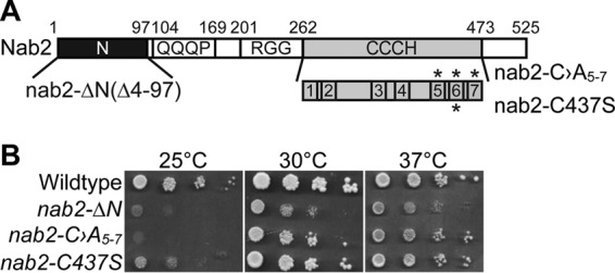
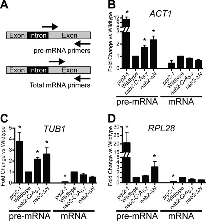
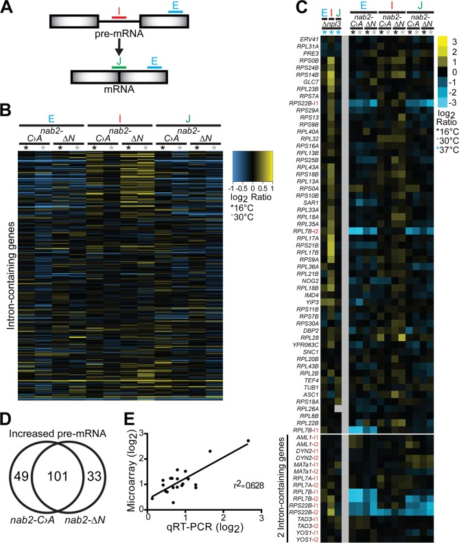
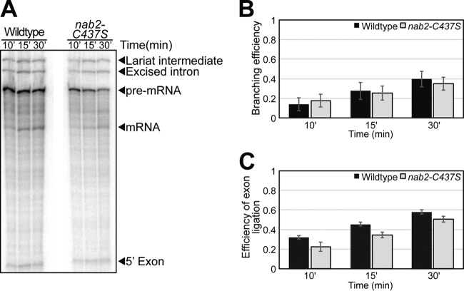
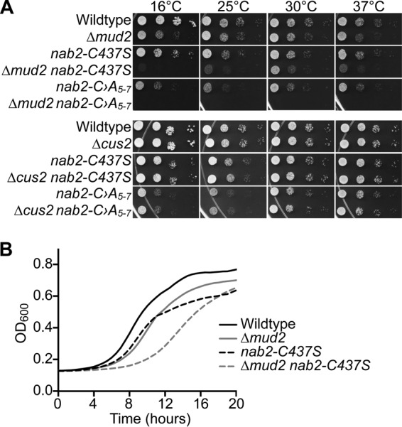
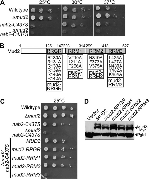
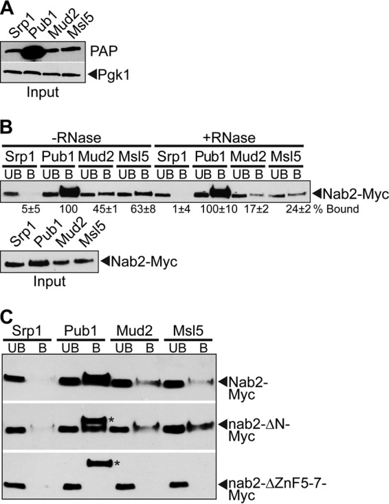
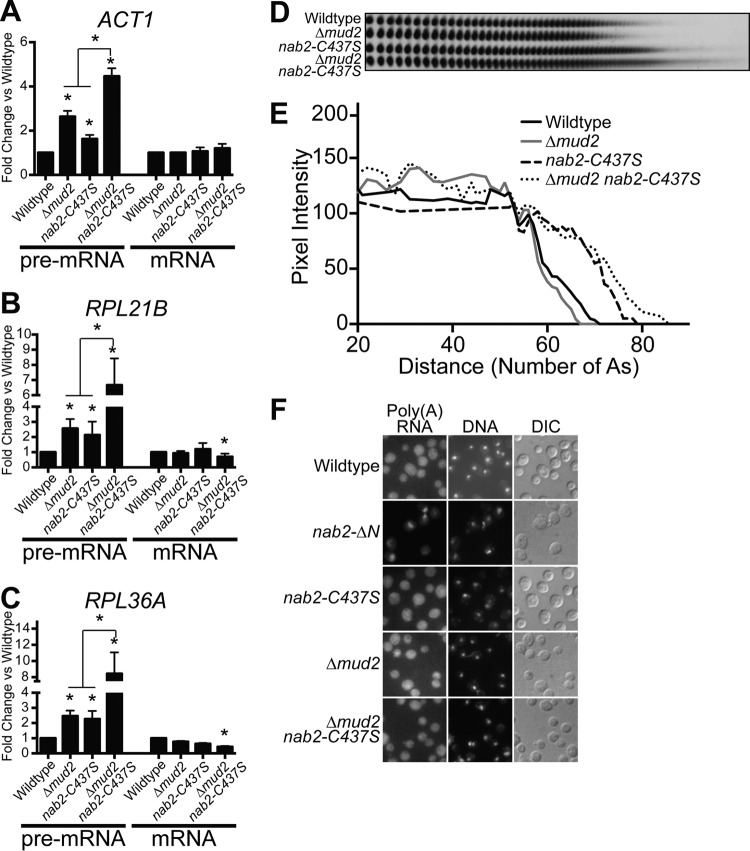
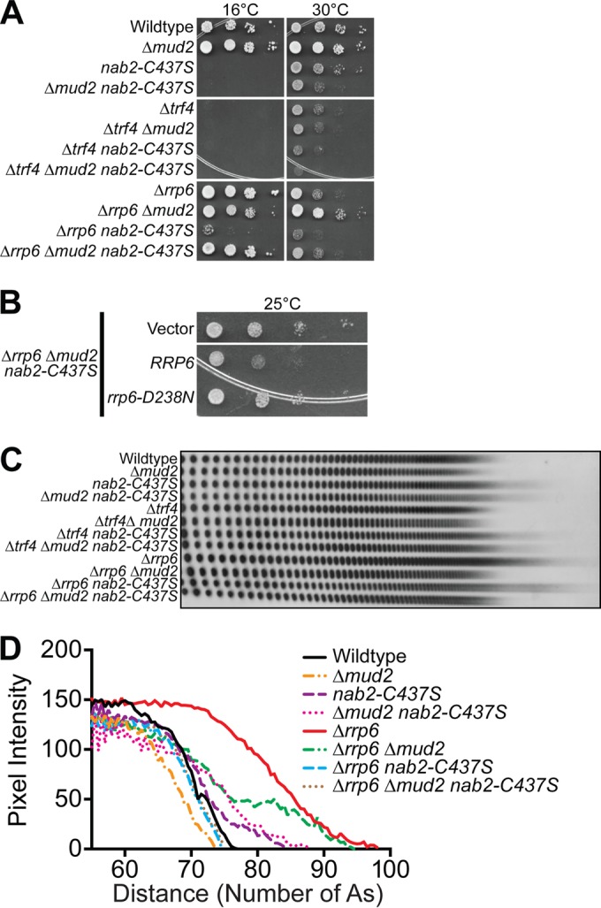
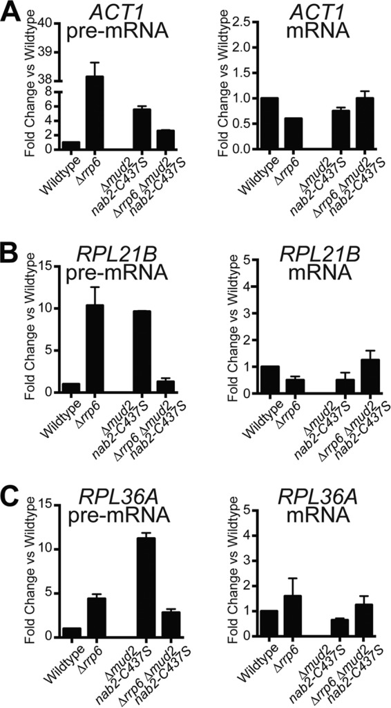
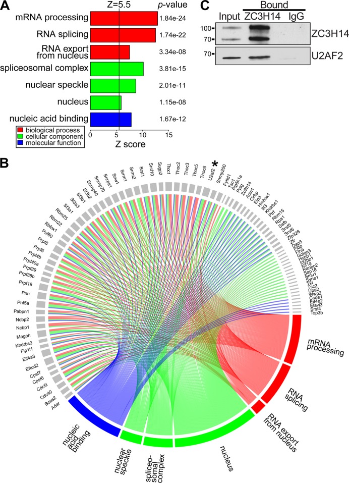
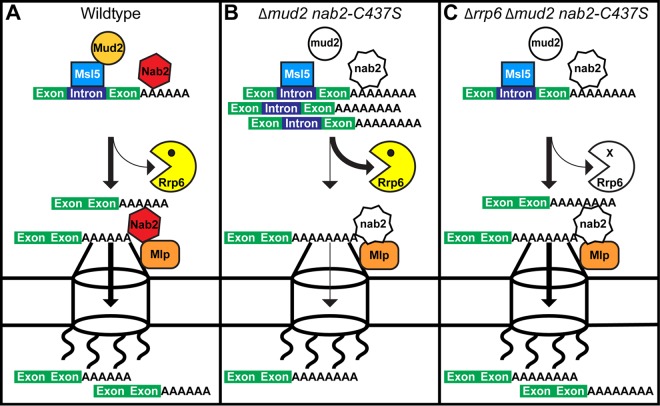
Numerous RNA binding proteins are deposited onto an mRNA transcript to modulate post-transcriptional processing events ensuring proper mRNA maturation. Defining the interplay between RNA binding proteins that couple mRNA biogenesis events is crucial for understanding how gene expression is regulated. To explore how RNA binding proteins control mRNA processing, we investigated a role for the evolutionarily conserved polyadenosine RNA binding protein, Nab2, in mRNA maturation within the nucleus. This work reveals that nab2 mutant cells accumulate intron-containing pre-mRNA in vivo We extend this analysis to identify genetic interactions between mutant alleles of nab2 and genes encoding the splicing factor, MUD2, and the RNA exosome, RRP6, with in vivo consequences of altered pre-mRNA splicing and poly(A) tail length control. As further evidence linking Nab2 proteins to splicing, an unbiased proteomic analysis of vertebrate Nab2, ZC3H14, identifies physical interactions with numerous components of the spliceosome. We validated the interaction between ZC3H14 and U2AF2/U2AF65 Taking all the findings into consideration, we present a model where Nab2/ZC3H14 interacts with spliceosome components to allow proper coupling of splicing with subsequent mRNA processing steps contributing to a kinetic proofreading step that allows properly processed mRNA to exit the nucleus and escape Rrp6-dependent degradation.


Figures

FIG 1
FIG 1
nab2 mutant cells show growth defects. (A) The Nab2 protein consists of four domains: a proline-tryptophan-isoleucine (PWI)-like N-terminal domain, a glutamine-rich domain (QQQP), an arginine-glycine-glycine (RGG) domain required for nuclear localization, and a tandem cysteine-cysteine-cysteine-histidine (CCCH) zinc finger domain comprised of seven zinc fingers (70). Shown below the CCCH zinc finger domain are zinc fingers 1 to 7 with zinc fingers 5 to 7 critical for high-affinity binding to polyadenosine RNA (71). The asterisks above zinc fingers 5, 6, and 7 denote the first cysteine residues (C415, C437, and C458) altered to alanine to generate nab2-C›A5-7. The asterisk below zinc finger 6 denotes the first cysteine residue (C437) altered to serine to generate nab2-C437S. The residues removed (i.e., 4 to 97) to generate the N-terminal nab2 mutant, nab2-ΔN, are also indicated. (B) Wild-type cells and the integrated nab2 mutants nab2-ΔN, nab2-C›A5-7, and nab2-C437S were grown at 30°C, serially diluted, spotted onto YEPD plates, and incubated at the indicated temperatures.
FIG 2
FIG 2
Unspliced transcripts accumulate in nab2 mutant cells. (A) Schematic of primer locations used to amplify unspliced pre-mRNA and total mRNA. For each transcript, unspliced pre-mRNA was detected with primers that span intron-exon boundaries, and total mRNA was detected with primers within the exon. (B to D) Total RNA was prepared from wild-type, nab2-C›A5-7, and nab2-ΔN cells, as well as a control splicing mutant (prp2-1) (72, 73). Quantitative RT-PCR was performed to measure both the pre-mRNA and the total mRNA of the ACT1 (B), TUB1 (C), and RPL28A (D) transcripts. The results were normalized to the PDA1 transcript, and values were plotted relative to wild-type cells, where the value for each transcript analyzed was set to 1.0. Experiments were performed in triplicate, error bars indicate the standard errors of the mean (SEM), and statistical significance (indicated by the asterisks) was calculated for both pre-mRNA and mRNA using one-way ANOVA.
FIG 3
FIG 3
A splicing microarray reveals a modest accumulation of unspliced transcripts in nab2 mutants. (A) The splicing microarray contains probes located within the intron (I) that detect pre-mRNA, located at the exon-exon junction (J) that detect mature mRNA, and located within an exon (E) that detect total mRNA for each intron-containing gene. (B) Splicing profile of nab2-C›A5-7 (C>A) and nab2-ΔN mutants compared to wild-type cells grown at 16 and 30°C for all 249 different intron-containing genes present on the microarray (see Table S3 in the supplemental material). Intron-containing pre-mRNA levels are elevated in nab2-C›A5-7 mutant at 16°C and in nab2-ΔN mutant at both temperatures. The cDNA from total RNA of nab2 mutants grown at 16 and 30°C was hybridized to the splicing array as described in Materials and Methods. Each horizontal line shows the behavior of a single transcript. (C) A closeup view of the splicing profile in nab2 mutants reveals that many ribosomal protein (RPL/RPS) intron-containing genes (e.g., RPL28) and some two-intron-containing genes (e.g., RPL7A) show elevated pre-mRNA levels. Two ribosomal protein two-intron-containing genes, RPL7B and RPS22B, show decreased total and mature mRNA levels. For comparison, the splicing profile of an SR protein mutant (Δnpl3) at 37°C is depicted, which was generated at the same time as the nab2 mutants and has previously been shown to cause pre-mRNA accumulation (112). (D) A Venn diagram illustrates the number of shared pre-mRNA transcripts that accumulate in nab2-C›A5-7 and nab2-ΔN cells. (E) The relative levels of expression of 20 transcripts measured by qRT-PCR and the splicing microarray show a positive correlation (r2 = 0.628). The relative levels of expression of the transcripts assayed by qRT-PCR and microarray were paired, plotted, and analyzed by linear regression.
FIG 4
FIG 4
In vitro splicing assay shows lysate prepared from nab2 mutant cells is competent for splicing. (A) Radiolabeled ACT1 pre-mRNA was incubated with lysates isolated from wild-type or nab2-C437S cells. Radiolabeled splicing products were analyzed at 10, 15, and 30 min of incubation by separation on an acrylamide gel. The positions of the pre-mRNA, lariat intermediate, 5′ exon, excised intron, and mature mRNA are indicated. (B and C) The branching efficiency (B) and the efficiency of exon ligation (C) were quantitated from five independent experiments and graphed with error bars that represent the SEM. The branching efficiency was calculated as (LI + mRNA)/(LI + mRNA + pre), and the efficiency of exon ligation was calculated as mRNA/(LI + mRNA), where “pre” represents pre-mRNA, and “LI” represents lariat intermediates.
FIG 5
FIG 5
Growth assays to examine genetic interactions between nab2 mutants and alleles of splicing factors. (A) As described in Materials and Methods, a plasmid shuffle assay was used to analyze the growth of the splicing factor mutants in combination with nab2 mutants. Δnab2, Δnab2 Δmud2, and Δnab2 Δcus2 cells maintained by a URA3 plasmid encoding wild-type NAB2 were transformed with LEU2 plasmids encoding wild-type NAB2, nab2-C›A5-7, or nab2-C437S. Cells were selected on 5-fluoroorotic acid (5-FOA) to eliminate the NAB2 maintenance plasmid and grown, serially diluted, and spotted onto plates lacking leucine to assess growth at 16, 25, 30, and 37°C. (B) Growth curves were generated for Δnab2 or Δnab2 Δmud2 cells carrying wild-type NAB2, nab2-C›A5-7, or nab2-C437S mutant plasmids in liquid media. The cells were grown to saturation and diluted, and their optical density at 600 nm (OD600) was measured at A600 for 20 h at 25°C. The growth curves are representative of three independent experiments.
FIG 6
FIG 6
Interaction between Mud2 and Msl5 is required when Nab2 function is impaired. (A) Isogenic integrated strains were generated by deleting MUD2 in wild-type and nab2-C437S cells to produce Δmud2 and Δmud2 nab2-C437S cells. Wild-type, Δmud2, nab2-C437S, and Δmud2 nab2-C437S cells were grown, serially diluted, and spotted onto YEPD plates at 25, 30, and 37°C. (B) Schematic of Mud2 protein showing the position of the tandem RRGR motif domain and three RNA recognition motifs (RRMs): RRM1, RRM2, and RRM3. Amino acid substitutions designed to impair the function of each domain are illustrated. The amino acid substitutions were as follows: the tandem RRGR domain (57) (R130A, R131A, R133A, R139A, R140A, and R142A), RRM1 (R208A, V210A, I211A, and F266A), RRM2 (N310A, I311A, and F373A), and RRM3 (L425A, L427A, N480A, Y482A, and Y484A). (C) Wild-type cells and integrated mutant Δmud2, nab2-C437S, and Δmud2 nab2-C437S cells were transformed with a vector plasmid and are shown as controls. Δmud2 nab2-C437S cells were transformed with plasmids expressing wild-type MUD2 or mud2 mutants in the RRGR, RRM1, RRM2, and RRM3 domains illustrated in panel B. Cells were grown, serially diluted, and spotted onto plates lacking uracil to assess growth at 25°C. (D) The Mud2 variants were all tagged with a Myc epitope to assess the relative expression levels by immunoblotting yeast cell lysates expressing the indicated Mud2 variants with anti-Myc antibody. Control cells that do not express Mud2-Myc (Vector) are shown as a negative control. Pgk1 was used as a loading control.
FIG 7
FIG 7
Nab2 physically interacts with the commitment complex in an RNA-dependent manner. (A) An immunoblot shows the expression of the TAP-tagged proteins Srp1, Pub1, Mud2, and Msl5 (all of which are approximately the same molecular weight). Immunoblotting was performed with a peroxidase-antiperoxidase (PAP) antibody to detect TAP-tagged protein expression and with an anti-Pgk1 antibody to detect 3-phosphoglycerate kinase as a loading control. (B) Cells expressing the C-terminal TAP-tagged proteins Srp1, Pub1, Mud2, or Msl5 were transformed with a Nab2-Myc plasmid. The TAP-tagged proteins were purified, and Nab2-Myc was detected by immunoblotting in the input, unbound (UB), and bound (B) fractions. Samples were treated with (+RNase) or without RNase (−RNase) to assess the RNA dependence of the interaction. The percentage of bound Nab2 protein relative to the amount of input protein and Nab2 bound to Pub1 (%Bound) is indicated below the bound fractions. Experiments were performed in triplicate, and the standard deviations were calculated. (C) To map the domain of Nab2 required for interaction with Mud2/Msl5, cells expressing TAP-tagged Srp1, Pub1, Mud2, or Msl5 were transformed with full-length Nab2-Myc, nab2-ΔN-Myc, or nab2-ΔZnF5-7-Myc plasmids. The TAP-tagged proteins were precipitated from yeast lysates, and immunoblotting was performed with anti-Myc antibody to detect Nab2 in the unbound (UB) and bound (B) lanes. The additional bands observed in the Pub1 bound lane in the nab2-ΔN-Myc and nab2-ΔZnF5-7-Myc immunoblot panels (indicated by asterisks) are cross-reacting bands similar in size to the Nab2 protein.
FIG 8
FIG 8
nab2 and mud2 mutants show defects in poly(A) tail length control and mRNA splicing. (A to C) Total RNA was isolated from wild-type and integrated mutant Δmud2, nab2-C437S, and Δmud2 nab2-C437S cells grown at 30°C. Quantitative real-time PCR analysis was performed to amplify ACT1 (A), RPL21B (B), and RPL36A (C) intron-containing transcripts. For each transcript, unspliced pre-mRNA was detected with primers that span intron-exon boundaries, and total mRNA was detected with primers within the exon. The mRNA levels were normalized to the PDA1 transcript and plotted relative to wild-type cells, which were set to 1.0. Experiments were performed in triplicate, error bars represent the SEM, and statistical significance (indicated by the asterisks) was calculated for both pre-mRNA and mRNA using one-way ANOVA. (D) Total RNA isolated from wild-type, Δmud2, nab2-C437S, and Δmud2 nab2-C437S cells grown at 30°C was analyzed for bulk poly(A) tail length as described in Materials and Methods. (E) Poly(A) tails were quantified using ImageJ by calculating pixel intensity along the length of the poly(A) tail and plotted relative to the number of A's, as described in Materials and Methods. (F) To examine poly(A) RNA export, wild-type, nab2-ΔN, nab2-C437S, Δmud2, and Δmud2 nab2-C437S cells grown at 30°C were examined by FISH with an oligo(dT) probe to detect poly(A) RNA and with DAPI to detect DNA. DIC images are also shown.
FIG 9
FIG 9
Genetic interactions between the nuclear exosome subunit, RRP6, and NAB2. (A) Wild-type cells and integrated mutant Δmud2 (ACY2270), nab2-C437S (ACY2202), and Δmud2 nab2-C437S (ACY2273) cells were combined with deletion of the TRAMP poly(A) polymerase, TRF4, or nuclear exosome component, RRP6, gene. The cells were grown, serially diluted, and spotted onto YEPD plates at 25, 30, and 37°C. (B) Δrrp6 Δmud2 nab2-C437S (ACY2313) cells were transformed with vector plasmid and are shown as a control. Δrrp6 Δmud2 nab2-C437S cells were transformed with Rrp6 plasmids expressing wild-type RRP6 or rrp6-D238N catalytic mutant. The cells were grown, serially diluted, and spotted onto plates lacking uracil to assess growth at 16°C. (C) Total yeast RNA was isolated from wild-type, Δmud2, nab2-C437S, and Δmud2 nab2-C437S cells combined with Δtrf4 or Δrrp6 mutant and analyzed for bulk poly(A) tail length as described in Materials and Methods. (D) Poly(A) tails were quantified using ImageJ by calculating pixel intensity along the length of the poly(A) tail and plotted relative to the number of A's, as described in Materials and Methods.
FIG 10
FIG 10
Deletion of RRP6 reduces the accumulation of pre-mRNA in Δmud2 nab2-C437S cells. To assess how deletion of RRP6 impacts both pre-mRNA and mRNA levels, total RNA was isolated from wild-type and Δmud2 nab2-C437S cells combined with Δrrp6 mutant. Quantitative real-time PCR analysis was performed to amplify ACT1 (A), RPL21B (B), and RPL36A (C) intron-containing transcripts (pre-mRNA) with primers that span the intron-exon boundary or total mRNA (mRNA) using primers within the exon. The results were normalized to the PDA1 transcript, and values were plotted relative to the wild type, which was set to 1.0.
FIG 11
FIG 11
ZC3H14 interacts with spliceosome components. As described in Materials and Methods, endogenous ZC3H14 was precipitated from mouse brain lysate using a polyclonal ZC3H14 antibody (48), and bound proteins were identified by on bead mass spectrometry (see Table S7 in the supplemental material). Three independent biological replicates were performed. (A) The gene ontology (GO) terms for the proteins that are mostly highly enriched with ZC3H14 compared to an IgG control are illustrated. The inclusion criteria for this analysis were a Z-score of ≥5.5, a P value of <0.00001, and ≥5 genes per GO term. (B) The genes that cluster into the listed GO terms in panel A are depicted. The corresponding GO terms are shown by a connecting line in the color corresponding to the GO term. The U2AF2 protein is indicated by an asterisk. (C) We validated the interaction between ZC3H14 and U2AF2 by coimmunoprecipitation from mouse brain lysate. The input and bound samples for anti-ZC3H14 or control IgG immunoprecipitation are shown. Samples were blotted to detect ZC3H14, which is alternatively spliced to produce protein isoforms of ∼100 and ∼75 kDa (48), and U2AF2. The results shown are typical of three independent experiments.
FIG 12
FIG 12
Kinetic proofreading model for Nab2 and Mud2 in splicing and mRNA surveillance. (A) In wild-type cells, Mud2 and Nab2 work together to promote efficient splicing, polyadenylation, and export of mRNA (denoted by thick lines). Nab2 interacts with Mlp proteins at the inner face of the nuclear pore to facilitate the efficient export of processed transcripts. (B) In mud2Δ nab2-C437S mutant cells which lack functional MUD2 and NAB2, transcript processing is impaired (thin arrows), triggering degradation by the nuclear exosome subunit, Rrp6 (thick arrow). Although some transcripts may escape nuclear surveillance and exit the nucleus, they may be improperly processed, resulting in extended poly(A) tails and/or missplicing. (C) In Δrrp6 Δmud2 nab2-C437S mutant cells lacking functional MUD2, NAB2, and RRP6, RNA processing is still inefficient, but more time is available in the nucleus to produce mature transcripts (thick arrows) in the absence of Rrp6-mediated degradation (thin arrow). As a result, the transcript is eventually processed properly and exported to the cytoplasm.

Similar articles

Cited by

References

    1. Moore MJ. 2005. From birth to death: the complex lives of eukaryotic mRNAs. Science 309:1514–1518. doi:10.1126/science.1111443. - DOI - PubMed
    1. Singh G, Pratt G, Yeo GW, Moore MJ. 2015. The clothes make the mRNA: past and present trends in mRNP fashion. Annu Rev Biochem 84:325–354. doi:10.1146/annurev-biochem-080111-092106. - DOI - PMC - PubMed
    1. Dreyfuss G, Kim VN, Kataoka N. 2002. Messenger-RNA-binding proteins and the messages they carry. Nat Rev Mol Cell Biol 3:195–205. doi:10.1038/nrm760. - DOI - PubMed
    1. Niwa M, Berget SM. 1991. Mutation of the AAUAAA polyadenylation signal depresses in vitro splicing of proximal but not distal introns. Genes Dev 5:2086–2095. doi:10.1101/gad.5.11.2086. - DOI - PubMed
    1. Nesic D, Cheng J, Maquat LE. 1993. Sequences within the last intron function in RNA 3′-end formation in cultured cells. Mol Cell Biol 13:3359–3369. doi:10.1128/MCB.13.6.3359. - DOI - PMC - PubMed

LinkOut - more resources