. 2016 Aug 15;44(4):1091-100.
doi: 10.1042/BST20160095.

Modulating mitochondrial quality in disease transmission: towards enabling mitochondrial DNA disease carriers to have healthy children

Affiliations

Modulating mitochondrial quality in disease transmission: towards enabling mitochondrial DNA disease carriers to have healthy children

Alan Diot et al. Biochem Soc Trans. .

Abstract

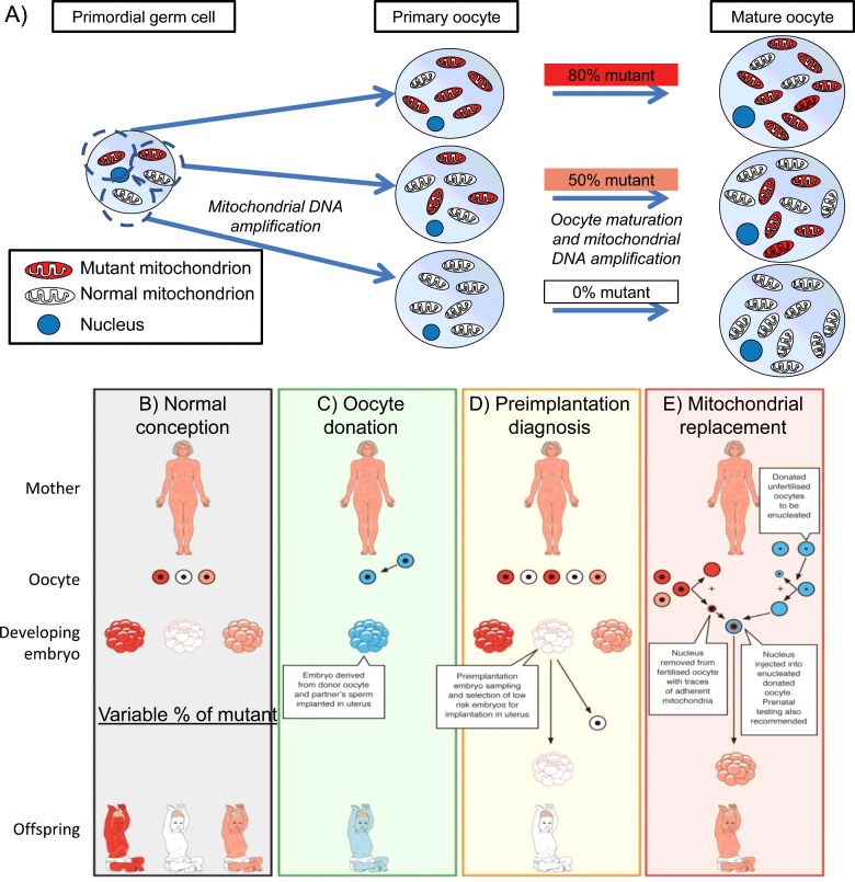
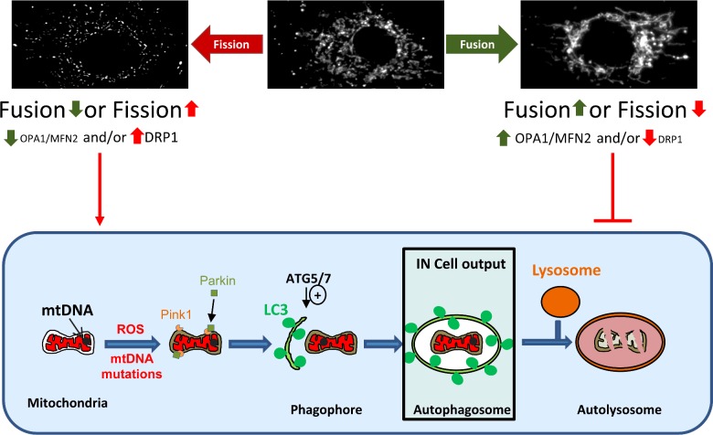
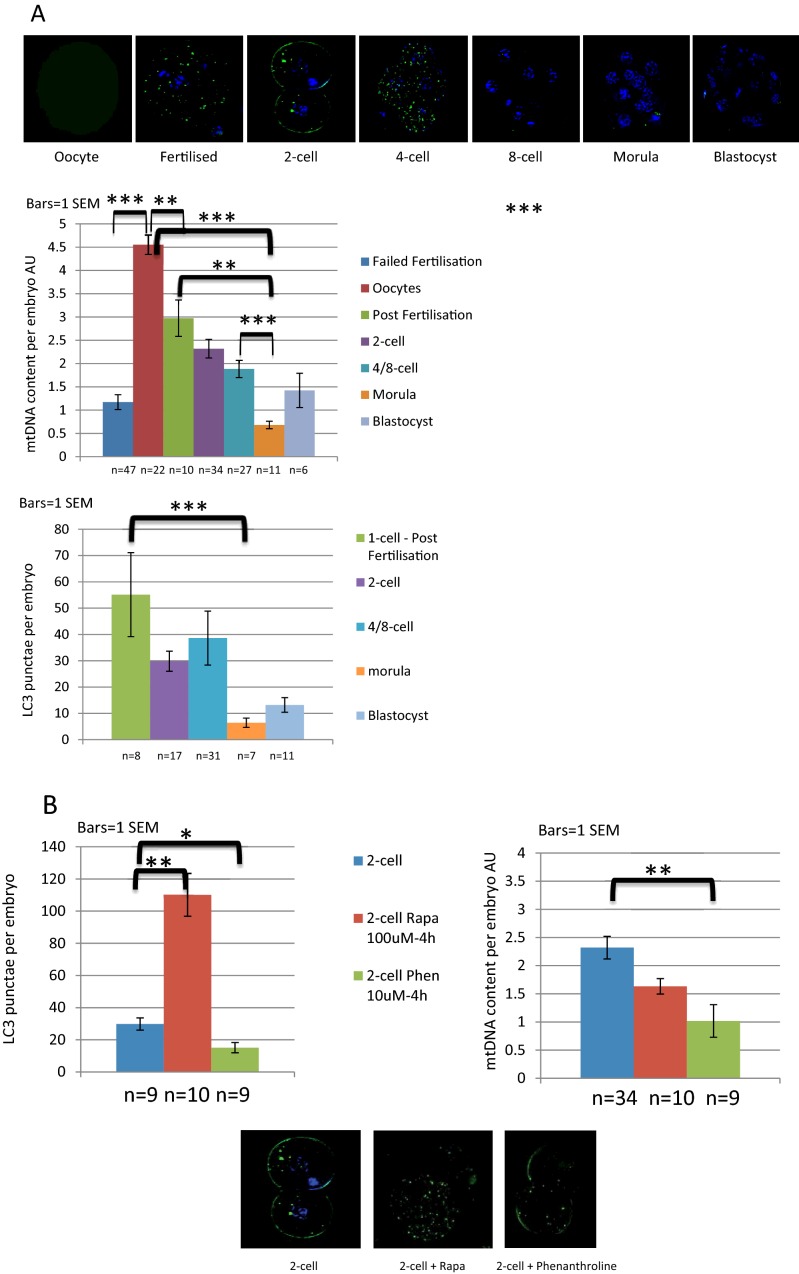
One in 400 people has a maternally inherited mutation in mtDNA potentially causing incurable disease. In so-called heteroplasmic disease, mutant and normal mtDNA co-exist in the cells of carrier women. Disease severity depends on the proportion of inherited abnormal mtDNA molecules. Families who have had a child die of severe, maternally inherited mtDNA disease need reliable information on the risk of recurrence in future pregnancies. However, prenatal diagnosis and even estimates of risk are fraught with uncertainty because of the complex and stochastic dynamics of heteroplasmy. These complications include an mtDNA bottleneck, whereby hard-to-predict fluctuations in the proportions of mutant and normal mtDNA may arise between generations. In 'mitochondrial replacement therapy' (MRT), damaged mitochondria are replaced with healthy ones in early human development, using nuclear transfer. We are developing non-invasive alternatives, notably activating autophagy, a cellular quality control mechanism, in which damaged cellular components are engulfed by autophagosomes. This approach could be used in combination with MRT or with the regular management, pre-implantation genetic diagnosis (PGD). Mathematical theory, supported by recent experiments, suggests that this strategy may be fruitful in controlling heteroplasmy. Using mice that are transgenic for fluorescent LC3 (the hallmark of autophagy) we quantified autophagosomes in cleavage stage embryos. We confirmed that the autophagosome count peaks in four-cell embryos and this correlates with a drop in the mtDNA content of the whole embryo. This suggests removal by mitophagy (mitochondria-specific autophagy). We suggest that modulating heteroplasmy by activating mitophagy may be a useful complement to mitochondrial replacement therapy.

Keywords: mitochondrial replacement therapy; mitophagy; mtDNA bottleneck.


Figures

Figure 1
Figure 1. Transmission of mtDNA disease and strategies to prevent transmission of mtDNA mutations
(A) Cartoon of the mitochondrial bottleneck: heteroplasmy of mtDNA in primordial germ cells may segregate during the ∼50-fold increase in mtDNA content as they develop into primary oocytes, resulting in different mutant loads (0–80% in this illustration). Although a major component of the trans-generation switching in mutant load has occurred by the oocyte stage, further segregation occurs during embryonic and fetal life. Three available ways to reduce the risk of transmitting mitochondrial DNA disease: oocyte donation, pre-implantation genetic diagnosis and mitochondrial replacement therapy. Red represents mutant mitochondrial DNA, pink and white represent successively higher proportions of normal mitochondrial DNA. Blue represents genetic material from an unrelated donor. (B) No intervention: offspring's mutant mitochondrial DNA load will vary greatly. (C) Oocyte donation: current availability in the United Kingdom is limited by the availability of oocyte donors. (D) Pre-implantation genetic diagnosis: is available in the United Kingdom for most mitochondrial DNA diseases. (E) MRT nuclear transfer: being developed in the United Kingdom, first cases likely this year, not yet available in the United States.
Figure 2
Figure 2. The mitophagy pathway
The mitochondrial network is dynamic with events of membrane fusion and fission within the network. The morphology of the mitochondrial network observed reflects the balance between these events. An increase in fission or a decrease of fusion is favourable to mitophagy when an increase of fusion or decreased fission inhibits the mitophagy. A simplified view of mitophagy is represented in the cartoon; briefly a dysfunctional mitochondria is targeted, potentially by Parkin and Pink1 proteins, to a forming autophagosome, the phagophore. The phagophore formation is under the control of Atg5/7 proteins notably. The autophagosome matures and engulfs the mitochondria before fusing with a lyososome for degradation. Our IN Cell system is able to measure the co-localization between mitochondria and autophagosome.
Figure 3
Figure 3. Evidence of autophagy in mouse oocytes and pre-implantation embryos
(A) Autophagosomes of oocytes and pre-implantation embryos of mice with GFP-tagged LC3 were visualized by fluorescence microscopy and autophagosomes per embryo counted. Relative mtDNA content (AU for arbitrary units) was assessed by single embryo qPCR. The mtDNA of these mice is wild type C57BL/6N and each embryo was individually lysed using an alkaline lysis protocol. After neutralization using tricine, the Taqman quantitative PCR was run for mtDNA along with standards to quantify the mtDNA content per embryo. The results show that mtDNA declines during active autophagy. (B) Two-cell embryos have been treated with rapamicin (red bars), phenanthroline (green bars) or not (blue bars) for 4 h to activate mitophagy; rapamycin 100 nM increased autophagosomes number per embryo and mtDNA dropped after exposure to 10 uM phenanthroline. Key *P<0.05, **P<0.01, ***P<0.001 t test. t tests where distribution was normal, Mann–Whitney elsewhere.

References

    1. Manwaring N., Jones M.M., Wang J.J., Rochtchina E., Howard C., Mitchell P., Sue C.M. Population prevalence of the MELAS A3243G mutation. Mitochondrion. 2007;7:230–733. doi: 10.1016/j.mito.2006.12.004. - DOI - PubMed
    1. Steffann J., Frydman N., Gigarel N., Burlet P., Ray P.F., Fanchin R., Feyereisen E., Kerbrat V., Tachdjian G., Bonnefont J.P., et al. Analysis of mtDNA variant segregation during early human embryonic development: a tool for successful NARP preimplantation diagnosis. J. Med. Genet. 2006;43:244–247. doi: 10.1136/jmg.2005.032326. - DOI - PMC - PubMed
    1. Monnot S., Gigarel N., Samuels D.C., Burlet P., Hesters L., Frydman N., Frydman R., Kerbrat V., Funalot B., Martinovic J., et al. Segregation of mtDNA throughout human embryofetal development: m.3243A>G as a model system. Hum. Mutat. 2011;32:116–125. doi: 10.1002/humu.21417. - DOI - PMC - PubMed
    1. Bouchet C., Steffann J., Corcos J., Monnot S., Paquis V., Rotig A., Lebon S., Levy P., Royer G., Giurgea I., et al. Prenatal diagnosis of MELAS syndrome: contribution to understanding mitochondrial DNA segregation during human embryo fetal development. J. Med. Genet. 2006;43:788–792. doi: 10.1136/jmg.2005.034140. - DOI - PMC - PubMed
    1. Bredenoord A.L., Pennings G., Smeets H.J., de Wert G. Dealing with uncertainties: ethics of prenatal diagnosis and preimplantation genetic diagnosis to prevent mitochondrial disorders. Hum. Reprod. Update. 2008;14:83–94. doi: 10.1093/humupd/dmm037. - DOI - PubMed

MeSH terms

Substances