Review
. 2016:36:138-46.

Pediatric stress fractures: a pictorial essay

Review

Pediatric stress fractures: a pictorial essay

Nirav H Shelat et al. Iowa Orthop J. 2016.

Abstract

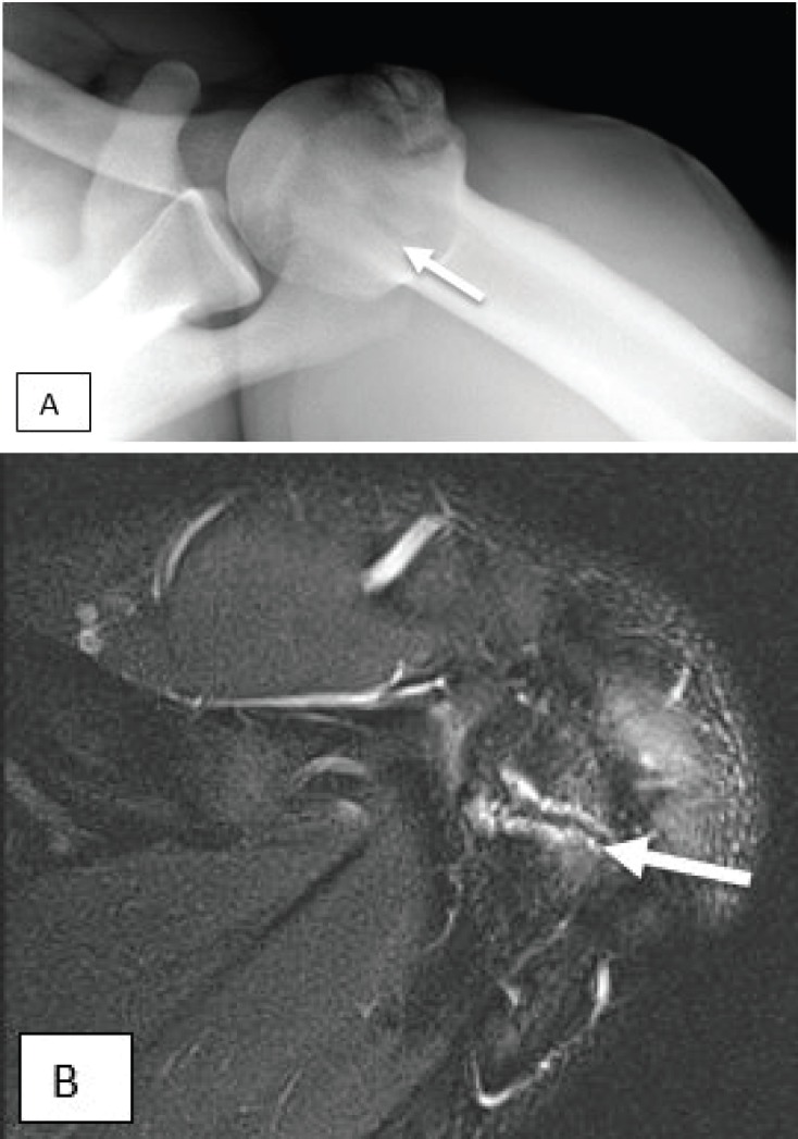
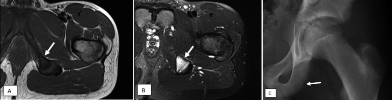
More children are participating in organized and recreational athletics at a younger age. It has been well documented that increased athletic specialization and year-round activities have resulted in higher incidences of overuse injuries, including stress fractures and stress reactions. Initially, stress fractures can be radiographically occult. Continued stress on the injured bone or cartilage can lead to progressive radiographic changes. Because of the prevalence of these injuries, both orthopedic surgeons and radiologists should be aware of the radiographic and magnetic resonance imaging (MRI) features of common stress fractures in children. This article reviews frequently encountered stress fractures involving various bones in the pediatric population.


Figures

Figure 1.
Figure 1.
13-year-old basketball player complaining of low back pain. (A) Axial T1 image demonstrates transversely oriented low signal (arrow) through the L5 pedicle and pars. (B) Axial T2 image demonstrates corresponding edema (arrow), confirmed on sagittal T1 (C) and STIR (D) sequences (arrows). Findings are consistent with unilateral stress fracture through the left pars interarticularis.
Figure 2.
Figure 2.
16-year-old baseball pitcher with recent onset of low back pain. (A) Axial T1, (B) T2 and (C) sagittal STIR images demonstrates bone marrow edema (arrows) in the left L3 pedicle, consistent with non-displaced stress fracture.
Figure 3.
Figure 3.
Collegiate American football player with shoulder pain and known lesser tuberosity avulsion. (A) Axillary shoulder radiograph performed to follow up lesser tuberosity avulsion demonstrates an unfused apophysis (arrow) at the acromion. (B) Follow up MRI demonstrates edema at the apophysis (arrow), consistent with acromial apophysiolysis.
Figure 4.
Figure 4.
13-year-old baseball pitcher. AP radiograph of the (A) right shoulder shows diffuse widening of the right proximal humeral physis (arrow). (B) For comparison, the left shoulder demonstrates normal width of the physis.
Figure 5.
Figure 5.
14-year-old baseball pitcher with medial elbow pain for 1 month duration. (A) AP radiograph demonstrates asymmetric widening of the right medial epicondyle physis (arrow). (B) The left elbow, submitted for comparison, is unremarkable. (C,D) MRI performed the following day demonstrates edema within the medial condyle epiphysis (arrow) and the adjacent metaphysis of the humerus. The ulnar collateral ligament (not fully shown) was intact.
Figure 6.
Figure 6.
11-year-old gymnast just days away from a championship meet, with ongoing wrist pain. (A) AP radiograph of the wrist is essentially unremarkable. Specifically, there is no evidence of physeal widening, irregularity, or fraying. (B, C) T1 and T2FS coronal MRI images demonstrate marrow edema through the distal metaphyses of the radius and ulna (white arrows), and to a lesser extent, the radial and ulnar styloids (red arrows).
Figure 7.
Figure 7.
18-year-old female cross country runner with gradually worsening low back pain. (A) Coronal T1, (B) coronal STIR, and (C) oblique coronal T2 fat saturated images demonstrate a stress fracture of the left sacral ala extending to the sacral foramen.
Figure 8.
Figure 8.
15-year-old female cross country runner with pubic pain, referred for MRI to “rule out sports hernia.” (A) Axial T1 and (B) T2FS images demonstrate a healing fracture (arrows) through the left inferior pubic ramus. This was radiographically occult.
Figure 9.
Figure 9.
13-year-old baseball player (shortstop) with gradually worsening groin pain. (A) Axial T1 and (B) axial T2FS images demonstrate bone marrow edema at the left ischial tuberosity (arrows). The hamstring tendons appeared normal. (C) Retrospective review of the radiograph shows subtle cortical irregularity along the lateral aspect of the left ischium (arrow).
Figure 10.
Figure 10.
9-year-old boy with history of renal transplant on chronic immunosuppressive therapy presenting with thigh pain. (A, B) Frontal and lateral radiographs demonstrate a linear band of sclerosis (arrow in A) through the distal femoral metaphysis and periosteal reaction (arrow in B), consistent with healing stress fracture. Incidental note is made of fracture progression through a non-ossifying fibroma along the medial cortex.
Figure 11.
Figure 11.
16-year-old female runner with prior history of pelvic stress fracture, now complaining of tibial pain. Sagittal STIR image demonstrates edema at the insertion of the patellar ligament (arrow), consistent with active Osgood Schlatter disease. There is mild pretibial edema in the soft tissues anterior to the tuberosity. There is minimally increased signal within the ligament itself.
Figure 12.
Figure 12.
3-year-old girl with a limp. Lateral radiograph demonstrates an area of uninterrupted periosteal reaction along the posteromedial aspect of the left tibia at the middle third of the tibia, consistent with stress fracture.
Figure 13.
Figure 13.
Two different patients. The patient in (A) is a 12-year old-girl who had been immobilized following a Lisfranc injury. The second patient in (B) is a 12-year-old girl who had been immobilized following medial cuneiform osteotomy. (A) Lateral radiograph demonstrates a vertically oriented sclerotic line in the calcaneal tuberosity (arrow). (B) AP radiograph demonstrates periosteal reaction (arrow) surrounding the second metatarsal, consistent with stress fracture.
Figure 14.
Figure 14.
14-year-old boy involved in multiple sports presents with worsening ankle pain. (A) Sagittal T1 and (B) T2FS images of the ankle demonstrate linear signal abnormality in the distal tibia consistent with stress fracture (white arrow). Multiple additional areas of signal abnormality (red arrows) are also consistent with stress reaction.
Figure 15.
Figure 15.
8-year-old girl with foot pain and a limp. (A) Lateral radiograph demonstrates subtle sclerosis in the cuboid (arrow). (B) Follow up MRI demonstrates a linear low T1 signal intensity focus (arrow) through the lateral aspect of the cuboid with (C) corresponding edema on T2 (arrow), consistent with stress fracture.

References

    1. Caine D, DiFiori J, Maffulli N. Physeal injuries in children’s and youth sports: reasons for concern? Br. J Sports Med. 2006 Sep;40(9):749–60. - PMC - PubMed
    1. Anderson MW, Greenspan A. Stress fractures. Radiology. 1996 Apr;199(1):1–12. - PubMed
    1. Jaimes C, Jimenez M, Shabshin N, Laor T, Jaramillo D. Taking the stress out of evaluating stress injuries in children. Radiographics. 2012 Mar-Apr;32(2):537–55. - PubMed
    1. Jones BH, Harris JM, Vinh TN, Rubin C. Exercise induced stress fractures and stress reactions of bone: epidemiology, etiology, and classification. Exerc Sport Sci Rev. 1989;17:379–422. - PubMed
    1. Laor T, Wall EJ, Vu LP. Physeal widening in the knee due to stress injury in child athletes. AJR Am J Roentgenol. 2006;186(5):1260–4. - PubMed

MeSH terms

LinkOut - more resources