Review
. 2016 Aug 15:4:52.
doi: 10.1186/s40560-016-0174-z. eCollection 2016.

Role of upper airway ultrasound in airway management

Affiliations
Review

Role of upper airway ultrasound in airway management

Adi Osman et al. J Intensive Care. .

Abstract

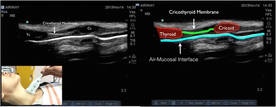
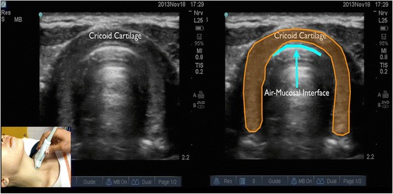
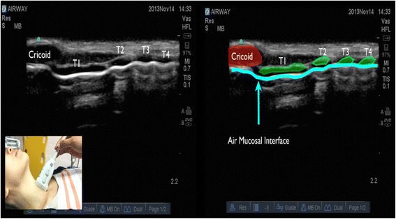
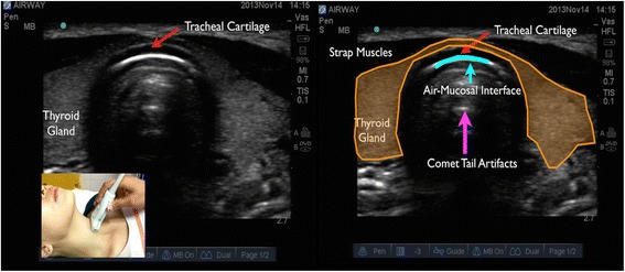
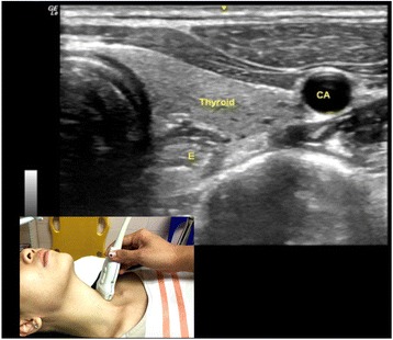
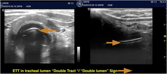
Upper airway ultrasound is a valuable, non-invasive, simple, and portable point of care ultrasound (POCUS) for evaluation of airway management even in anatomy distorted by pathology or trauma. Ultrasound enables us to identify important sonoanatomy of the upper airway such as thyroid cartilage, epiglottis, cricoid cartilage, cricothyroid membrane, tracheal cartilages, and esophagus. Understanding this applied sonoanatomy facilitates clinician to use ultrasound in assessment of airway anatomy for difficult intubation, ETT and LMA placement and depth, assessment of airway size, ultrasound-guided invasive procedures such as percutaneous needle cricothyroidotomy and tracheostomy, prediction of postextubation stridor and left double-lumen bronchial tube size, and detecting upper airway pathologies. Widespread POCUS awareness, better technological advancements, portability, and availability of ultrasound in most critical areas facilitate upper airway ultrasound to become the potential first-line non-invasive airway assessment tool in the future.

Keywords: Airway management; Point of care ultrasound; Upper airway ultrasound.


Figures

Fig. 1
Fig. 1
Cricoid cartilage, thyroid cartilage, and cricothyroid membrane in longitudinal plane. Cc cricoid cartilage, Tc thyroid cartilage
Fig. 2
Fig. 2
Cricoid cartilage in transverse plane
Fig. 3
Fig. 3
Cricoid cartilage and tracheal cartilage in longitudinal plane is seen as a “string of beads.” T1 first tracheal cartilage, T2 second tracheal cartilage, T3 third tracheal cartilage, T4 fourth tracheal cartilage
Fig. 4
Fig. 4
Tracheal cartilage in transverse plane
Fig. 5
Fig. 5
Vocal cord seen in transverse view. Sm sternocleidomastoid muscle, Tc thyroid cartilage, VM vocalis muscle, VL vocalis ligament, AC arytenoid cartilage
Fig. 6
Fig. 6
Esophagus is seen using transverse plane at the level of the first and second tracheal cartilage. E esophagus, CA carotid artery
Fig. 7
Fig. 7
Transverse and longitudinal view of ETT in trachea, seen as “double tract”/“double lumen” sign

References

    1. Adi O, Chuan TW, Rishya M. A feasibility study on bedside upper airway ultrasonography compared to waveform capnography for verifying endotracheal tube location after intubation. Crit Ultrasound J. 2013;5(1):7. doi: 10.1186/2036-7902-5-7. - DOI - PMC - PubMed
    1. Chou EH, Dickman E, Tsou PY, Tessaro M, Tsai YM, Ma MH, Lee CC, Marshall J. Ultrasonography for confirmation of endotracheal tube placement: a systematic review and meta-analysis. Resuscitation. 2015;90:97–103. doi: 10.1016/j.resuscitation.2015.02.013. - DOI - PubMed
    1. Lakhal K, Delplace X, Cottier JP, Tranquart F, Sauvagnac X, Mercier C, Fusciardi J, Laffon M. The feasibility of ultrasound to assess subglottic diameter. Anesth Analg. 2007;104(3):611–614. doi: 10.1213/01.ane.0000260136.53694.fe. - DOI - PubMed
    1. Sustic A, Miletic D, Protic A, Ivancic A, Cicvaric T. Can ultrasound be useful for predicting the size of a left double-lumen bronchial tube? Tracheal width as measured by ultrasonography versus computed tomography. J Clin Anesth. 2008;20(4):247–252. doi: 10.1016/j.jclinane.2007.11.002. - DOI - PubMed
    1. Shibasaki M, Nakajima Y, Ishii S, Shimizu F, Shime N, Sessler DI. Prediction of pediatric endotracheal tube size by ultrasonography. Anesthesiology. 2010;113(4):819–824. doi: 10.1097/ALN.0b013e3181ef6757. - DOI - PubMed