

doi: 10.1002/smll.201601540.
Epub 2016 Aug 16.
G Protein-Coupled Receptors Incorporated into Rehydrated Diblock Copolymer Vesicles Retain Functionality
Affiliations
- PMID: 27529518
- PMCID: PMC5148614
- DOI: 10.1002/smll.201601540
Item in Clipboard
G Protein-Coupled Receptors Incorporated into Rehydrated Diblock Copolymer Vesicles Retain Functionality
Small.
2016 Oct.
Abstract
G protein-coupled receptor (GPCR) is incorporated into polymeric vesicles made up of diblock copolymer bilayers. Successfully incorporated GPCRs exhibit correct biased physiological orientation and respond to various ligands. After extended dehydrated storage via lyophilization and subsequent rehydration, diblock copolymer polymersomes retain their shape and incorporated GPCR retains its function.
Keywords: GPCR; bilayer vesicles; block copolymers; lyophilization; polymersomes.
© 2016 WILEY-VCH Verlag GmbH & Co. KGaA, Weinheim.
Figures
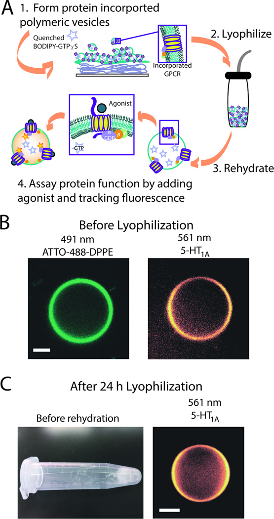
GPCR incorporation into diblock copolymer bilayer vesicles, pGUPs. (A) Schematic of pGUP formation and protein incorporation. Films of protein, agarose, and polymer are made on a coverslip and rehydrated with a sucrose buffer solution containing BODIPY-GTPγS. pGUPs formed of diblock copolymer bilayers can be lyophilized and the GPCR retains its function (steps 2–4); for enlarged image see Supporting Information (Figure S1). (B) Confocal micrographs of pGUPs prior to lyophilization. The left micrograph shows the polymer bilayer tagged with ATTO-488-DPPE. The right micrograph shows that rhodamine antibody-tagged 5-HT1AR is evenly distributed throughout the polymer bilayer. (C) The left image shows a pGUP sample after lyophilization. Upon rehydration, pGUPs can still be detected as shown in the right micrograph. All scale bars represent 5 µm.
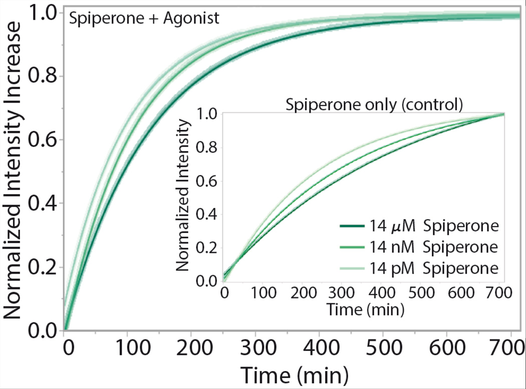
5-HT1AR in pGUPS display response to increasing antagonist concentration while keeping agonist concentration constant. Fluorescence unquenching due to the irreversible binding of BODIPY-GTPγS to G proteins was tracked for 12 hours for pGUPs formed with increasing amount of the antagonist (spiperone) and constant amount of agonist. Increasing the amount of antagonist in the system decreases the protein functional rate (See Table 1, for change in intensity rates). The inset shows control curves for the pGUPs that were incubated without agonist. 5-HT1AR basal activity is captured in the pGUPs lacking agonist, spiperone only (control).
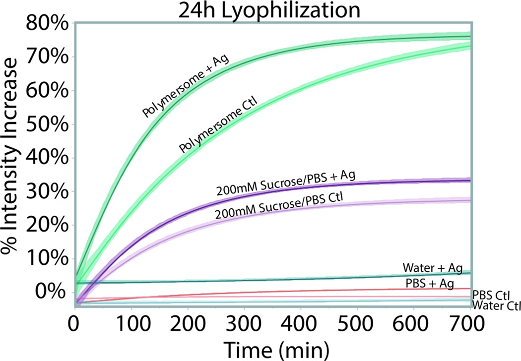
Functional rates of 5-HT1AR in polymersomes (pGUPs) versus various solutions. Controls (Ctl), no agonist pGUPs, are plotted alongside agonist-exposed samples (+Ag). The percent intensity increase of the samples indicates the population of functional protein. In DI water and PBS, the 5-HT1AR displays no fluorescence activity. In 200 mM sucrose in PBS (pH 7.4) 5-HT1AR displays weaker fluorescence intensity increase compared to pGUPs. Furthermore there is no difference in rate between the Ctl and +Ag protein in 200 mM sucrose in PBS.
Similar articles
-
Durable vesicles for reconstitution of membrane proteins in biotechnology.Biochem Soc Trans. 2017 Feb 8;45(1):15-26. doi: 10.1042/BST20160019. Biochem Soc Trans. 2017. PMID: 28202656 Free PMC article. Review.
-
Physicochemical properties of L-alpha dipalmitoyl phosphatidylcholine large unilamellar vesicles: Effect of hydrophobic block (PLA/PCL) of amphipathic diblock copolymers.Chem Phys Lipids. 2020 Aug;230:104927. doi: 10.1016/j.chemphyslip.2020.104927. Epub 2020 May 23. Chem Phys Lipids. 2020. PMID: 32454007
-
The Functional Activity of the Human Serotonin 5-HT1A Receptor Is Controlled by Lipid Bilayer Composition.Biophys J. 2016 Jun 7;110(11):2486-2495. doi: 10.1016/j.bpj.2016.04.042. Biophys J. 2016. PMID: 27276266 Free PMC article.
-
Human serotonin receptor 5-HT(1A) preferentially segregates to the liquid disordered phase in synthetic lipid bilayers.J Am Chem Soc. 2014 Oct 1;136(39):13530-3. doi: 10.1021/ja507221m. Epub 2014 Sep 19. J Am Chem Soc. 2014. PMID: 25211019 Free PMC article.
-
Solubilization of G protein-coupled receptors: a convenient strategy to explore lipid-receptor interaction.Methods Enzymol. 2015;557:117-34. doi: 10.1016/bs.mie.2015.01.001. Epub 2015 Mar 18. Methods Enzymol. 2015. PMID: 25950962 Review.
Cited by
-
Recent advances in permeable polymersomes: fabrication, responsiveness, and applications.Chem Sci. 2023 Jun 21;14(27):7411-7437. doi: 10.1039/d3sc01707a. eCollection 2023 Jul 12. Chem Sci. 2023. PMID: 37449076 Free PMC article. Review.
-
Durable vesicles for reconstitution of membrane proteins in biotechnology.Biochem Soc Trans. 2017 Feb 8;45(1):15-26. doi: 10.1042/BST20160019. Biochem Soc Trans. 2017. PMID: 28202656 Free PMC article. Review.
-
Photolithographic patterned surface forms size-controlled lipid vesicles.APL Bioeng. 2018 Jan 2;2(1):016104. doi: 10.1063/1.5002604. eCollection 2018 Mar. APL Bioeng. 2018. PMID: 31069289 Free PMC article.
References
MeSH terms
Substances
Grants and funding
LinkOut - more resources
Full Text Sources
Other Literature Sources
