. 2016 Oct;89(1066):20160546.
doi: 10.1259/bjr.20160546. Epub 2016 Aug 16.

Superb microvascular imaging in diagnosis of breast lesions: a comparative study with contrast-enhanced ultrasonographic microvascular imaging

Affiliations

Superb microvascular imaging in diagnosis of breast lesions: a comparative study with contrast-enhanced ultrasonographic microvascular imaging

Xiao-Yun Xiao et al. Br J Radiol. 2016 Oct.

Abstract

Objective: To evaluate the diagnostic performance of superb microvascular imaging (SMI) in breast lesions, comparing with contrast-enhanced ultrasonographic microvascular imaging (MVI).

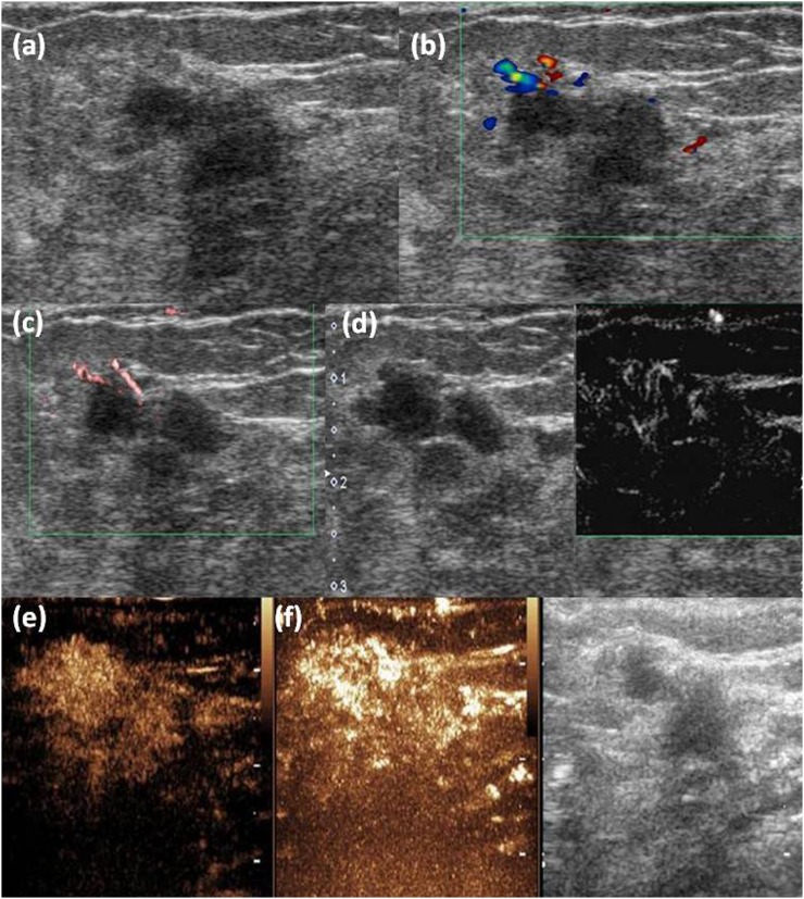
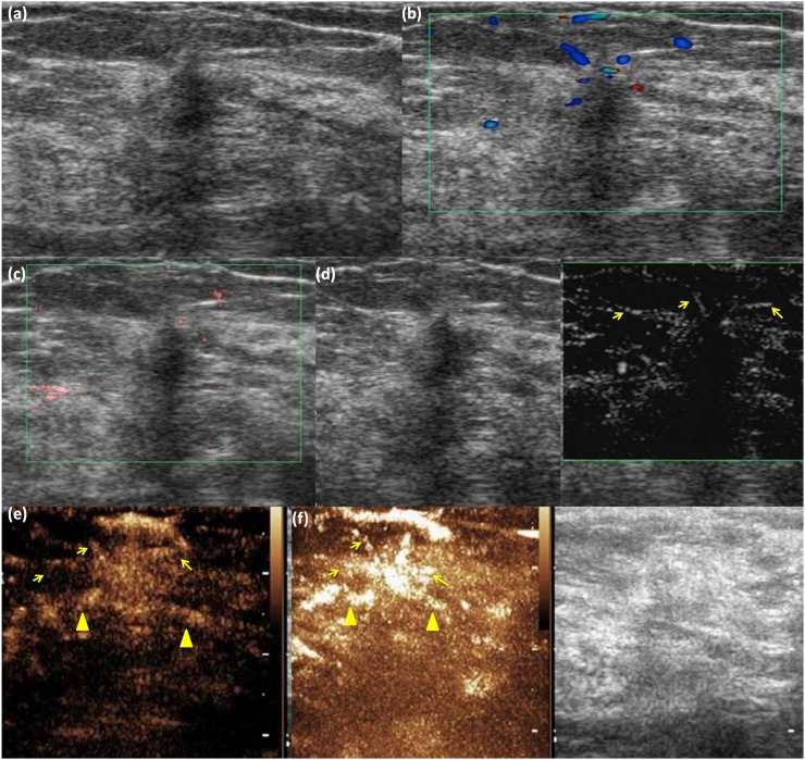
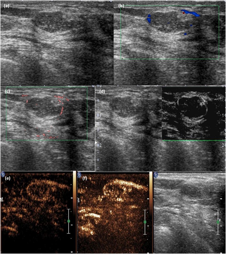
Methods: From April to November 2015, 132 patients (with 132 breast lesions) were enrolled in the retrospective study. All lesions were evaluated with colour Doppler flow imaging (CDFI), colour SMI (cSMI), monochrome SMI (mSMI) and contrast-enhanced ultrasonographic MVI. Receiver-operating characteristic curve analysis was performed to compare the diagnostic performance of SMI and MVI for discrimination between benign and malignant breast lesions.

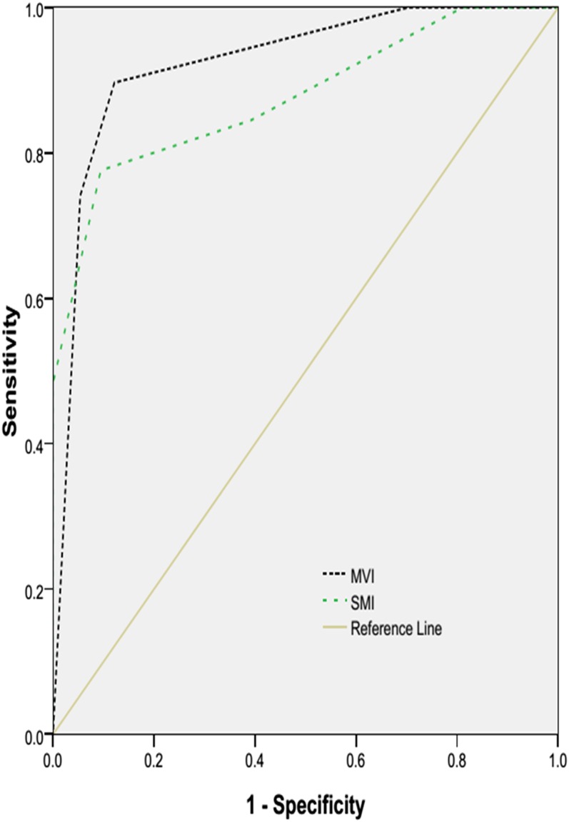
Results: Histological analysis showed 58 malignant and 74 benign lesions. mSMI was more sensitive in detecting blood flow signals in breast lesions than CDFI (p < 0.001) and cSMI (p < 0.001). Differences of vessels inside breast lesions and morphologic features of vessels between benign and malignant lesions were statistically significant on mSMI (p < 0.001). Using root hair-like and crab claw-like patterns as the criteria for malignant lesions, the sensitivity, specificity and accuracy for differentiation based on the microvascular architecture patterns were 77.6, 90.5 and 84.8% for mSMI and 89.6, 87.8 and 88.6% for MVI. Areas under curve of mSMI and MVI were not significantly different (p = 0.129).

Conclusion: mSMI can increase blood flow detection and depict the microvascular architecture of breast lesions. The diagnostic performance of mSMI was not significantly different from MVI. SMI has potential in the differential diagnosis of breast lesions.

Advances in knowledge: mSMI is a non-invasive technique for vascularity evaluation of breast tumours and it is beneficial for breast tumour differentiation.


Figures

Figure 1.
Figure 1.
(a) A 59-year-old female with a lesion in the upper outer quadrant of the right breast. (b, c) Colour Doppler flow imaging and colour superb microvascular imaging are showing vessels adjunct to the upper right of the lesion; (d) monochrome superb microvascular imaging (mSMI) is showing more detailed twisted vessels inside the lesion, especially in the upper right part, which has manifested as a root-hair like pattern; (e) contrast-enhanced ultrasound of the same lesion; (f) microvascular imaging is also showing the twisted vessels inside the lesions, but vessel courses are not seen as clearly as on mSMI. This lesion was pathologically proven to be invasive ductal carcinoma.
Figure 2.
Figure 2.
(a) A 50-year-old female with a lesion in the upper inner quadrant of the left breast. (b, c) Colour Doppler flow imaging and colour superb microvascular imaging are showing blood signals in the peripheral region of the lesion; (d) monochrome superb microvascular imaging (mSMI) is showing no vessels inside the lesion but radial vessels in the peripheral (arrows), which has manifested as a crab claw-like pattern; (e) contrast-enhanced ultrasound (CEUS) of the same lesion; (f) microvascular imaging (MVI) is also showing crab claw-like enhancement. Both CEUS and MVI are showing more radial vessels (arrowheads) than mSMI. This lesion was pathologically proven to be invasive ductal carcinoma.
Figure 3.
Figure 3.
(a) A 50-year-old female with a lesion in the lower inner quadrant of the left breast. (b, c) Colour Doppler flow imaging and colour superb microvascular imaging are showing blood signals around the lesion; (d) monochrome superb microvascular imaging is showing that the peripheral annular vessel was discontinuously observed; (e) contrast-enhanced ultrasound of the same lesion; (f) microvascular imaging is also showing peripheral annular vessel. This lesion was pathologically proved to be fibroadenoma.
Figure 4.
Figure 4.
The performances of monochrome superb microvascular imaging (mSMI) and microvascular imaging in the discrimination between benign and malignant breast lesions according to microvascular architecture are shown in the receiver-operating characteristic curve. CEUS, contrast-enhanced ultrasound.

References

    1. Tirona MT, Sehgal R, Ballester O. Prevention of breast cancer (part I): epidemiology, risk factors, and risk assessment tools. Cancer Invest 2010; 28: 743–50. doi: 10.3109/07357907.2010.494321 - DOI - PubMed
    1. Berg WA, Blume JD, Cormack JB, Mendelson EB, Lehrer D, Bohm-Velez M, et al. . Combined screening with ultrasound and mammography vs mammography alone in women at elevated risk of breast cancer. JAMA 2008; 299: 2151–63. doi: 10.1001/jama.299.18.2151 - DOI - PMC - PubMed
    1. Chala L, Endo E, Kim S, de Castro F, Moraes P, Cerri G, et al. . Gray-scale sonography of solid breast masses: diagnosis of probably benign masses and reduction of the number of biopsies. J Clin Ultrasound 2007; 35: 9–19. doi: 10.1002/jcu.20298 - DOI - PubMed
    1. Weidner N, Semple JP, Welch WR, Folkman J. Tumor angiogenesis and metastasis—correlation in invasive breast carcinoma. N Engl J Med 1991; 324: 1–8. doi: 10.1056/NEJM199101033240101 - DOI - PubMed
    1. Less JR, Skalak TC, Sevick EM, Jain RK. Microvascular architecture in a mammary carcinoma: branching patterns and vessel dimensions. Cancer Res 1991; 51: 265–73. - PubMed

MeSH terms