

Recombinant adenovirus of human p66Shc inhibits MCF-7 cell proliferation
- PMID: 27530145
- PMCID: PMC4987618
- DOI: 10.1038/srep31534
Recombinant adenovirus of human p66Shc inhibits MCF-7 cell proliferation
Abstract
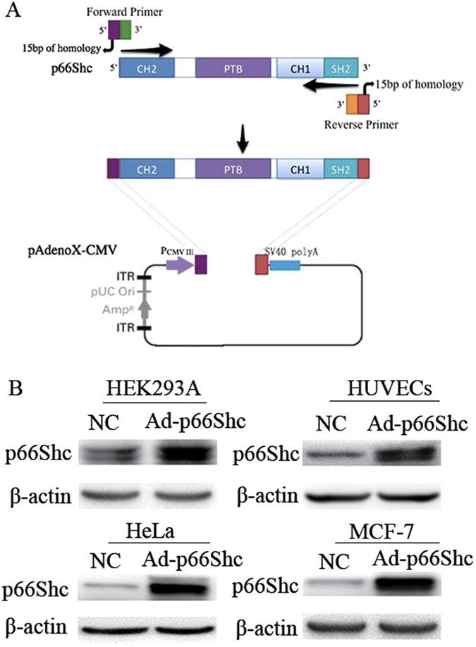
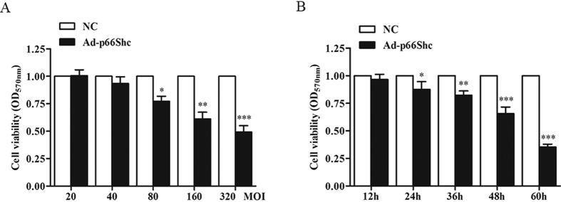
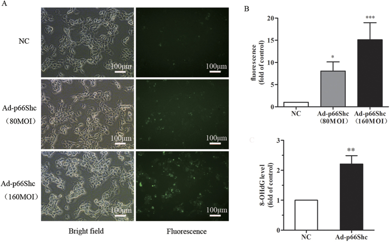
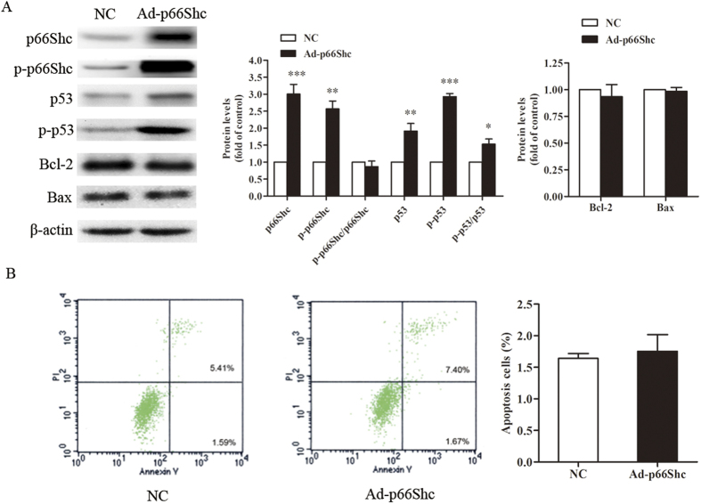
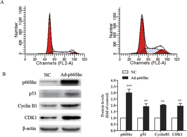
The aim of this work was to construct a human recombinant p66Shc adenovirus and to investigate the inhibition of recombinant p66Shc adenovirus on MCF-7 cells. The recombinant adenovirus expression vector was constructed using the Adeno-X Adenoviral System 3. Inhibition of MCF-7 cell proliferation was determined by MTT. Intracellular ROS was measured by DCFH-DA fluorescent probes, and 8-OHdG was detected by ELISA. Cell apoptosis and the cell cycle were assayed by flow cytometry. Western blot were used to observe protein expression. p66Shc expression was upregulated in 4 cell lines after infection. The inhibitory effect of p66Shc recombinant adenovirus on MCF-7 cells was accompanied by enhanced ROS and 8-OHdG. However, no significant differences were observed in the cell apoptosis rate. The ratio of the cell cycle G2/M phase showed a significant increase. Follow-up experiments demonstrated that the expressions of p53, p-p53, cyclin B1 and CDK1 were upregulated with the overexpression of p66Shc. The Adeno-X Adenoviral System 3 can be used to efficiently construct recombinant adenovirus containing p66Shc gene, and the Adeno-X can inhibit the proliferation of MCF-7 cells by inducing cell cycle arrest at the G2/M phase. These results suggested that p66Shc may be a key target for clinical cancer therapy.
Figures
Similar articles
-
Gambogic Acid Inhibits Malignant Melanoma Cell Proliferation Through Mitochondrial p66shc/ROS-p53/Bax-Mediated Apoptosis.Cell Physiol Biochem. 2016;38(4):1618-30. doi: 10.1159/000443102. Epub 2016 Apr 28. Cell Physiol Biochem. 2016. PMID: 27119348
-
Propofol attenuates high glucose-induced P66shc expression in human umbilical vein endothelial cells through Sirt1.Acta Biochim Biophys Sin (Shanghai). 2019 Feb 1;51(2):197-203. doi: 10.1093/abbs/gmy167. Acta Biochim Biophys Sin (Shanghai). 2019. PMID: 30590376
-
Construction of p66Shc gene interfering lentivirus vectors and its effects on alveolar epithelial cells apoptosis induced by hyperoxia.Drug Des Devel Ther. 2016 Aug 16;10:2611-22. doi: 10.2147/DDDT.S84820. eCollection 2016. Drug Des Devel Ther. 2016. PMID: 27574400 Free PMC article.
-
Role of adaptor protein p66Shc in renal pathologies.Am J Physiol Renal Physiol. 2018 Feb 1;314(2):F143-F153. doi: 10.1152/ajprenal.00414.2017. Epub 2017 Oct 4. Am J Physiol Renal Physiol. 2018. PMID: 28978535 Free PMC article. Review.
-
The interplay between p66Shc, reactive oxygen species and cancer cell metabolism.Eur J Clin Invest. 2015 Jan;45 Suppl 1:25-31. doi: 10.1111/eci.12364. Eur J Clin Invest. 2015. PMID: 25524583 Review.
Cited by
-
The Role of p66Shc in Cancer: Molecular Mechanisms and Therapeutic Implications.J Cell Mol Med. 2025 Jul;29(14):e70737. doi: 10.1111/jcmm.70737. J Cell Mol Med. 2025. PMID: 40690563 Free PMC article. Review.
-
The oxidoreductase p66Shc acts as tumor suppressor in BRAFV600E-transformed cells.Mol Oncol. 2018 Jun;12(6):869-882. doi: 10.1002/1878-0261.12199. Epub 2018 May 5. Mol Oncol. 2018. PMID: 29624862 Free PMC article.
-
Rigosertib-Activated JNK1/2 Eliminate Tumor Cells through p66Shc Activation.Biology (Basel). 2020 May 15;9(5):99. doi: 10.3390/biology9050099. Biology (Basel). 2020. PMID: 32429320 Free PMC article.
References
-
- Luzi L., Confalonieri S., Di Fiore P. P. & Pelicci P. G. Evolution of Shc functions from nematode to human. Curr Opin Genet Dev. 10, 668–674 (2000). - PubMed
-
- Napoli C. et al.. Deletion of the p66Shc longevity gene reduces systemic and tissue oxidative stress, vascular cell apoptosis, and early atherogenesis in mice fed a high-fat diet. Proceedings of the National Academy of Sciences of the United States of America 100, 2112–2116, doi: 10.1073/pnas.0336359100 (2003). - DOI - PMC - PubMed
Publication types
MeSH terms
Substances
LinkOut - more resources
Full Text Sources
Other Literature Sources
Research Materials
Miscellaneous
