. 2016 Sep 30;44(17):8465-78.
doi: 10.1093/nar/gkw714. Epub 2016 Aug 16.

Structure of a human pre-40S particle points to a role for RACK1 in the final steps of 18S rRNA processing

Affiliations

Structure of a human pre-40S particle points to a role for RACK1 in the final steps of 18S rRNA processing

Natacha Larburu et al. Nucleic Acids Res. .

Abstract

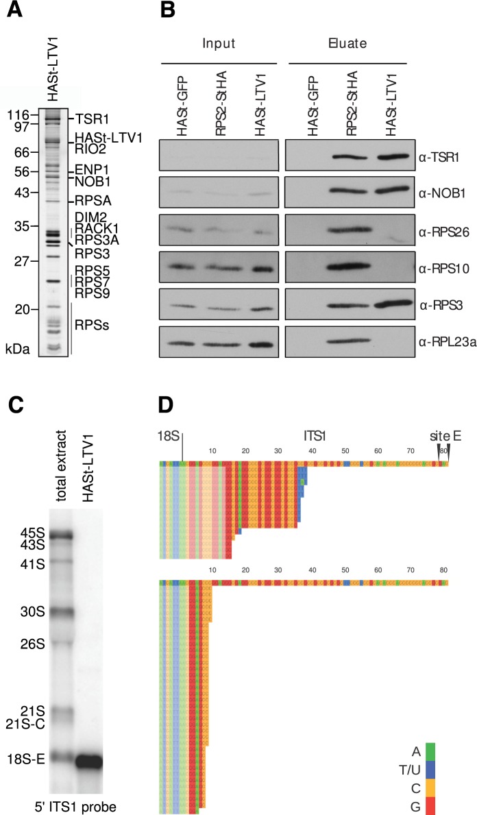
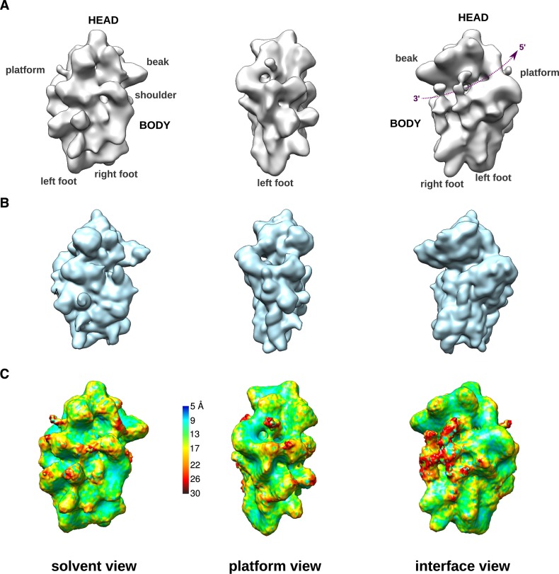
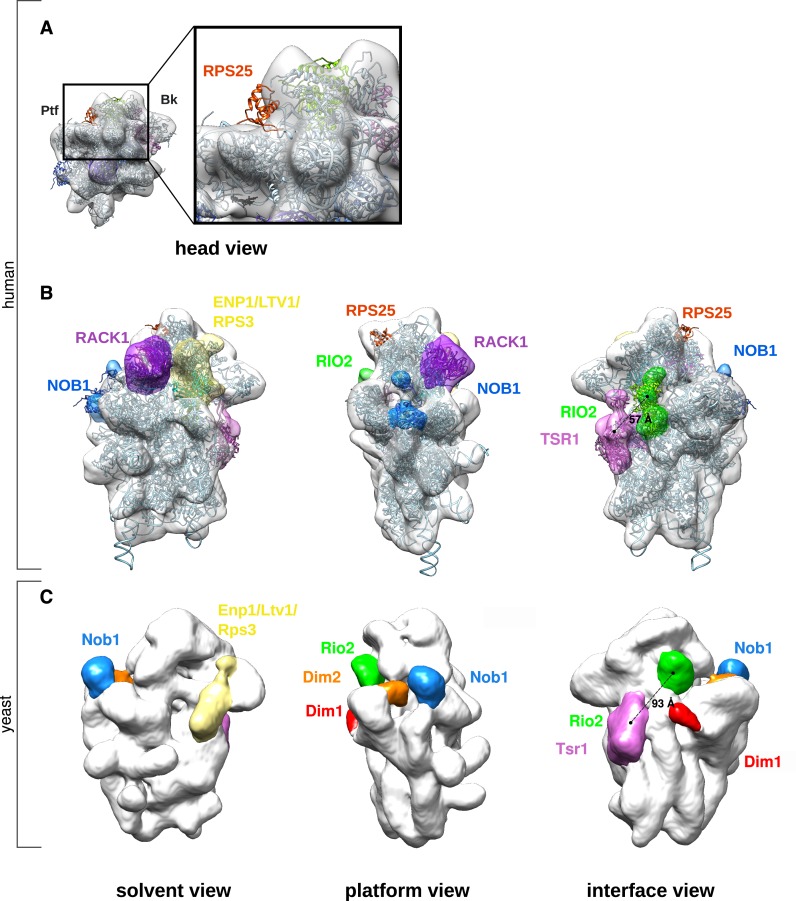
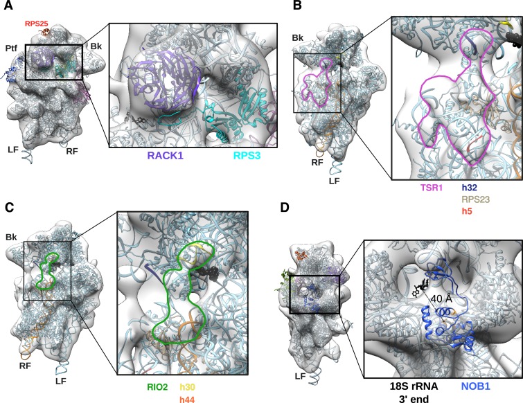
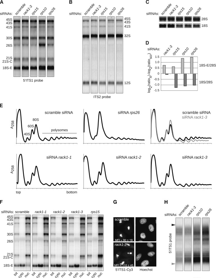
Synthesis of ribosomal subunits in eukaryotes is a complex and tightly regulated process that has been mostly characterized in yeast. The discovery of a growing number of diseases linked to defects in ribosome biogenesis calls for a deeper understanding of these mechanisms and of the specificities of human ribosome maturation. We present the 19 Å resolution cryo-EM reconstruction of a cytoplasmic precursor to the human small ribosomal subunit, purified by using the tagged ribosome biogenesis factor LTV1 as bait. Compared to yeast pre-40S particles, this first three-dimensional structure of a human 40S subunit precursor shows noticeable differences with respect to the position of ribosome biogenesis factors and uncovers the early deposition of the ribosomal protein RACK1 during subunit maturation. Consistently, RACK1 is required for efficient processing of the 18S rRNA 3'-end, which might be related to its role in translation initiation. This first structural analysis of a human pre-ribosomal particle sets the grounds for high-resolution studies of conformational transitions accompanying ribosomal subunit maturation.


Figures

Figure 1.
Figure 1.
Biochemical characterization of human HASt-LTV1 pre-40S particles. (A), HASt-LTV1 purification product analyzed by silver staining. The bands from the SDS-PAGE gel were annotated according to (26). (B), Western blot analysis of RBFs and RPs in HASt-LTV1 pre-40S particles. Proteins copurifying with HASt tagged RPS2 or GFP are shown as controls. Total cell extracts (Input) and the corresponding purification products (Eluate) are displayed on the left and right panels, respectively. (C), Northern blot analysis of the RNA content of the HASt-LTV1 pre-40S particles revealed with the 5′ ITS1 probe (0.2μg/lane). Total RNAs were loaded for comparison (1.5 μg/lane). (D), 3′ RACE analyses of the RNAs extracted from HASt-LTV1 pre-40S particles. Sequences were aligned relative to the 3′ end of 18S rRNA followed by the first 81 nt of the ITS1, which are left by endonucleolytic cleavage at site E (top sequences). PCR was performed with two different forward primers spanning 13 nt (top panel) or 2 nt (bottom panel) into the ITS1, as indicated by semi-transparent blocks.
Figure 2.
Figure 2.
Cryo-EM 3D structure of human HASt-LTV1 pre-40S particles. (A), Surface views of the cryo-EM 3D structure of HASt-LTV1 pre-40S particles, solved at 19 Å resolution. On the interface view, the mRNA groove located between the head and the body has been outlined by a purple dotted line arrow. (B), Surface views, displayed at identical angles, of the 3D structure of the human mature 40S subunit (PDB code: 4V6X) devoid of RPS10 and RPS26. This model was scaled to the same pixel size as in (A) and low-pass filtered at 21-Å resolution. (C), Surface views of the unfiltered final map of HASt-LTV1 pre-40S particles colored according to local resolution calculated with ResMap (32).
Figure 3.
Figure 3.
Interpretation of the cryo-EM structure of human HASt-LTV1 pre-40S particles. (A), The electron density map of HASt-LTV1 pre-40S particles (white density) was fitted with the quasi-atomic 3D structure of the mature human 40S subunit devoid of RPS10 and RPS26 (ribbon style, pale blue unless otherwise stated). The view from the head (left panel) and the close-up of the region framed by a black rectangle (right panel) show that RPS25 in its mature conformation (red ribbon) cannot be fitted in the pre-40S density map. (B), Ribosome biogenesis factors associated to HASt-LTV1 pre-40S particles were positioned on the human pre-40S 3D structure according to CRAC data (8,34) and cryo-EM structures (10,25) obtained in yeast. Segmentation of the HASt-LTV1 pre-40S map was performed with the ‘Segger’ command (75) from Chimera (33). Available atomic models of ribosome biogenesis factors were fitted into their corresponding densities zones by rigid body docking. (C), As a comparison, the electron density map of yeast Rio2-TAP pre-40S particle (EMDB accession code: 1927) was aligned to the human HASt-LTV1 pre-40S particle, by using the ‘Fit’ command of Chimera. The density zones attributed to pre-ribosomal factors are displayed in similar colors in (B) and (C).
Figure 4.
Figure 4.
Details of the 3D model of human HASt-LTV1 pre-40S particle. (A), Solvent view of the 3D model (left panel) and close-up of the framed head region (right panel). (B-C), Interface views (left panel) and close-ups (right panel) on the electron density map domain of the HASt-LTV1 pre-40S particle attributed to TSR1 (B, pink contour) and RIO2 (C, green contour). Both domains have been left empty to see the contacts with the components of the mature 40S subunit, i.e. for TSR1 (B): rRNA segments of h32 (dark blue), and h5 (red) and RPS23 (light grey). (C), Interactions between RIO2 and rRNA: h30 (yellow), and upper part of h44 (orange). Cross-linking site of yeast Rio2 on h31 as found by CRAC analyses (34) is represented by black beads. (D), Platform view of the HASt-LTV1 pre-40S 3D model (left panel) and close-up (right panel). Atoms of the 3′ end of 18S rRNA are represented in black. The NMR structure of P. horikoshii Nob1 (PDB code: 2LCQ) is in medium blue, while atoms of its catalytic site are displayed in orange.
Figure 5.
Figure 5.
Analysis of the role of RACK1 in ribosome biogenesis. (A–C) Northern blot analysis of total RNAs extracted from HeLa cells treated for 48 h with the indicated siRNAs. Precursors to the 18S and 28S rRNAs were successively revealed with the 5′ ITS1 (A) and ITS2 probes (B), before visualizing the 18S and 28S rRNAs (C). (D) After quantification, the levels of 18S-E pre-rRNA and 18S rRNA relative to 28S rRNAs were assessed for each sample (76) and expressed relatively to the control (scr). (E), Cytoplasmic fractions prepared from siRNA-treated cells were separated on 10–50% sucrose gradients in order to evaluate the impact of the corresponding knockdown upon ribosomal subunit synthesis and assembly. (F), After treatment with different siRNAs for 48 h, HeLa cells were subjected to sub-cellular fractionation, and the 18S-E pre-rRNA precursors contained in each fraction were evidenced by northern blot. (G) The intracellular localization of the precursors to the 18S rRNA was also assessed by FISH experiments conducted with a 5′ ITS1 probe. The mean fluorescence intensity (MFI) in the cytoplasm was measured in 316 pixel (∼1.3 μm2) regions distributed over 18–20 cells taken with the same exposure time. The MFI values displayed in the picture correspond to the mean of 48 measurements (Student's t-test: P = 0.0005). (H) RNA samples displayed in (A) were hybridized to a primer complementary to the 3′ end of the 18S rRNA, and subjected to RNase H treatment prior to separation on a denaturing 12% polyacrylamide gel. After transfer to a nylon membrane, the 3′ extremities of the various 18S-E species were revealed with a 5′ ITS1 probe. The position of the longest 18S-E precursors, corresponding to an 81-nt ITS1 extension, is indicated by a black arrowhead. The shortest 18S-E pre-rRNAs revealed by the 5′ ITS1 probe corresponded to ITS1 extensions around 20 nt (gray arrowhead).

Similar articles

Cited by

References

    1. Anger A.M., Armache J.-P., Berninghausen O., Habeck M., Subklewe M., Wilson D.N., Beckmann R. Structures of the human and Drosophila 80S ribosome. Nature. 2013;497:80–85. - PubMed
    1. Zemp I., Kutay U. Nuclear export and cytoplasmic maturation of ribosomal subunits. FEBS Lett. 2007;581:2783–2793. - PubMed
    1. Woolford J.L., Baserga S.J. Ribosome biogenesis in the yeast Saccharomyces cerevisiae. Genetics. 2013;195:643–681. - PMC - PubMed
    1. Rouquette J., Choesmel V., Gleizes P.-E. Nuclear export and cytoplasmic processing of precursors to the 40S ribosomal subunits in mammalian cells. EMBO J. 2005;24:2862–2872. - PMC - PubMed
    1. Zemp I., Wild T., O'Donohue M.-F., Wandrey F., Widmann B., Gleizes P.-E., Kutay U. Distinct cytoplasmic maturation steps of 40S ribosomal subunit precursors require hRio2. J. Cell Biol. 2009;185:1167–1180. - PMC - PubMed

MeSH terms

LinkOut - more resources